R E S E A R C H A R T I C L E
Open Access
Emphysematous change with
scleroderma-associated interstitial lung disease: the
potential contribution of vasculopathy?
Hideaki Yamakawa
1,6*, Tamiko Takemura
2, Tae Iwasawa
3, Yumie Yamanaka
1,6, Satoshi Ikeda
1, Akimasa Sekine
1,
Hideya Kitamura
1,6, Tomohisa Baba
1, Shinichiro Iso
4, Koji Okudela
5, Kazuyoshi Kuwano
6and Takashi Ogura
1Abstract
Background:Pulmonary emphysema combined with systemic sclerosis (SSc)-associated interstitial lung disease (ILD) occurs more often in smokers but also in never-smokers. This study aimed to describe a new finding characterized by peculiar emphysematous change with SSc-associated ILD (SSc-ILD).
Methods:We conducted a retrospective review of 21 consecutive patients with SSc-ILD diagnosed by surgical lung biopsy and focused on the radio-pathological correlation of the emphysematous change.
Results:Pathological pulmonary emphysema (p-PE) with SSc-ILD was the predominant complication in 16 patients (76. 2%) with/without a smoking history, of whom 62.5% were never-smokers. A low attenuation area (LAA) within interstitial abnormality on high-resolution computed tomography (HRCT) was present in 31.3%. Diffusing capacity of the lung for carbon monoxide (DLCO) was lower, disease extent on HRCT higher, and intimal/medial thickening in muscular pulmonary arteries more common in the patients with p-PE with SSc-ILD. However, forced vital capacity (FVC) was well preserved regardless of whether p-PE was observed. Most SSc-ILD patients had pulmonary microvasculature changes in arterioles (90.5%), venules (85.7%), and interlobular veins (81.0%).
Conclusions:Pulmonary emphysematous changes (LAA within interstitial abnormalities on HRCT and destruction of fibrously thickened alveolar walls) are specific and novel radio-pathological features of SSc-ILD. Peripheral vasculopathy may help to destroy the fibrously thickened alveolar walls, resulting in emphysematous change in SSc-ILD.
Keywords:Systemic sclerosis, Emphysematous change, Vasculopathy
Background
Interstitial lung disease (ILD) is common in patients with systemic sclerosis (SSc), and a minority of patients evolve to end-stage respiratory insufficiency [1]. Ground-glass opacity, reticular intralobular interstitial thickening, and traction bronchiectasis are the most common findings on high-resolution computed tomography (HRCT) [2, 3]. The HRCT features of ILD in patients with SSc closely re-semble those in patients with idiopathic nonspecific inter-stitial pneumonia (NSIP) [3]. In terms of pathological
findings, NSIP is increasingly regarded as the more preva-lent histologic pattern in patients with SSc-associated ILD (SSc-ILD) and in those with other connective tissue dis-eases (CTD) [4–8].
We previously reported that SSc-related autoantibody-positive ILD, but not CTD, is a different disease entity from that of SSc or mixed CTD associated with ILD [9]. In that report, pathological pulmonary emphysema (p-PE) with ILD was observed more frequently (56%) in the pa-tients with SSc or mixed CTD associated with ILD. Inter-estingly, a high proportion (64.3%) of these patients were never-smokers [9], and another recent report found that pulmonary emphysema combined with SSc-ILD was also present in some never-smokers [10]. In contrast, Over-beek et al. attached importance to the correlation between vasculopathy and ILD in the patients with SSc [11]. * Correspondence:[email protected]
1Department of Respiratory Medicine, Kanagawa Cardiovascular and
Respiratory Center, 6-16-1 Tomioka-higashi, Kanazawa-ku, Yokohama 236-0051, Japan
6Department of Respiratory Medicine, Tokyo Jikei University Hospital, Tokyo,
Japan
Full list of author information is available at the end of the article
Detailed radiological and pathological features of pulmon-ary emphysema combined with SSc-ILD have not yet been described. Therefore, this study aimed to clarify the clinico-radio-pathological characteristics of SSc-ILD, fo-cusing on pulmonary emphysema and vasculopathy.
Methods
This retrospective cohort study was approved by the in-stitutional review board of Kanagawa Cardiovascular and Respiratory Center (no. 28–11). Because of the retro-spective nature of the study, the review board waived the need for written informed consent from the patients. The study population consisted of patients who had been diagnosed as having SSc-ILD, which was proven by surgical lung biopsy, at Kanagawa Cardiovascular and Respiratory Center between March 1997 and April 2016. The patients fulfilled the criteria for SSc of the American College of Rheumatology/European League Against Rheumatism classification [12]. One patient with SSc-rheumatoid arthritis overlap was excluded from this study, whereas 3 patients who developed manifestations of SSc during their follow-up period (0.42–2.17 years) were included. Baseline clinical measurements, radio-logical finding of HRCT, and right ventricular systolic pressure of echocardiography as indicator of pulmonary hypertension were obtained within 3 months of the histological diagnosis of ILD by surgical lung biopsy. The definition of chronic kidney disease were not a GFR below 60 mL/min/1.73m2for three months or more or a GFR 60 mL/min/1.73m2 with kidney damage (i.e. marked of high levels of albumin in urine) [13]. Two ra-diologists (T. I. and S. I.) reviewed the HRCT scans for consensus of the diagnosis of ILD without knowledge of the patients’clinical data. Patients were classified as pre-senting a HRCT pattern either “suggestive or consistent with NSIP”or“suggestive of usual interstitial pneumonia (UIP)” [14, 15]. The HRCT scans were analyzed for the following characteristics: honeycombing, ground-glass opacity, consolidation, reticulation, emphysema, cyst, traction bronchiectasis, bronchial wall thickening, mo-saic attenuation, pulmonary artery dilation, enlarged me-diastinal lymph nodes, micro-nodules, pleural thickening or effusion, and volume loss. These features were se-lected on the basis of previous studies [9,16]. Moreover, low attenuation areas (LAA) within the interstitial ab-normalities were also evaluated. This term was defined as low attenuation within an area such as a reticular shadow or traction bronchiectasis/bronchiolectasis, and the LAA itself had an apparent wall thickness of not more than 1 mm. Any disagreements between the two radiologists after the first assessment were resolved by discussion. The extent (%) of pulmonary abnormalities on HRCT was calculated with computer-aided 3D quan-titative analysis [9, 17]. We also categorized the study
patients by the easily applied disease staging of Goh’s cri-teria: l) limited disease or 2) extensive disease [18]. The surgical lung biopsy slides were reviewed by two pulmon-ary pathologists (K. O. and T. T.) who were blinded to the patients’ clinical and radiologic information. Histologic patterns were classified according to the current classifica-tion of idiopathic interstitial pneumonia [15]. The patho-logical features of lung parenchyma, airway, and pleural lesions were semi-quantitatively graded as 0 (absent), 1 (mild), 2 (moderate), or 3 (severe) [9,19–21]. Pathological emphysema was defined as grade based on the number of alveolar pores due to alveolar wall destruction and the de-gree of enlarged alveolar lumina [22].“Cicatricial emphy-sema,” defined as permanent enlargement of air spaces distal to terminal bronchioles caused by severe fibrosis (e.g. idiopathic pulmonary fibrosis), was excluded from the definition of emphysema [23]. Pulmonary vasculopa-thies, which include 1) muscular pulmonary arteries (from 200 to 300μm in diameter) by the Heath-Edwards scoring system [24], 2) cellular and/or fibrotic intimal thickening and muscularisation in the interlobular veins and venules, and 3) changes such as medial hypertrophy and/or intimal thickening in the arterioles along the alveolar ducts (< 100 μm in diameter), were also evaluated based on previous reports [11, 25]. Any disagreements between the two pa-thologists were discussed until consensus was reached.
Data analysis
Categorical baseline characteristics are summarized by fre-quency and percentage, and continuous characteristic are reported as mean ± SD. To detect differences between groups, the Wilcoxon test, Fisher’s exact test, or Mann-Whitney U test was used as appropriate. We consideredP < 0.05 to represent statistical significance. All data were an-alyzed with SPSS version 22.0 (IBM Japan, Tokyo, Japan).
Results
Patient characteristics
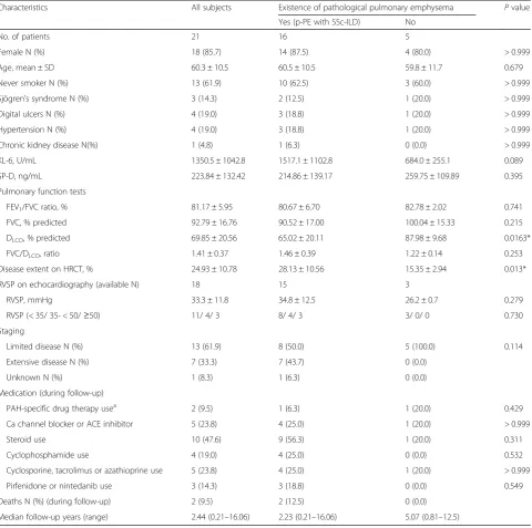
Clinical characteristics of the 21 patients with SSc-ILD are summarized in Table 1. The proportion of patients who had smoke was 38.1%, and 3 patients (14.3%) had Sjögren syndrome. The median follow-up period was 2.44 years, and only 2 patients (9.5%) died during this period.
preserved in the patients regardless of whether p-PE was observed. The radiological and pathological characteris-tics of the patients are summarized in Tables 2, 3. The major HRCT pattern was NSIP in 19 patients (90.5%). The frequency of diffuse distribution of reticulation was more common in the patients with p-PE with SSc-ILD than in those without p-PE (P= 0.009). The association of vasculopathy with SSc-ILD patients are summarized
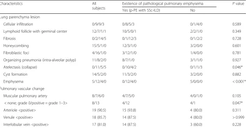
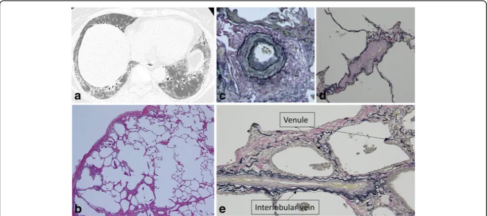
in Table4. LAA within the interstitial abnormalities was seen only in patients with p-PE with SSc-ILD (31.3%) (Figs.1a,2a, 3a,4a). Fibrotic NSIP was the major histo-logic pattern seen in the SSc-ILD patients (85.7%). Of the SSc-ILD patients with p-PE, 12 (75.0%) had grade 1 emphysema, and 4 (25.0%) had grade 2 emphysema (Figs.1b,2b,3b,4b). Notably, emphysematous change in the SSc-ILD patients seemed to occur in the fibrotic Table 1Patient characteristics at surgical lung biopsy between the existence and non-existence of pathological emphysema with SSc-associated ILD
Characteristics All subjects Existence of pathological pulmonary emphysema Pvalue
Yes (p-PE with SSc-ILD) No
No. of patients 21 16 5
Female N (%) 18 (85.7) 14 (87.5) 4 (80.0) > 0.999
Age, mean ± SD 60.3 ± 10.5 60.5 ± 10.5 59.8 ± 11.7 0.679
Never smoker N (%) 13 (61.9) 10 (62.5) 3 (60.0) > 0.999
Sjögren’s syndrome N (%) 3 (14.3) 2 (12.5) 1 (20.0) > 0.999
Digital ulcers N (%) 4 (19.0) 3 (18.8) 1 (20.0) > 0.999
Hypertension N (%) 4 (19.0) 3 (18.8) 1 (20.0) > 0.999
Chronic kidney disease N(%) 1 (4.8) 1 (6.3) 0 (0.0) > 0.999
KL-6, U/mL 1350.5 ± 1042.8 1517.1 ± 1102.8 684.0 ± 255.1 0.089
SP-D, ng/mL 223.84 ± 132.42 214.86 ± 139.17 259.75 ± 109.89 0.395
Pulmonary function tests
FEV1/FVC ratio, % 81.17 ± 5.95 80.67 ± 6.70 82.78 ± 2.02 0.741
FVC, % predicted 92.79 ± 16.76 90.52 ± 17.00 100.04 ± 15.33 0.215
DLCO, % predicted 69.85 ± 20.56 65.02 ± 20.11 87.98 ± 9.68 0.0163*
FVC/DLCO, ratio 1.41 ± 0.37 1.46 ± 0.39 1.22 ± 0.14 0.253
Disease extent on HRCT, % 24.93 ± 10.78 28.13 ± 10.56 15.35 ± 2.94 0.013*
RVSP on echocardiography (available N) 18 15 3
RVSP, mmHg 33.3 ± 11.8 34.8 ± 12.5 26.2 ± 0.7 0.279
RVSP (< 35/ 35- < 50/≥50) 11/ 4/ 3 8/ 4/ 3 3/ 0/ 0 0.730
Staging
Limited disease N (%) 13 (61.9) 8 (50.0) 5 (100.0) 0.114
Extensive disease N (%) 7 (33.3) 7 (43.7) 0 (0.0)
Unknown N (%) 1 (8.3) 1 (6.3) 0 (0.0)
Medication (during follow-up)
PAH-specific drug therapy usea 2 (9.5) 1 (6.3) 1 (20.0) 0.429
Ca channel blocker or ACE inhibitor 5 (23.8) 4 (25.0) 1 (20.0) > 0.999
Steroid use 10 (47.6) 9 (56.3) 1 (20.0) 0.311
Cyclophosphamide use 4 (19.0) 4 (25.0) 0 (0.0) 0.532
Cyclosporine, tacrolimus or azathioprine use 5 (23.8) 4 (25.0) 1 (20.0) > 0.999
Pirfenidone or nintedanib use 3 (14.3) 3 (18.8) 0 (0.0) 0.549
Deaths N (%) (during follow-up) 2 (9.5) 2 (12.5) 0 (0.0)
Median follow-up years (range) 2.44 (0.21–16.06) 2.23 (0.21–16.06) 5.07 (0.81–12.5)
Data are presented as mean ± SD, unless otherwise stated.SScsystemic sclerosis,ILDinterstitial lung disease,p-PEpathological pulmonary emphysema,KL-6Krebs von den Lungen-6,SP-Dsurfactant protein-D,FEV1forced expiratory volume in 1 s,FVCforced vital capacity,DLCOdiffusing capacity of the lung for carbon monoxide,
HRCThigh-resolution computed tomography,RVSPright ventricular systolic pressure,PAHpulmonary arterial hypertension.a
Table 2Comparison of HRCT findings in patients with SSc-associated ILD
Characteristics All
subjects
Existence of pathological pulmonary emphysema Pvalue
Yes (p-PE with SSc-ILD) No
HRCT pattern N (%)
Suggestive of UIP 2 (9.5) 2 (12.5) 0 (0.0) > 0.999
Suggestive or consistent with NSIP 19 (90.5) 14 (87.5) 5 (100.0)
HRCT findings N (%)
GGO 21 (100.0) 16 (100.0) 5 (100.0) N/A
Consolidation 2 (9.5) 1 (6.3) 1 (20.0) 0.429
Reticulation 21 (100.0) 16 (100.0) 5 (100.0) N/A
Honeycombing 1 (4.8) 1 (6.3) 0 (0.0) > 0.999
Traction bronchiectasis 20 (95.2) 16 (100.0) 4 (80.0) 0.238
Bronchial wall thickening 10 (47.6) 7 (43.8) 3 (60.0) 0.635
Micro-nodules 3 (14.3) 3 (18.7) 0 (0.0) 0.549
Emphysema (LAA around no fibrotic appearance) 4 (19.0) 3 (18.7) 1 (20.0) > 0.999
LAA within interstitial abnormalities 5 (23.8) 5 (31.3) 0 (0.0) 0.278
Cyst (non-honeycombing, emphysema) 5 (23.8) 4 (25.0) 1 (20.0) > 0.999
Mosaic attenuation (air trapping) 7 (33.3) 4 (25.0) 3 (60.0) 0.280
Enlarged mediastinal lymph node 4 (19.0) 3 (18.7) 1 (20.0) > 0.999
Pleural thickening or effusion 3 (14.3) 3 (18.7) 0 (0.0) 0.549
Pulmonary artery dilatation 6 (28.6) 5 (31.3) 1 (20.0) > 0.999
Volume loss 17 (81.0) 14 (87.5) 3 (60.0) 0.228
Data are presented as mean ± SD, unless otherwise stated.SScsystemic sclerosis,ILDinterstitial lung disease,p-PEpathological pulmonary emphysema,
HRCThigh-resolution computed tomography,UIPusual interstitial pneumonia,NSIPnonspecific interstitial pneumonia,GGOground glass opacity
Table 3Comparison of pathological findings (grade 0/1/2/3) in patients with SSc-associated ILD
Characteristics All
subjects
Existence of pathological pulmonary emphysema Pvalue
Yes (p-PE with SSc-ILD) No
Lung parenchyma lesion
Cellular infiltration 0/9/9/3 0/8/5/3 0/1/4/0 0.589
Lymphoid follicle with germinal center 12/7/1/1 10/5/0/1 2/2/1/0 0.349
Fibrosis 0/2/14/5 0/1/12/3 0/1/2/2 0.728
Honeycombing 15/5/1/0 12/3/1/0 3/2/0/0 0.601
Fibroblastic foci 4/16/1/0 3/12/1/0 1/4/0/0 0.781
Organizing pneumonia (intra-alveolar polyp) 11/8/2/0 8/7/1/0 3/1/1/0 0.927
Atelectasis (collapse) 0/11/5/5 0/10/4/2 0/1/1/3 0.046*
Cyst formation 14/5/2/0 11/3/2/0 3/2/0/0 0.882
Emphysema 5/12/4/0 0/12/4/0 5/0/0/0 < 0.001*
Pulmonary vascular change
Muscular pulmonary artery 8/7/6/0 4/7/5/0 4/0/1/0 0.105
< none; grade 0/positive = grade 1–3> 8/13 4/12 4/1 0.047*
Arteriole <positive> 19 (90.5) 15 (93.8) 4 (80.0) 0.311
Venule <positive> 18 (85.7) 14 (87.5) 4 (80.0) > 0.999
Interlobular vein <positive> 17 (81.0) 14 (87.5) 3 (60.0) 0.228
Table 4Characteristics of the 21 patients with SSc-associated ILD
Patient Sex HRCT findings Pathological findings Pulmonary vascular change
Disease extent, %
LAA within the interstitial abnormalities
Classification Emphysema, (grading score)
Muscular type, (grading score)
Arteriole Venule Interlobular vein
1 Female 41.37 + fNSIP 2 1 + + +
2 Female 38.24 None fNSIP 2 1 + + +
3 Female 42.37 + fNSIP 1 2 + + +
4 Female 17.54 + fNSIP 1 1 + + +
5 Female 13.65 None Unclassifiable 1 1 + + +
6 Female 33.2 None fNSIP 1 0 + + +
7 Female 13.47 None fNSIP 1 0 + + –
8 Male 25.77 + fNSIP 2 0 + – +
9 Female 18.86 None fNSIP 1 0 + + +
10 Male 29.37 None fNSIP 1 1 – + +
11 Female N/A None fNSIP 1 2 + + +
12 Female 42.31 + fNSIP 2 1 + – +
13 Female 16.7 None fNSIP 1 1 + + –
14 Female 23.1 None fNSIP 1 2 + + +
15 Female 36.22 None Unclassifiable 1 2 + + +
16 Female 29.78 None fNSIP 1 2 + + +
17 Female 19.58 None fNSIP 0 0 + + +
18 Female 12.03 None fNSIP 0 0 – + –
19 Male 13.3 None fNSIP 0 2 + – +
20 Female 15.27 None Unclassifiable 0 0 + + +
21 Female 16.55 None fNSIP 0 0 + + –
SScsystemic sclerosis,ILDinterstitial lung disease,HRCThigh-resolution computed tomography,LAAlow attenuation area,fNSIPfibrotic nonspecific interstitial pneumonia,N/Anot applicable
alveolar walls and mainly presented as the destruction of fibrous thickened alveolar walls (Fig.1c).
Although the severity of changes in muscular pulmon-ary arteries in the patients with p-PE with SSc-ILD was not significantly different from that in the patients without p-PE (P= 0.105), its incidence was significantly higher (P = 0.047) in the patients with p-PE. Histological analysis found that 61.9% of the muscular pulmonary arteries were of Heath-Edwards grade 1–2 (Figs. 1d, 2c, 4c). Heath-Edwards grading of muscular pulmonary arteries of more than grade 2 (e.g. plexiform lesions) was not detected in our subjects. The incidences of changes in other pulmon-ary microvasculature were as follows: 90.5% showing
arteriolar changes of intimal fibrosis and musculisation (Figs.1e,2d,3c,4d), 85.7% showing venular intimal thick-ening (Fig. 1f, 2e, 3d, 4e), and 81.0% showing intimal thickening of the interlobular veins (Fig.4e). Most of the patients with SSc-ILD had arteriolar and venous-venular changes in their pulmonary microvasculature whether pathological emphysema coexist or not.
Discussion
We investigated characteristic radiological and patho-logical features of 21 patients with SSc-ILD. This study presented that pulmonary emphysematous changes (LAA within interstitial abnormalities on HRCT and
Fig. 2HRCT and surgical lung biopsy of a 70-year-old never-smoking woman.aComputed tomography shows reticulation and a low attenuation area within a ground glass opacity and reticulation (circle).bPathologic specimen (H & E staining, × 40) shows fibrotic nonspecific interstitial pneumonia with emphysematous change (grade 1).cVascular intimal and medial thickening of a muscular pulmonary artery (grade 2) (Elastica van Gieson staining, × 100).dIntimal fibrosis of alveolar interstitial vessels (arteriole) ande) (venule) (Elastica van Gieson staining, × 200)
destruction of fibrously thickened alveolar walls) are spe-cific and novel radio-pathological features of SSc-ILD. We provided two important findings. First, among pa-tients with SSc-ILD, the complication of p-PE was fre-quently present in patients regardless of whether they had ever smoked. Surprisingly, 62.5% of the patients with p-PE with SSc-ILD were never-smokers. Historic-ally, the syndrome of combined pulmonary fibrosis and emphysema (CPFE) has been individualized within the spectrum of smoking-induced chronic lung disease [26]. Later, Cottin et al. reported that the syndrome of CPFE warrants recognition as a novel distinct pulmonary manifestation within the spectrum of lung diseases asso-ciated with CTD, especially in patients with rheumatoid arthritis and SSc, whether current- or ex-smokers; how-ever, 11.8% of their patients with CTD complicated by CPFE never smoked. [27] Antoniou et al. recently re-ported a prevalence of emphysema in patients with SSc-ILD of 12%; emphysema was present more often in current or former smokers, but it was also present in 7.5% of the never-smokers [10]. In both reports, al-though emphysema was analyzed as a radiological find-ing, the patients with CPFE had a lower DLCO, preserved FVC, which were also found in our study, and wider dis-ease extent. However, their definition of CPFE was based on the presence of both emphysema predominant in the upper lobes and pulmonary fibrosis predominant in the lower lung zones [10, 27]. Because the emphysema in our study was analyzed pathologically, it is unclear whether these disease entities were evaluated from the
same viewpoint in the previous studies. Our analysis em-phasizes that the emphysematous change occurring with SSc-ILD is different from the usual form of smoking-related emphysema, which presents as destructive holes in the center of the secondary lobules and is generally well demarcated from the adjacent parenchyma and which also centers around the respiratory bronchioles and alveolar ducts [28]. In contrast, the emphysematous change in SSc-ILD mainly presents as destruction of fibrously thick-ened alveolar walls, resulting in abnormal dilatation of the alveolar lumina and alveolar ducts. Obviously, as men-tioned above [23], “cicatricial emphysema” together with bronchiolectasis presents a characteristic gross appearance known as microscopic honeycombing that reflects end-stage fibrosis that is different from the emphysematous change in SSc-ILD. We believe that the presence of p-PE with SSc-ILD is more commonly seen in SSc patients than previously realized, even if they have never smoked, and that this is a new finding in SSc-ILD.
The second important finding was that LAA within interstitial abnormalities on HRCT might be a specific radiological finding in patients with SSc-ILD. Although LAAs are also shown on HRCT in a variety of fibrotic ILDs (in the extreme, honeycombing is composed of cys-tic spaces) [29], this finding refers to the fusing of discrete cysts with walls, whereas LAAs in our analysis were closely adjacent to interstitial abnormalities, and the LAAs themselves did not have an apparent wall thickness of more than 1 mm. This radiological finding was only seen in some of the patients with p-PE with
SSc-ILD, and therefore, it might correlate with the pathological emphysema of SSc-ILD, though all our hy-potheses were not corroborated.
In the vast majority of people, smoking is the cause of emphysema [30]. A previous radiological analysis showed that pulmonary microvascular blood flow is re-duced in mild chronic obstructive pulmonary disease (COPD) [31]. In the aspects of the pathological analysis, pulmonary vascular changes in COPD or CPFE are char-acterized by a thickening of the vessel wall that begins early in the process of emphysematous change [25]. Thickening of the intima is the first structural change and is followed by an increase in smooth muscle cells and infiltration of the vessel wall by inflammatory cells [32]. In contrast, pulmonary vascular abnormalities are common in SSc patients, and the pulmonary hyperten-sive vascular changes manifest as concentric intimal thickening by fibromyxoid tissue and medial hyper-trophy leading to thickened and stenotic pulmonary ar-terioles [33]. Most SSc patients with or without pulmonary artery hypertension (PAH) have intimal fi-brosis and medial hypertrophy of the pulmonary arteries with an external diameter between 101 and 200 μm, which lie beside bronchioles [34]. Overbeek et al. re-ported that PAH in SSc patients is characterized by in-timal fibrosis in the small vessels (arterioles, venules, and interlobular veins) [11]. Because changes were seen in 61.9% of the muscular pulmonary arteries in our pa-tients with SSc-ILD, and in 90.5% of the arterioles and 85.7% of the venules in the microvasculature, these find-ings could be caused by reduced pulmonary microvascu-lar blood flow, which might be one factor contributing to the emphysematous changes occurring in ILD.
Our study has several limitations. First, it was a single-institution, retrospective study with a small sample size. Second, there was selection bias because only the pa-tients who could undergo surgical lung biopsy were in-cluded. Additionally, only two patients received PAH-specific drug therapy during the follow-up period, and most patients could not be evaluated for PAH by right heart catheterization. Therefore, more severe cases might present different radiological and pathological findings. Third, although this study reveals characteristic radio-pathological features in SSc-ILD, a comparable analysis was not performed for other etiologies of ILD (e.g. CTD-ILD). Fourth, although previous research showed that increased matrix metalloproteinases expres-sion may play a role in accelerating the process of de-struction as emphysema [35], immunohistochemical analysis could not be performed in our analysis and would be an issue in the future. Fifth, we used several types of HRCT scanning with the times. Therefore, im-aging protocol may lead to a little different interpret-ation of radiological finding.
Conclusions
Emphysematous changes, i.e. LAA within the interstitial abnormalities on HRCT and destruction of the fibrously thickened alveolar walls, are specific and novel radio-pathological features of SSc-ILD. Historadio-pathological ana-lysis suggested that peripheral vasculopathy may partici-pate in the destruction of the fibrously thickened alveolar walls, resulting in the emphysematous changes seen in pa-tients with SSc-ILD. Our results might help to provide more information for patients who do not fulfil the diag-nosis of SSc (such as patients classified as having intersti-tial pneumonia with autoimmune features [36]) and suffer from a specific CTD diagnosis from the pulmonology viewpoint. Further studies are needed to clarify the quanti-tative loss of alveolar septa in the peripheral vascular bed that occurs with the emphysematous changes of SSc-ILD.
Abbreviations
COPD:Chronic obstructive pulmonary disease; CPFE: Combined pulmonary emphysema with fibrosis; CTD: Connective tissue disease; DLCO: Diffusing
capacity of the lung for carbon monoxide; FVC: Forced vital capacity; HRCT: High-resolution computed tomography; ILD: Interstitial lung disease; LAA: Low attenuation area; MCTD: Mixed connective tissue disease; NSIP: Nonspecific interstitial pneumonia; PAH: Pulmonary artery hypertension; PMBF: Pulmonary microvascular blood flow; p-PE: Pathological pulmonary emphysema; SSc: Systemic sclerosis
Acknowledgments
We offer our sincerest thanks to Yosuke Sasaki of Satista Co., Ltd. for his advice on statistical analysis. The authors would like to thank Rise Japan LLC for the professional English language review.
Funding Not applicable.
Availability of data and materials
The data will not be shared with participant confidentiality.
Authors’contributions
HY was the primary author of the manuscript, had full access to all of the data in the study, and takes responsibility for the integrity of the data and the accuracy of the data analysis. TT, AS, HK, TB, and TO contributed to the study design; YY, SaI, AS, HK, TB, and TO contributed to collecting samples and carrying out experiments; AS, TI, and KK contributed to data analysis; TT, SaI, ShI, KO, and TO contributed to data interpretation; TT, TI, KO, and TO contributed to writing the manuscript. All authors read and approved the final version of the manuscript.
Ethics approval and consent to participate
This retrospective cohort study was approved by the institutional review board of Kanagawa Cardiovascular and Respiratory Center (no. 28–11). The patients’approval or informed consent was not required for a retrospective review of their records, pursuant to the ethical guidelines of the Japanese Ministry of Health, Labor, and Welfare.; however, the present retrospective study was carried out by the opt-out method of our hospital website.
Consent for publication Not applicable.
Competing interests
The authors declare that they have no competing interests.
Publisher’s Note
Author details
1Department of Respiratory Medicine, Kanagawa Cardiovascular and
Respiratory Center, 6-16-1 Tomioka-higashi, Kanazawa-ku, Yokohama 236-0051, Japan.2Department of Pathology, Japanese Red Cross Medical Center, Tokyo, Japan.3Department of Radiology, Kanagawa Cardiovascular
and Respiratory Center, Yokohama, Japan.4Department of Radiology,
Yokohama Rousai Hospital for Labour Welfare Corporation, Yokohama, Japan.
5
Department of Pathobiology, Yokohama City University Graduate School of Medicine, Yokohama, Japan.6Department of Respiratory Medicine, Tokyo
Jikei University Hospital, Tokyo, Japan.
Received: 15 September 2017 Accepted: 22 January 2018
References
1. Bussone G, Mouthon L. Interstitial lung disease in systemic sclerosis. Autoimmun Rev. 2011;10:248–55.
2. Goldin JG, Lynch DA, Strollo DC, Suh RD, Schraufnagel DE, Clements PJ, et al. High-resolution CT scan findings in patients with symptomatic scleroderma-related interstitial lung disease. Chest. 2008;134:358–67. 3. Desai SR, Veeraraghavan S, Hansell DM, Nikolakopolou A, Goh NS, Nicholson
AG, et al. CT features of lung disease in patients with systemic sclerosis: comparison with idiopathic pulmonary fibrosis and nonspecific interstitial pneumonia. Radiology. 2004;232:560–7.
4. Fujita J, Yoshinouchi T, Ohtsuki Y, Tokuda M, Yang Y, Yamadori I, et al. Non-specific interstitial pneumonia as pulmonary involvement of systemic sclerosis. Ann Rheum Dis. 2001;60:281–3.
5. Kim DS, Yoo B, Lee JS, Kim EK, Lim CM, lee SD, et al. the major histopathologic pattern of pulmonary fibrosis in scleroderma is nonspecific interstitial pneumonia. Sarcoidosis Vasc Diffuse Lung Dis. 2002;19:121–7.
6. Bouros D, Wells AU, Nicholson AG, Colby TV, Polychronopoulos V, Pantelidis P, et al. Histopathologic subsets of fibrosing alveolitis in patients with systemic sclerosis and their relationship to outcome. Am J Respir Crit Care Med. 2002;165:1581–6.
7. Katzenstein AL, Fiorelli RF. Nonspecific interstitial pneumonia/fibrosis. Histologic features and clinical significance Am J Surg Pathol. 1994;18:136–47.
8. Nicholson AG, Colby TV, wells AU. Histopathological approach to patterns of interstitial pneumonia in patient with connective tissue disorders. Sarcoidosis Vasc Diffuse Lung Dis. 2002;19:10–7.
9. Yamakawa H, Hagiwara E, Kitamura H, Yamanaka Y, Ikeda S, Sekine A, et al. Clinical features of idiopathic interstitial pneumonia with systemic sclerosis-related autoantibody in comparison with interstitial pneumonia with systemic sclerosis. PLoS One. 2016;11:e0161908.
10. Antoniou KM, Margaritopoulos GA, Goh NS, Karagiannis K, Desai SR, Nicholson AG, et al. Combined pulmonary fibrosis and emphysema in scleroderma-related lung disease has a major confounding effect on lung physiology and screening for pulmonary hypertension. Arthritis Rheumatol. 2016;68:1004–12.
11. Overbeek MJ, Vonk MC, Boonstra a, Voskuyl AE, Vonk-Noordegraaf a, smit EF, et al. pulmonary arterial hypertension in limited cutaneous systemic sclerosis: a distinctive vasculopathy. Eur Respir J. 2009;34:371–9.
12. van den Hoogen F, Khanna D, Fransen J, Johnson SR, Baron M, Tyndall A, et al. 2013 classification criteria for systemic sclerosis: an American College of Rheumatology/European league against rheumatism collaborative initiative. Arthritis Rheum. 2013;65:2737–47.
13. Lopez-Giacoman S, Madero M. Biomarkers in chronic kidney disease, from kidney function to kidney damage. World J Nephrol. 2015;4:57–73. 14. American Thoracic Society. European Respiratory Society. American Thoracic
Society/European Respiratory Society international multidisciplinary consensus classification of the idiopathic interstitial pneumonias. This joint statement of the American Thoracic Society (ATS), and the European Respiratory Society (ERS) was adopted by the ATS board of directors, June 2001 and by the ERS executive committee, June 2001. Am J Respir Crit Care Med. 2002;166:426. 15. Travis WD, Costabel U, Hansell DM, King TE Jr, Lynch DA, Nicholson AG, et
al. ATS/ERS Committee on idiopathic interstitial pneumonias. An official American Thoracic Society/European Respiratory Society statement: update of the international multidisciplinary classification of the idiopathic interstitial pneumonias. Am J Respir Crit Care Med. 2013;188:733–48. 16. Hansell DM, Bankier AA, MacMahon H, McLoud TC, Müller NL, Remy J.
Fleischner society: glossary of terms for thoracic imaging. Radiology. 2008; 246:697–722.
17. Iwasawa T, Kato S, Ogura T, Kusakawa Y, Iso S, Baba T, et al. Low-normal lung volume correlates with pulmonary hypertension in fibrotic idiopathic interstitial pneumonia: computer-aided 3D quantitative analysis of chest CT. AJR Am J Roentgenol. 2014;203:W166–73.
18. Goh NS, Desai SR, Veeraraghavan S, Hansell DM, Copley SJ, Maher TM, et al. Interstitial lung disease in systemic sclerosis: a simple staging system. Am J Respir Crit Care Med. 2008;177:1248–54.
19. Song JW, Do KH, Kim MY, Jang SJ, Colby TV, Kim DS. Pathologic and radiologic differences between idiopathic and collagen vascular disease-related usual interstitial pneumonia. Chest. 2009;136:23–30.
20. Enomoto Y, Takemura T, Hagiwara E, Iwasawa T, Okudela K, Yanagawa N, et al. Features of usual interstitial pneumonia in patients with primary Sjögren’s syndrome compared with idiopathic pulmonary fibrosis. Respir Investig. 2014;52:227–35.
21. Flaherty KR, Colby TV, Travis WD, Toews GB, Mumford J, Murray S, et al. Fibroblastic foci in usual interstitial pneumonia: idiopathic versus collagen vascular disease. Am J Respir Crit Care Med. 2003;167:1410–5.
22. Wright JL. The importance of ultramicroscopic emphysema in cigarette smoke-induced lung disease. Lung. 2001;179:71–81.
23. Corrin, B, Nicholson AG: Pathology of the lungs. 3rd ed. Philadelphia, Elsevier, 2011,pp 91–134.
24. Heath D, Edwards JE. The pathology of hypertensive pulmonary vascular disease; a description of six grades of structural changes in the pulmonary arteries with special reference to congenital cardiac septal defects. Circulation. 1958;18:533–47.
25. Awano N, Inomata M, Ikushima S, Yamada D, Hotta M, Tsukuda S, et al. Histological analysis of vasculopathy associated with pulmonary hypertension in combined pulmonary fibrosis and emphysema: comparison with idiopathic pulmonary fibrosis and emphysema alone. Histopathology. 2017;70:896–905. 26. Cottin V, Nunes H, Brillet PY, Delaval P, Devouassoux G, Tillie-Leblond I, et al.
Groupe d'Etude et de Recherche sur les maladies Orphelines Pulmonaires (GERM O P): combined pulmonary fibrosis and emphysema: a distinct underrecognised entity. Eur Respir J. 2005;26:586–93.
27. Cottin V, Nunes H, Mouthon L, Gamondes D, Lazor R, Hachulla E, et al. Groupe d'Etudes et de Recherche sur les maladies "Orphelines" Pulmonaires: combined pulmonary fibrosis and emphysema syndrome in connective tissue disease. Arthritis Rheum. 2011;63:295–304.
28. Wright JL, Tazelaar HD, Churg A. Fibrosis with emphysema. Histopathology. 2011;58:517–24.
29. Raoof S, Bondalapati P, Vydyula R, Ryu JH, Gupta N, Raoof S, et al. cystic lung diseases: algorithmic approach. Chest. 2016;150:945–65.
30. Wilson JS, Galvin JR. Normal diffusing capacity in patients with PiZ alpha(1)-antitrypsin deficiency, severe airflow obstruction, and significant radiographic emphysema. Chest. 2000;118:867–71.
31. Hueper K, Vogel-Claussen J, Parikh MA, Austin JH, Bluemke DA, Carr J, et al. pulmonary microvascular blood flow in mild chronic obstructive pulmonary disease and emphysema. The MESA COPD study. Am J Respir Crit Care Med. 2015;192:570–80.
32. Seimetz M, Parajuli N, Pichl A, Veit F, Kwapiszewska G, Weisel FC, et al. Inducible NOS inhibition reverses tobacco-smoke-induced emphysema and pulmonary hypertension in mice. Cell. 2011;147:293–305.
33. Vivero M, Padera RF. Histopathology of lung disease in the connective tissue diseases. Rheum Dis Clin N Am. 2015;41:197–211.
34. Yousem SA. The pulmonary pathologic manifestations of the CREST syndrome. Hum Pathol. 1990;21:467–74.
35. Cardoso WV, Sekhon HS, Hyde DM, Thurlbeck WM. Collagen and elastin in human pulmonary emphysema. Am Rev Respir Dis. 1993;147:975–81. 36. Fischer A, Antoniou KM, Brown KK, Cadranel J, Corte TJ, du bois RM, et al.