© 2019 by the Serbian Biological Society How to cite this article: Nikolić B, Vasilijević B, Ćirić A, Mitić-Ćulafić D, Cvetković S, 235 Džamić A, Knežević-Vukčević J. Bioactivity of Juniperus communis essential oil and post-distillation waste: Assessment of selective toxicity against food contaminants. Arch Biol Sci. 2019;71(2):235-44.
Bioactivity of
Juniperus communis
essential oil and post-distillation waste: assessment of
selective toxicity against food contaminants
Biljana Nikolić1,*, Bojana Vasilijević1, Ana Ćirić2, Dragana Mitić-Ćulafić1, Stefana Cvetković1, Ana Džamić1 and Jelena Knežević-Vukčević1
1Faculty of Biology, University of Belgrade, Studentski trg 16, 11000 Belgrade, Serbia
2Institute for Biological Research “Siniša Stanković”, University of Belgrade, Bulevar Despota Stefana 142, 11000 Belgrade,
Serbia
*Corresponding author: [email protected]
Received: December 17, 2018; Revised: January 31, 2019; Accepted: February 1, 2019; Published online: February 21, 2019
Abstract: Previously chemically characterized Juniperus communis essential oil (EO) and post-distillation waste (PDW) were tested for cytotoxicity and antimicrobial activity against food contaminants. Microdilution assay showed that PDW induced moderate antifungal (minimum inhibitory concentration (MIC) and minimum fungicidal concentration (MFC)
values, ranging between 0.118-0.900 mg mL-1), and an antibacterial effect against Listeria monocytogenes (MIC and
mini-mum bactericidal concentration (MBC) were 0.39 and 0.74 mg mL-1, respectively). Combinations of EO/PDW with selected
antibiotics induced synergistic antilisterial activity in the checkerboard assay. The MTT assay determined that cytotoxicity
against colon cancer cells was high for the EO but negligible for PDW (IC50 values were 0.087-0.106 and 1.450-6.840 mg
mL-1, respectively). The selectivity indices indicated high selectivity of PDW against tested fungi and L. monocytogenes. In
the adhesion-inhibition assay, PDW reduced in vitro adhesion of L. monocytogenes to colon cells (29-62% of inhibition).
In conclusion, PDW exhibited an antimicrobial effect against important food spoilage and poisoning fungi and L.
monocy-togenes, and also reduced in vitro adhesion of L. monocytogenes to colon cells. The results indicate that J. communis PDW could be considered as natural preservative against food spoilage and poisonous fungi, and as an adjuvant to conventional therapy of listeriosis.
Keywords:Juniperus communis; essential oil; post-distillation waste; selective antimicrobial effect; adhesion-inhibition properties
INTRODUCTION
The search for new antimicrobial agents has become a necessity because growing resistance to existing an-tibiotics is posing a serious problem to global public health [1]. Similarly, the increased use of antifungal agents has resulted in the rapid development of fungal resistance and made it a major clinical problem, due to the limited arsenal of available systemic antifungal agents [2].
Plants are considered as an extremely important source of antimicrobials and many species used in traditional medicine are currently under investigation [3]. Junipers (Juniperus spp.), which contain numer-ous active compounds, are among the most
wide-spread species in the Northern Hemisphere. The use of these plants, especially their seed cones (‘berries’), in folk medicine and manufacturing is extensive [4]. In traditional medicine they are used as diuretics, appetizers, carminatives, stomachics, anticonvul-sants and antihypertensive agents, as well as in the treatment of headaches, fever, bronchitis, asthma and some gynecological disorders [5,6]. Literature data also indicate remarkable antioxidant, antimicrobial and hypoglycemic activities of these berries [7-10]. Furthermore, juniper extracts have found wide ap-plications in pharmaceutical industry, perfumery and aromatherapy [11].
extracts are used as a flavoring agent in food and alco-holic beverage industries. The most famous alcoalco-holic beverage containing juniper is gin but locally manufac-tured juniper brandies are also very popular [13,14]. In addition, juniper berries are used in European and particularly in Scandinavian cuisine. They are used in meat preparation, especially wild birds and game, and to flavor dishes prepared with pork, cabbage and sauerkraut [15,16].
In recent years, interest in plant-derived food additives is in great expansion, since they possess different health-promoting properties [17]. Further-more, particular adverse effects such as immunologic hypersensitivity have been reported for some synthetic food additives [18], and this has additionally stimulated the search for natural replacements. Among natural compounds that could be used as alternatives to anti-microbial synthetic food additives, essential oils (EOs) are considered as effective candidates [19]. In addition, various plant waste materials, including post-distillation waste (PDW), are receiving increased attention since they contain numerous bioactive compounds available for further extraction [20].
The aim of this work was to study the antimi-crobial effect of J. communis EO and PDW against selected food-borne pathogenic and spoilage bacteria, as well as food-poisoning and pathogenic fungi. The antibacterial effect was determined for EO and PDW alone or in combination with common antibiotics. To estimate the selective toxicity to microbial strains, the cytotoxic potential against colon cancer cells was also determined and selectivity indices were calculated. Finally, the inhibitory potential of PDW against bac-terial adhesion to colon cells was examined in vitro using the most sensitive bacterial strain.
MATERIALS AND METHODS
Plant material, EO and PDW preparation
Plant material (seed cones of Juniperus communis L. var. saxatilis Pall.) was collected on Mt. Stara Planina, Serbia. The voucher specimen (No. 16693) was pre-pared, identified by Nemanja Rajčević (PhD in botany), and deposited at the Herbarium of the University of Belgrade, Faculty of Biology, Institute of Botany and
Botanical Garden “Jevremovac” (BEOU Herbarium). Air-dried and finely ground seed cones were submitted to hydrodistillation in a Clevenger-type apparatus, as previously described [21]. The EO was dissolved in dimethyl sulfoxide (DMSO) for all performed bioas-says. After distillation of the EO, the residual aqueous solution was evaporated in vacuum at 45°C, resulting in a dry PDW extract. Distilled water (dH2O) was used as a solvent for the PDW extract.
Bacterial and fungal strains and human cell cultures
The antimicrobial effect was determined against: (i) Gram-positive bacteria Staphylococcus aureus (ATCC 25923), methicillin-resistant S. aureus MRSA (ATCC43300), Enterococcus faecalis (ATCC 29212), Lis-teria monocytogenes (ATCC 19111), (ii) Gram-negative bacteria Escherichia coli (ATCC 8739), Shigella flexneri (ATCC 9199), Salmonella enteritidis (ATCC 13076), Pseudomonas aeruginosa (ATCC 15442) and (iii) fungi Aspergillus fumigatus (human isolate), Aspergillus ver-sicolor (ATCC 11730), Aspergillus ochraceus (ATCC 12066), Aspergillus niger (ATCC 6275), Trichoderma viride (IAM 5061), Penicillium funiculosum (ATCC 36839), Penicillium ochrochloron (ATCC 9112), and Penicillium verrucosum var. cyclopium (food isolate). The human cell lines used in cytotoxicity and adhesion-inhibition assays were colorectal carcinoma cells HT-29 (ATCC HTB-38) and HCT116 (ATCC CCL-247).
Chemicals, media and growth conditions
Ampicillin sodium salt (Amp, Cas No. 69-52-3, Sigma-Aldrich, St. Louis, USA), streptomycin sulfate salt (Str, Cas No. 3810-74-0, Sigma-Aldrich) and azithromycin dihydrate (Azm, Cas No. 117772-70-0, Sigma-Aldrich, St. Louis, USA) were used in antibacterial microdilution and checkerboard assays. Stock solutions of antibiotics were 1 mg mL-1, prepared in sterile dH
Louis, USA, stock solution 0,675 mg mL-1 in sterile dH2O) was used as a growth indicator in antibacterial microdilution and checkerboard assays.
Bacteria were cultivated at 37°C in brain hart infusion broth (BHI, LAB M, Lancashire, UK) and brain heart agar (BHA) for L. monocytogenes and E. faecalis, or in Müller-Hinton broth (MHB, Himedia, Mumbai, India) and Müller-Hinton Agar (MHA) for S. aureus, MRSA, E. coli, S. flexneri, S. enteritidis and P. aeruginosa. Fungal strains were cultivated at 28°C in malt broth (MB, Institute of Immunology and Vi-rology, Torlak, Belgrade, Serbia) and malt agar (MA). All solid media (BHA, MHA and MA) contained 1.5% (w/w) agar (LAB M, Lancashire, UK).
The human cells (HT-29 and HCT116) were grown in Dulbecco’s modified Eagle’s medium (DMEM) with 4.5% glucose and 2 mM L-glutamine, supplemented with 10% fetal bovine serum (FBS) and a penicillin/ streptomycin cocktail. Cells were maintained in an incubator at 37ºC with 5.0% CO2 in a humidified atmo-sphere. The cells growing attached to the surface were subcultured at 90% confluence twice a week. Single cell suspensions for subculturing and for experiments were obtained using 0.1% trypsin (from porcine pancreas). Cell viability in suspensions was inspected by the trypan blue dye exclusion method. In the cytotoxicity assay, 3-(4,5-dimethylthiazol-2-yl)-2,5-diphenyltetrazolium bromide (MTT, Cas. No 298-93-1), 5-fluorouracil (5-FU, Cas No. 51-21-8) and phosphate buffered saline (PBS) were used as indicators of cell viability, positive control and to wash cells, respectively. All media and reagents used to grow and manipulate human cells were purchased from Sigma-Aldrich, St. Louis, USA.
Microdilution assay
The antimicrobial properties of the EO and PDW were determined in microdilution assay performed in 96-well microtiter plates. The serial two-fold dilu-tions of test substances were made in corresponding medium (BHI for L. monocytogenes and E. faecalis, MHB for other bacteria, and MB for fungi). Suspen-sions of indicator strains were adjusted to 106 CFU mL-1 by preparing an exponential bacterial culture or by washing fungal spores with sterile 0.85% saline containing 0.1% Tween 80 (v/v) from the surface of MA
plates. EO was tested in concentration range 0.39-50 mg mL-1 for both bacteria and fungi, while PDW was tested in the ranges 0.195-25 mg mL-1 and 7-900 µg mL-1 for bacteria and fungi, respectively. Assays were performed in triplicate in three individual experiments.
A slightly modified resazurin-incorporated mi-crodilution assay, performed as previously described [22], was used to evaluate the antibacterial properties. Briefly, test substances were serially two-fold diluted along the columns. Bacterial inoculum (105 CFU mL-1) was added to each well except the sterility control. The growth indicator resazurin (final concentration 0.067 mg mL-1) was added to the wells and after 24 h of incubation at 37°C, MICs were determined as the lowest concentrations that did not induce color change. After plating by inoculation loop from each well with-out visible growth on solid media (BHA/MHA) and incubation (24 h at 37°C), MBCs were determined. As positive controls, conventional antibiotics Str, Amp and Azm were applied in a concentration range of 0.78-100 µg mL-1. Sterilized solvent (5% DMSO and dH2O for EO and PDW, respectively) was used as a negative control.
The microdilution assay, performed as previously described [23], was used to evaluate the antifungal potential. Briefly, the fungal spore suspensions (1.0 × 10⁵ CFU mL-1) were added to each well containing graded concentrations of test substances. After 72 h of incubation at 28°C, MICs were determined with a binocular microscope as the lowest concentrations without visible growth in seeded wells. The MFCs were determined by the same procedure after serial subcultivation from each well without visible growth into microtiter plates. Standard fungicides Bfz and Kcz, both applied in the concentration range 4-512 µg mL-1, served as positive controls, while a solvent (sterile 0.85% saline containing 0.1% Tween 80 and dH2O for EO and PDW, respectively) was used as a negative control.
Checkerboard assay
serially two-fold diluted along the vertical, while the second one was serially two-fold diluted along the horizontal line of 96-well microtiter plates. In combina-tions prepared with PDW, the concentracombina-tions ranged between (1/32)×MIC-4×MIC, while in combinations prepared with EO the concentrations ranged between (1/64)×MIC-2×MIC. The MIC values of combina-tions were determined by adding resazurin (final concentration 0.067 mg mL-1) and inspecting for color changes. Combinations that did not induce color change of resazurin were used for the calculation of the fractional inhibitory concentration index (FICI) for two antimicrobials in combination. The FICI was calculated according to equation (1), where substance A was EO or PDW, and substance B was the antibiotic (Str, Amp, Azm).
(1)
FICI was used to distinguish between the mode of inter-actions as follows: FICI≤0.5 – synergistic; 0.5<FICI≤1 – additive; 1<FICI≤4 – indifferent; FICI<4 – antagonistic effect [24]. The checkerboard assay was performed in triplicate in two individual experiments.
Cytotoxicity assay
Cytotoxicity was determined by the MTT reduction assay, performed as previously described [23]. Briefly, the assay was performed on HT-29 and HCT116 cells inoculated in 96-well plates at a density of 5x10⁴ cells/ well and incubated until they formed a monolayer; EO and PDW were serially two-fold diluted in tested con-centration ranges (0.016-0.500 and 0.313-20 mg mL-1, respectively). After 24 h of incubation, the medium was removed and replaced with the MTT solution (final concentration 0.5 mg mL-1 in DMEM); the plates were additionally incubated for 3 h to allow for mitochon-drial reduction of MTT into formazan, performed in viable cells. After this step, the medium was carefully removed and the formazan crystals were dissolved in DMSO. Cell viability was determined by measuring the absorbance at 570 nm, using a microplate reading spectrophotometer (Multiskan FC, Thermo Fisher Scientific, Shanghai, China). The cytotoxic activity was evaluated by comparing the absorbance of the wells containing the test substances to that containing the
vehicle (DMSO or dH2O for EO and PDW, respectively). 5-fluorouracil (5-FU) was used as a positive control. For each test substance, two independent experiments with six wells per treatment point were performed.
Selectivity index
In order to estimate the selective toxicity of test sub-stances, a relationship between cytotoxic and antimi-crobial effects was determined through the selectivity index (SI). The SI was calculated as previously described [25] using the following equation (2):
SI = log IC50/MIC (2)
Positive values of SI indicate higher toxicity to bacteria or fungi, while negative values indicate higher toxicity to colon cells.
In vitro adhesion-inhibition assay
percentage of adhering bacteria in the medium with and without the test substance. For each cell line, two independent experiments in triplicate were performed.
Statistical analysis
Experimental data were analyzed by Student’s t-Test. The level of statistical significance was defined as p<0.05.
RESULTS
Antimicrobial properties
The antimicrobial potential of J. communis EO and PDW was determined against selected bacterial and fungal strains. While the antifungal effect of EO was
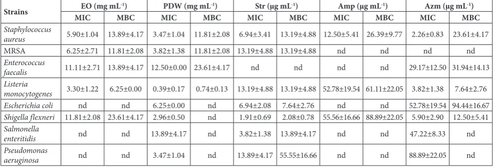
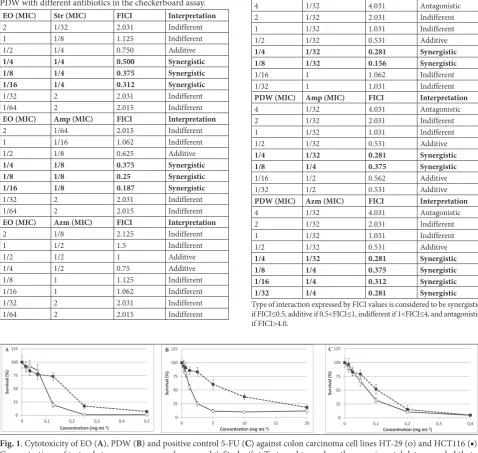
negligible, PDW demonstrated remarkable activity, with MICs and MFCs ranging between 119-900 µg mL-1 and 250-900 µg mL-1, respectively (Table 1). The highest activity was recorded against A. versicolor. On the other hand, the antibacterial effect of both EO and PDW was weak, with MICs mainly above 3 mg mL-1 (Table 2). The most sensitive bacterium was L. monocytogenes with MIC and MBC values of 3.30 mg mL-1 and 6.25 mg mL-1 for EO, respectively, and 0.39 mg mL-1 and 0.74 mg mL-1 for PDW, respectively. The effect of EO and PDW against L. monocy-togenes was further monitored in the checkerboard assay, where they were combined with conventional antibiotics Str, Amp and Azm. The results showed that certain combinations of EO with Str and Amp, as well as of PDW with all three tested antibiotics, induced a synergistic antimicrobial effect (Table 3).
Table 1. Antifungal activity of Juniperus communis EO, PDW and conventional fungicides.
Strains EO (mg mL-1) PDW (µg mL-1) Bfz (µg mL-1) Kcz (µg mL-1)
MIC MFC MIC MFC MIC MFC MIC MFC
Aspergillus fumigatus 11.11±2.76 nd 425.00±75.00 900.00±0.00 156.44±56.44 213.33±64.00 8.89±2.67 17.78±5.33
Aspergillus versicolor 11.81±2.08 nd 119.13±47.05 250.00±75.00 106.67±32.00 199.11±67.46 4.00±0.00 7.11±1.76
Aspergillus ochraceus 12.50±0.00 nd 400.00±99.22 850.00±150.00 142.22±42.67 184.89±67.46 9.78±3.53 19.56±7.06
Aspergillus niger 13.89±4.17 nd nd nd 156.44±56.44 199.11±67.46 8.00±0.00 19.56±7.06
Trichoderma viride 11.81±2.08 nd 225.00±0.00 550.00±198.43 142.22±42.67 213.33±64.00 19.56±7,06 39.11±14.11
Penicillium ochrochloron 11.81±2.08 nd 250.00±75.00 450.00±0.00 199.11±67.46 241.78±42.67 4.44±1.33 9.78±3.53
Penicillium funiculosum 12.50±0.00 nd 425.00±75.00 900.00±0.00 213.33±64.00 241.78±42.67 4.44±1.33 15.11±2.67
Penicillium verrucosum var.
cyclopium 11.11±2.76 nd 900.00±0.00 nd 184.89±67.46 284.47±85.33 5.78±2.11 17.78±5.33
nd – not determined in the applied concentration range
Table 2. Antibacterial activity of Juniperus communis EO, PDW and conventional antibiotics.
Strains MICEO (mg mLMBC-1) MICPDW (mg mLMBC-1) MICStr (µg mLMBC-1) MICAmp (µg mLMBC-1) MICAzm (µg mLMBC-1)
Staphylococcus
aureus 5.90±1.04 13.89±4.17 3.47±1.04 11.81±2.08 6.94±3.41 13.19±4.88 12.50±5.41 26.39±9.77 2.26±0.83 23.61±4.17
MRSA 6.25±2.71 11.81±2.08 3.82±1.38 11.81±2.08 13.19±4.88 13.19±4.88 nd nd nd nd
Enterococcus
faecalis 11.11±2.71 13.89±4.17 12.50±0.00 23.61±4.17 nd nd nd nd 29.17±12.50 31.94±14.13
Listeria
monocytogenes 3.30±1.22 6.25±0.00 0.39±0.17 0.74±0.13 13.19±4.88 13.19±4.88 52.78±19.54 61.11±22.05 3.82±1.38 7.64±2.76
Escherichia coli nd nd 6.25±0.00 nd 6.94±2.08 7.64±2.76 nd nd 52.78±19.54 94.44±16.67
Shigella flexneri 11.81±2.08 23.61±4.17 2.96±0.50 nd 1.91±0.69 2.08±0.78 55.56±16.66 88.89±22.05 5.90±2.90 12.50±5.41
Salmonella
enteritidis nd nd 13.89±4.17 nd 3.82±1.38 13.89±4.17 nd nd 47.22±8.33 nd
Pseudomonas
aeruginosa nd nd 3.47±1.04 nd 13.89±4.17 55.55±16.66 nd nd 88.89±22.05 nd
Cytotoxic potential
Evaluation of cytotoxicity, performed on colon carci-noma HT-29 and HCT116 cells, revealed that both EO and PDW possess a cytotoxic potential (Fig. 1). The estimated IC50 values (concentrations that decreased cell viability to 50%) pointed at a significantly higher cytotoxicity of EO, comparable to the common
cy-tostatic 5-FU (Table 4). PDW induced much lower cytotoxicity, with IC50 values approximately 20- and 60-fold higher than those of 5-FU in HT-29 and HCT116 cells, respectively. The results also showed higher sensitivity of HT-29 cells to all test substances, with the most pronounced discrepancy between cell lines determined for the effect of PDW.
Table 3. Antilisterial effects of combinations of J. communis EO/ PDW with different antibiotics in the checkerboard assay.
EO (MIC) Str (MIC) FICI Interpretation
2 1/32 2.031 Indifferent
1 1/8 1.125 Indifferent
1/2 1/4 0.750 Additive
1/4 1/4 0.500 Synergistic 1/8 1/4 0.375 Synergistic 1/16 1/4 0.312 Synergistic
1/32 2 2.031 Indifferent
1/64 2 2.015 Indifferent
EO (MIC) Amp (MIC) FICI Interpretation
2 1/64 2.015 Indifferent
1 1/16 1.062 Indifferent
1/2 1/8 0.625 Additive
1/4 1/8 0.375 Synergistic 1/8 1/8 0.25 Synergistic 1/16 1/8 0.187 Synergistic
1/32 2 2.031 Indifferent
1/64 2 2.015 Indifferent
EO (MIC) Azm (MIC) FICI Interpretation
2 1/8 2.125 Indifferent
1 1/2 1.5 Indifferent
1/2 1/2 1 Additive
1/4 1/2 0.75 Additive
1/8 1 1.125 Indifferent
1/16 1 1.062 Indifferent
1/32 2 2.031 Indifferent
1/64 2 2.015 Indifferent
PDW (MIC) Str (MIC) FICI Interpretation
4 1/32 4.031 Antagonistic
2 1/32 2.031 Indifferent
1 1/32 1.031 Indifferent
1/2 1/32 0.531 Additive
1/4 1/32 0.281 Synergistic 1/8 1/32 0.156 Synergistic
1/16 1 1.062 Indifferent
1/32 1 1.031 Indifferent
PDW (MIC) Amp (MIC) FICI Interpretation
4 1/32 4.031 Antagonistic
2 1/32 2.031 Indifferent
1 1/32 1.031 Indifferent
1/2 1/32 0.531 Additive
1/4 1/32 0.281 Synergistic 1/8 1/4 0.375 Synergistic
1/16 1/2 0.562 Additive
1/32 1/2 0.531 Additive
PDW (MIC) Azm (MIC) FICI Interpretation
4 1/32 4.031 Antagonistic
2 1/32 2.031 Indifferent
1 1/32 1.031 Indifferent
1/2 1/32 0.531 Additive
1/4 1/32 0.281 Synergistic 1/8 1/4 0.375 Synergistic 1/16 1/4 0.312 Synergistic 1/32 1/4 0.281 Synergistic
Type of interaction expressed by FICI values is considered to be synergistic if FICI≤0.5, additive if 0.5<FICI≤1, indifferent if 1<FICI≤4, and antagonistic if FICI>4.0.
Fig. 1. Cytotoxicity of EO (A), PDW (B) and positive control 5-FU (C) against colon carcinoma cell lines HT-29 (O) and HCT116 (■). Concentrations of test-substances are expressed as mg mL-1. Student’s t-Test used to analyze the experimental data, revealed that a
statistically significant difference (p<0.05) corresponding to the control was observed for the following concentrations: A – for EO at 0.125 and 0.0625 mg mL-1 and all lower ones for HT-29 and HCT116 cells, respectively; B – for PDW at 0.625 and 1.25 mg mL-1 and all
lower ones for HT-29 and HCT116 cells, respectively; (C) for 5-FU at 0.05 and 0.025 mg mL-1 and all lower ones for HT-29 and HCT116
Selective toxicity
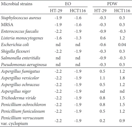
Calculation of the SI indicated the absence of selective toxicity of EO to all tested microorganisms, while SI values obtained for PDW were variable. They were almost all positive in respect to HCT116 cells (in-dicating selectivity against microorganisms), but in respect to HT-29, positive values were determined for L. monocytogenes and almost all fungal strains (Table 5).
Anti-adhesive properties
Taking into account that SI values for PDW indicated higher toxicity to L. monocytogenes than to both hu-man colon cells, we performed the in vitro adhesion-inhibition assay and monitored the potential of PDW to reduce the ability of L. monocytogenes to adhere to the colon cells. The obtained results indicated that PDW significantly decreased adhesion of L. mono-cytogenes to both HT-29 and HCT116 cells (Table 6). The inhibitory potential was more prominent in the case of HCT116 cells: 62% inhibition vs 29% inhibi-tion in HT-29 cells.
DISCUSSION
Previous studies indicated that plants from Juniperus genus possess antibacterial and antifungal potential [10,26,27]. Although the antimicrobial potential of J. communis extracts has been previously reported, this work specifically monitored their effect against food-borne pathogenic, spoilage and poisoning mi-croorganisms and additionally estimated their selective toxicity. The EO and PDW used in this work have been chemically characterized previously [21]. The main constituents determined in EO were α-pinene (23.61%), δ-cadinene (10.71%), sabinene (9.53%), germacrene D (7.25%), α-murolene (6.58%), γ-cadinene (5.87%), germacrene B (4.56%) and β-elemene (4.37%). Interest-ingly, all EO constituents were monoterpenoid (40.7%) and sesquiterpenoid (59.3%) hydrocarbons, with no oxygenated terpenes. On the other hand, only 3.2% of the total PDW content was identified, and rutin (12.2 mg/g), quinic acid (11.1 mg/g), catechin (5.53 mg/g) and epicatechin (1.74 mg/g) were the most abundant.
Concerning the antifungal properties tested in this work, a general observation was that EO induced
Table 4. IC50 concentrations (mg mL-1) of J. communis EO and
PDW and the positive control, 5-FU against colon carcinoma cell lines HT-29 and HCT116.
HT-29 HCT 116
EO 0.090 0.160
PDW 1.450 6.840
5-FU 0.075 0.110
Table 5. Selectivity Index (SI)* values of J. communis EO and PDW, corresponding to HT-29 and HCT116 cells.
Microbial strains EO PDW
HT-29 HCT116 HT-29 HCT116
Staphylococcus aureus -1.9 -1.6 -0.3 0.3
MRSA -1.9 -1.6 -0.3 0.3
Enterococcus faecalis -2.2 -1.9 -0.9 -0.3
Listeria monocytogenes -1.6 -1.3 0.6 1.2
Escherichia coli nd nd -0.6 0.04
Shigella flexneri -2.2 -1.9 -0.3 0.3
Salmonella enteritidis nd nd -0.9 -0.3
Pseudomonas aeruginosa nd nd -0.3 0.3
Aspergillus fumigatus -2.2 -1.9 0.5 1.2
Aspergillus versicolor -2.2 -1.9 1.1 1.8
Aspergillus ochraceus -2.2 -1.9 0.5 1.2
Aspergillus niger -2.2 -1.9 nd nd
Trichoderma viride -2.2 -1.9 0.8 1.5
Penicillium ochrochloron -2.2 -1.9 0.8 1.5
Penicillium funiculosum -2.2 -1.9 0.5 1.2
Penicillium verrucosum
var. cyclopium -2.2 -1.9 0.2 0.9
Nd – not determined; SI=log (IC50/MIC); the positive values indicate higher toxicity to bacteria or fungi, while negative values indicate higher toxicity to colon cells.
Table 6. Anti-adhesive properties of PDW against L. monocytogenes on HT-29 and HCT 116 cells.
HT-29 HCT116
Bacteria
added Adhered without PDW Adhered with PDW Bacteria added Adhered without PDW Adhered with PDW
262±18* 77±14* 54±5* 130±19* 18.5±2* 7±1*
% of adhesion 29.3% 20.7% 13.7% 5.2
Inhibition of
adhesion 29% 62%
only a weak, almost negligible effect, while the effect of PDW was remarkably higher. Literature data also indicates a weak antifungal effect of EO prepared from J. communis berries [28] and a slightly higher effect of oil prepared from needles [29]. To the best of our knowledge, the antifungal effect of PDW has not been previously tested, but literature data show the antifungal effect of the hydroalcoholic extract of J. communis [30]. According to Holetz et al. [31] who classified the antimicrobial agents according to MIC values into groups with high (MIC<0.1 mg mL-1), moderate (0.1<MIC<0.5 mg mL-1) and weak (0.5<MIC<1 mg mL-1) antimicrobial activities, the antifungal effect of PDW could be considered as mod-erate. Bearing in mind that toxigenic fungi associated with food spoilage and poisoning belong mainly to three genera, Aspergillus, Fusarium and Penicillium, and that Trichoderma species could also be important food contaminants [32, 33], the antifungal activity of PDW obtained in this work could be of special inter-est for food preservation. According to SI values, the cytotoxicity of PDW against human colon cells was markedly lower than its antifungal effect, especially in the case of the more resistant HCT116 cells. The differ-ent sensitivities of the two cell lines could be attributed to their intrinsic differences [34], and they show the importance of using more than one cell line for SI assessment. The generally high SI values determined for almost all tested fungi are particularly important because biocontrol of toxigenic fungi present in foods has to make it safe for human use.
The antibacterial effect of J. communis EO and PDW was generally weak. Similar to our results, Glišić et al. [28] revealed low antibacterial activity of J. com-munis EO, but higher activity of different preparations of EO fractions. However, good antibacterial properties of some J. communis EOs were also previously reported [10,35], indicating that the origin and distilling pro-cedure significantly influences the chemical composi-tion and consequently the biological activities of the oils. The low antimicrobial activity of our EO could be due to the unusually high content of hydrocarbon terpenes, which commonly possess a lower antimicro-bial potential than oxygenated terpenoids [36]. On the other hand, the higher antimicrobial activity of PDW, especially against micromycetes and L. monocytogenes, could be attributed to the antimicrobial properties of polyphenolics which are well established [37].
L. monocytogenes is an invasive food-borne patho-gen that commonly enters the host by consumption of contaminated food. In our experiments, L. monocyto-genes was the most sensitive bacterium to J. communis EO and especially PDW. Even more important is the finding that EO and PDW could act synergistically with the tested antibiotics against L. monocytogenes, lower-ing their MICs. While EO decreased the MICs of Str and Amp 4- and 8-fold, respectively, PDW decreased the MICs of all three antibiotics 32-fold. Considering the growing problem of antibiotic resistance, the use of natural compounds as adjuvants capable of increasing the efficacy of conventional antibiotics could be an important strategy to combat infections [38]. Amplified antibiotic activity could consequently decrease their therapeutic doses, which is additionally important in terms of their registered side effects [39].
J. communis PDW showed selective toxicity against L. monocytogenes and reduction of adhesion of this bacterium to intestinal cells in vitro. This is another important finding since L. monocytogenes and many other food-borne pathogens need to cross the epithelial barrier of the intestine to cause a systemic disease. The first step in this process is attachment to host intestinal cells, thus the search for natural products with anti-adhesive properties is encouraged [23,40]. The ability of J. communis products to inhibit adhe-sion of Campylobacter jejuni to polystyrene has been recently reported [41]. Moreover, in the same study, anti-adhesive properties were even increased in co-cultured C. jejuni and L. monocytogenes. Yet, to the best of our knowledge this is the first report indicating the potential of J. communis PDW to reduce adhesion of L. monocytogenes to intestinal cell lines.
for further study as a potential natural antimicrobial preservative, as well as an adjuvant in conventional therapy of listeriosis.
Funding: This work was supported by the Ministry of Education, Science and Technological Development of the Republic of Serbia, Projects 172058 and 173032.
Acknowledgments: We are grateful to Dragana Četojević-Simin, Oncology Institute of Vojvodina, Sremska Kamenica, Serbia and Snežana Marković, Laboratory of Cell and Molecular Biology, Faculty of Science, University of Kragujevac, Serbia, for providing HT-29 and HCT-116 cells, respectively.
Author contributions: BN provided the concept and design of the study and supervised it and wrote the manuscript; BV and SC performed the experiments examining cytotoxicity, antibac-terial and anti-adhesive properties; AĆ and ADž performed the experiments examining antifungal properties; DMĆ performed the literature data search and prepared the tables and figures; JKV critically reviewed the manuscript. All authors have read and ap-proved the final manuscript.
Conflict of interest disclosure: The authors declare that they have no conflict of interest.
REFERENCES
1. Blair JM, Webber MA, Baylay AJ, Ogbolu DO, Piddock LJ. Molecular mechanisms of antibiotic resistance. Nat Rev Microbiol. 2015;13(1):42-51.
2. Wiederhold NP. Antifungal resistance: current trends and future strategies to combat. Infect Drug Resist. 2017;10:249-59.
3. Savoia D. Plant-derived antimicrobial compounds: alterna-tives to antibiotics. Future microbial. 2012;7(8):979-90. 4. Adams RP. Junipers of the world: the genus Juniperus. 4th ed.
Waco, USA: Trafford Publishing; 2014. 422 p.
5. Khan M, Khan AU, Gilani AH. Pharmacological explana-tion for the medicinal use of Juniperus excelsa in hyperac-tive gastrointestinal and respiratory disorders. J Nat Med. 2012;66(2):292-301.
6. Leporatti ML, Ivancheva S. Preliminary comparative analysis of medicinal plants used in traditional medicine of Bulgaria and Italy. J Ethnopharmacol. 2003;87(2-3):123-42.
7. Elmastaş M, Gülçin İ, Beydemir Ş, İrfan Küfrevioğlu Ö, Aboul‐Enein HY. A study on the in vitro antioxidant activ-ity of juniper (Juniperus communis L.) fruit extracts. Anal Lett. 2006;39(1):47-65.
8. Kurti L, Jovanova B, Kelmendi A, Hamidi M, Kadifkova-Panovska T, Kulevanova S. Antioxidant activity of Macedo-nian Juniper (Juniperus communis L.) fruit extracts. Toxicol Lett. 2015;238(2):S89.
9. Orhan N, Aslan M, Demirci B, Ergun F. A bioactiv-ity guided study on the antidiabetic activbioactiv-ity of Juniperus
oxycedrus subsp. oxycedrus L. leaves. J Ethnopharmacol.
2012;140(2):409-15.
10. Zheljazkov VD, Semerdjieva IB, Dincheva I, Kacaniova M, Astatkie T, Radoukova T, Schlegel V. Antimicrobial and anti-oxidant activity of Junipergalbuli essential oil constituents eluted at different times. Ind Crops Prod. 2017;109:529-37. 11. Golebiowski M, Paszkiewicz M, Halinski L, Malinski E, Step-nowski P. Chemical composition of commercially available essential oils from Eucalyptus, Pine, Ylang, and Juniper.
ChemNat Compd. 2009;45(2):278-9.
12. Ciesla WM. Non-wood forest products from conifers. 1st
ed. Rome: Food and Agriculture Organization of the United Nations; 1998. 124 p.
13. Lesjak MM, Beara IN, Orčić DZ, Ristić JD, Anačkov GT, Božin BN, Mimica-Dukić NM. Chemical characterisation and biological effects of Juniperus foetidissima Willd. 1806. LWT-Food Sci Technol. 2013;53(2):530-9.
14. Uríčková V, Sádecká J, Májek P. Classification of Slovak juni-per-flavoured spirit drinks. J Food Nutr Res. 2015;54(4):298-307.
15. Brindza J, Toth D, Ostrovsky R, Kucelova L. Traditional Foods in Slovakia. In: Kristbergsson K, Oliveira J, editors. Traditional Foods. General and Consumer Aspects. New York, USA: Springer; 2016. p. 71-84.
16. Nybe EV, Mini Raj N, Peter KV. Spices. 1st ed. New Delhi: New India Publishing Agency; 2007. 316 p. (Horticulture science series; vol. 5).
17. Lobo V, Patil A, Phatak A, Chandra N. Free radicals, anti-oxidants and functional foods: Impact on human health. Pharmacogn Rev. 2010;4(8):118-26.
18. Wilson BG, Bahna SL. Adverse reactions to food additives. Ann Allergy Asthma Immunol. 2005;95(6):499-507. 19. Si W, Gong J, Tsao R, Zhou T, Yu H, Poppe C, Johnson R,
Du Z. Antimicrobial activity of essential oils and struc-turally related synthetic food additives towards selected pathogenic and beneficial gut bacteria. J Appl Microbiol. 2006;100(2):296-305.
20. Gavarić N, Kovač J, Kretschmer N, Kladar N, Možina SS, Bucar F, Bauer R, Božin B. Natural Products as Antibac-terial Agents — AntibacAntibac-terial Potential and Safety of Post-distillation and Waste Material from Thymus vulgaris L.,
Lamiaceae. In: Bobbarala V, editor. Concepts, Immunology
and Microbiology: Compounds and the Alternatives of Anti-bacterials. Rijeka, Croatia: InTech; 2015. p. 123-52. 21. Vasilijević B, Knežević-Vukčević J, Mitić-Ćulafić D, Orčić
D, Francišković M, Srdic-Rajic T, Jovanović M, Nikolić B. Chemical characterization, antioxidant, genotoxic and in vitro cytotoxic activity assessment of Juniperus communis var. saxatilis. Food Chem Toxicol. 2018;112:118-25. 22. Sarker SD, Nahar L, Kumarasamy Y. Microtitre plate-based
antibacterial assay incorporating resazurin as an indicator of cell growth, and its application in the in vitro antibacte-rial screening of phytochemicals. Methods, 2007;42:321-4. 23. Džamić AM, Nikolić BJ, Giweli AA, Mitić‐Ćulafić DS,
Soković MD, Ristić MS, Knežević-Vukčević JB, Marin PD. Libyan Thymus capitatus essential oil: antioxidant, antimi-crobial, cytotoxic and colon pathogen adhesion‐inhibition properties. J Appl Microbiol. 2015;119(2):389-99.
and 1, 8-cineole from the essential oil of Eucalyptus globulus against antibiotic-susceptible and antibiotic-resistant patho-gens. Phytomedicine. 2010;17(13):1061-6.
25. Nunes BC, Martins MM, Chang R, Morais SA, Nascimento EA, de Oliveira A, Cunha LCS, da Silva CV, Teixeira TL, Ambrósio MALV, Martins CHG, de Aquino FJT. Antimi-crobial activity, cytotoxicity and selectivity index of
Banis-teriopsis laevifolia (A. Juss.) B. Gates leaves. Ind Crops Prod.
2016;92:277-89.
26. Cavaleiro C, Pinto E, Gonçalves MJ, Salgueiro L. Anti-fungal activity of Juniperus essential oils against dermato-phyte, Aspergillus and Candida strains. J Appl Microbiol. 2006;100(6):1333-8.
27. Taviano MF, Marino A, Trovato A, Bellinghieri V, Melchini A, Dugo P, Cacciola F, Donato P, Mondello L, Güvenç A, De Pasquale R, Miceli N. Juniperus oxycedrus L. subsp. oxyce-drus and Juniperus oxycedrus L. subsp. macrocarpa (Sibth. & Sm.) Ball.“berries” from Turkey: Comparative evaluation of phenolic profile, antioxidant, cytotoxic and antimicrobial activities. Food Chem Toxicol. 2013;58:22-9.
28. Glišić SB, Milojević SŽ, Dimitrijević SI, Orlović AM, Skala DU. Antimicrobial activity of the essential oil and
differ-ent fractions of Juniperus communis L. and a
compari-son with some commercial antibiotics. J Serb Chem Soc.
2007;72(4):311-20.
29. Cabral C, Francisco V, Cavaleiro C, Gonçalves MJ, Cruz MT, Sales F, Batista MT, Salgueiro L. Essential oil of Juniperus
communis subsp. alpina (Suter) Čelak needles: chemical
composition, antifungal activity and cytotoxicity. Phytother Res. 2012;26(9):1352-7.
30. Fierascu I, Ungureanu C, Avramescu SM, Cimpeanu C, Georgescu MI, Fierascu RC, Ortan A, Sutan AN, Anuta V, Zanfirescu A, Dinu-Pirvu CE, Valescu BS. Genoprotective, antioxidant, antifungal and anti-inflammatory evaluation of hydroalcoholic extract of wild-growing Juniperus communis L. (Cupressaceae) native to Romanian southern sub-Car-pathian hills. BMC Complement Altern Med. 2018;18(1):3.
31. Holetz FB, Pessini GL, Sanches NR, Cortez DAG, Nakamura CV, Dias Filho BP. Screening of some plants used in the Bra-zilian folk medicine for the treatment of infectious diseases. Mem Inst Oswaldo Cruz. 2002;97(7):1027-31.
32. Adeyeye SA. Fungal mycotoxins in foods: A review. Cogent Food Agric. 2016;2:1213127.
33. Ashiq S. Natural occurrence of mycotoxins in food and feed: Pakistan perspective. Compr Rev Food Sci Food Saf. 2015;14(2):159-75
34. Ahmed D, Eide PW, Eilertsen IA, Danielsen SA, Eknaes M, Hektoen M, Lind GE, Lothe RA. Epigenetic and genetic features of 24 colon cancer cell lines. Oncogenesis. 2013;2(9):e71.
35. Filipowicz N, Kamiński M, Kurlenda J, Asztemborska M, Ochocka JR. Antibacterial and antifungal activity of juni-per berry oil and its selected components. Phytother Res. 2003;17(3):227-31.
36. Griffin SG, Wyllie SG, Markham JL, Leach DN. The role of structure and molecular properties of terpenoids in determining their antimicrobial activity. Flavour Frag J. 1999;14(5):322-32.
37. Daglia M. Polyphenols as antimicrobial agents. Curr Opin Biotechnol. 2012;23(2):174-81.
38. Milenković M, Stošović J, Slavkovska V. Synergy between Essential Oils of Calamintha Mill. Species (Lamiaceae) and Antibiotics. Nat Prod Commun. 2018;13(3):371-4.
39. Cunha BA. Antibiotic side effects. Med Clin North Am.
2001;85(1):149-85.
40. Lee JH, Shim JS, Chung MS, Lim ST, Kim KH. In vitro anti-adhesive activity of green tea extract against pathogen adhe-sion. Phytother Res. 2009;23(4):460-6.