1801
Introduction
Six inner ear sensory end organs mediate balance and hearing in mammals. Three vestibular canals and associated cristae respond to angular acceleration, while the utricle and saccule with their respective macula respond to gravity and linear acceleration (Goldberg and Hudspeth, 2000). The cochlea and its organ of Corti respond to auditory stimuli (Hudspeth, 2000). Together, these diverse end organs form a closed, continuous epithelium known as the membranous labyrinth. Sensory organs are composed of structural and sensory epithelia (crista, macula, or organ of Corti), the latter tissues comprising a unique arrangement of mechanosensory (hair) and support cells. Afferent innervation of all mechanosensory epithelia is provided by neurons of the VIIIth ganglion, a compound structure with auditory (spiral) and vestibular subdivisions.
Inner ear sensory organs and VIIIth ganglion neurons arise from the otocyst, an epithelial vesicle that forms by invagination of an ectodermal (otic) placode (Kiernan et al., 2002). A subset of otocyst epithelial cells express the Atonal-related basic helix-loop-helix (bHLH) genes neurogenin 1 (Ngn1) and NeuroD (Neurod1), delaminate into the mesenchyme, and coalesce with neural crest-derived glial progenitors to form a ganglion (Rubel and Fritzsch, 2002; D’Amico-Martel and Noden, 1983). Ngn1 is required for neural progenitor determination and formation of the VIIIth ganglion (Ma et al., 1998; Ma et al., 2000), while NeuroD requires Ngn1 activity for its expression and controls delamination and growth factor-mediated neuronal survival (Kim et al., 2001; Liu et al., 2000). Attainment of inner ear morphology by growth/remodeling of the otocyst follows a
major period of neurogenesis (Adam et al., 1998). However, during neurogenic stages, homeobox patterning genes such as Otx1, Pax2, Dlx5 and Hmx2/3 are regionally expressed in the otocyst, and their inactivation in mice causes selective loss or malformation of inner ear sensory organs (Morsli et al., 1999; Torres et al., 1996; Merlo et al., 2002; Wang et al., 2001; Wang et al., 1998; Hadrys et al., 1998). Other genes expressed during the period of neurogenesis and implicated in early sensory organ development include bone morphogenetic protein 4 (Bmp4) and the Notch signaling modulator lunatic fringe (Lfng) (Morsli et al., 1998; Cole et al., 2000).
Divergence of otocyst-derived cell lineages has been modeled as a series of binary fate choices (Fekete and Wu, 2002), however, no single factor or pathway has been identified that differentially influences neural and sensory organ fate specification in vivo. Fate specification in the otocyst could involve compartmentalization (Brigande et al., 2000; Kiernan et al., 1997), change in competence of a pluripotent progenitor field as a function of time (Fritzsch et al., 2002; Cole et al., 2000), or some combination of the two mechanisms.
Tbx1 was identified by sequence homology to the DNA binding domain of Brachyury (T) (Bollag et al., 1994). Brachyury–/– gastrulae generate insufficient mesoderm, and
posterior structures are absent at later embryonic stages (Chesley, 1935; Yanagisawa et al., 1981; Herrmann et al., 1990; Wilkinson et al., 1990). Other T-box genes such as Drosophila optomotor-blind and mouse Tbx6 also specify tissue fate and control posteriorization during development (Kopp and Duncan, 1997; Chapman and Papaioannou, 1998). T-box gene mutations cause a variety of human congenital malformations
Inner ear sensory organs and VIIIth cranial ganglion neurons of the auditory/vestibular pathway derive from an ectodermal placode that invaginates to form an otocyst. We show that in the mouse otocyst epithelium, Tbx1 suppresses neurogenin 1-mediated neural fate determination and is required for induction or proper patterning of gene expression related to sensory organ morphogenesis (Otx1 and Bmp4, respectively). Tbx1 loss-of-function causes dysregulation of neural competence in otocyst regions linked to the formation of either mechanosensory or structural sensory organ epithelia. Subsequently, VIIIth
ganglion rudiment form is duplicated posteriorly, while the inner ear is hypoplastic and shows neither a vestibular apparatus nor a coiled cochlear duct. We propose that Tbx1 acts in the manner of a selector gene to control neural and sensory organ fate specification in the otocyst.
Supplemental data available online
Key words: Tbx1, Ngn1, NeuroD, Bmp4, Otx1, Otocyst, Neurogenesis, Patterning, Mouse
Summary
Suppression of neural fate and control of inner ear morphogenesis
by Tbx1
Steven Raft1,*, Sonja Nowotschin2, Jun Liao2and Bernice E. Morrow2,†
1Department of Neuroscience, Albert Einstein College of Medicine, 1300 Morris Park Avenue, Bronx, NY 10461, USA 2Department of Molecular Genetics, Albert Einstein College of Medicine, 1300 Morris Park Avenue, Bronx, NY 10461, USA *Present address: Department of Cell and Molecular Biology, House Ear Institute, 2100 W 3rd Street, Los Angeles, CA 90057, USA
†Author for correspondence (e-mail: [email protected])
Accepted 22 December 2003 Development 131, 1801-1812
Published by The Company of Biologists 2004 doi:10.1242/dev.01067
(Bamshad et al., 1997; Basson et al., 1997; Li et al., 1997; Braybrook et al., 2001), and TBX1 haploinsufficiency is implicated in the etiology of Velocardiofacial/DiGeorge syndrome (Merscher et al., 2001; Lindsey et al., 2001). Inner ear and cranial ganglion dysplasias have been described in case reports of this syndrome (Adkins and Gussen, 1974; Black et al.,1975; Ohatni and Schuknecht, 1984), and in mice, Tbx1 null homozygous mutation causes otocyst hypoplasia and arrest of inner ear morphogenesis (Vitelli et al., 2003; Jerome and Papaioannou, 2001). We have previously reported an occurrence of middle and inner ear abnormalities in adult BAC-transgenic mice expressing TBX1, with the inner ear phenotype characterized by dysplastic and ectopic/supernumerary sensory organs (Funke et al., 2001). Here, we show that Tbx1 suppresses neural fate determination in the otocyst epithelium and is required for proper morphogenesis of inner ear sensory organs.
Materials and methods
Experimental animalsMice with a targeted disruption of the Tbx1 locus (Merscher et al., 2001) or with a transgenic insertion (Tg) of BAC 316 DNA (BAC line 316.23) (Funke et al., 2001) were genotyped as previously described and maintained in an FVB background. Wild-type, heterozygous and homozygous mutant embryos were derived from Tbx1+/– × Tbx1+/– matings; Tbx1+/+, Tbx1–/+, Tbx1–/–, Tg;Tbx1+/+, Tg;Tbx1+/– and Tg;Tbx1–/–embryos were derived from either Tg;+/–×Tbx1+/–matings or Tg;Tbx1+/+×Tbx1+/+matings. Wild-type morphology and marker expression were also analyzed in CD1 strain (Charles River) embryos; no differences in normal features were discerned between strains. All quantitative comparisons between mutants and wild-type embryos were characterized in the FVB strain. Embryos were staged according to somite number and the method of Theiler (Theiler, 1989).
Immunohistochemistry
Undiluted mAb4D5 (anti-islet1/2) or mAb2H3 (anti-neurofilament,
165kDa) supernatant (Developmental Studies Hybridoma Bank) was applied to tissue sections (for 3 hours at room temperature) and detected with a biotinylated horse anti-mouse IgG conjugate (1:200; Vectalab), avidin-biotin complex formation (Vectalab) and DAB reaction (Research Genetics). Tissue was prepared by Carnoy fixation of whole embryos, which were paraffin wax embedded and serially sectioned at a thickness of 7 µm. Sections were counterstained with either Eosin or Cresyl Violet. Affinity-purified rabbit anti-MATH1 (Helms and Johnson, 1998) was diluted 1:50 in PBS/0.2% Triton X-100/1% goat serum, incubated overnight at 4°C on 4% paraformaldehyde-fixed 12 µm serial cryosections, and detected with a TRITC-conjugated goat anti-rabbit IgG (Jackson Immunoresearch). Double-label immunofluorescence was performed by sequential incubation with anti-MATH1 (anti-Atoh1) and mAb2H3, followed by detection with goat anti-rabbit and goat anti-mouse secondary antibodies conjugated to TRITC and FITC (Jackson Immunoresearch).
Whole-mount in situ hybridization
Digoxigenin-labeled RNA probes for Tbx1, TBX1 (Funke et al., 2001), Ngn1 (Ma et al., 1998), NeuroD (Lee et al., 1995), Bmp4, Lfng (Morsli et al., 1998), Gata3 (George et al., 1994), Fgf3 (Riccomagno, 2002), Otx1 (PstI/SacI cDNA fragment), and Pax2 (554 bp coding fragment cloned by PCR) were prepared by standard methods. Whole-mount hybridization and anti-digoxigenin immunochemistry were performed according to the method of Wilkinson (Wilkinson, 1992), with minor modifications. Following enzymatic color development, embryos were post-fixed (3.7% formaldehyde in 0.1 M Mops, pH 7.5), dehydrated, and embedded in paraffin wax for serial sectioning at a thickness of 7 µm. Bmp4-, Lfng- and Tbx1-hybridized embryos were also sunk in 30% sucrose overnight, embedded in OCT compound (Tissue Tek) and cryosectioned at a thickness of 30 µm. Some Pax2-and Gata3-hybridized embryos were embedded in agarose (3.5%, 8% sucrose, PBS) and vibratome sectioned at a thickness of 40 µm.
Quantitative analyses
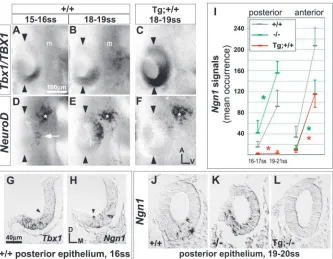
[image:2.612.227.560.71.330.2]Transverse 7 µm serial sections were digitized and imported into either Photoshop7.0 (Adobe) or Openlab3.1.1 (Improvision) at a Fig. 1. Tbx1/TBX1 suppresses expression of
neural bHLH genes in the otic placode. (A-F) Lateral surface views of whole mount hybridized embryos. Arrowheads mark the anterior and posterior poles of the invaginating otic placode. asterisk, facial ganglion
resolution of 0.32-0.64 µm/pixel. NeuroD and Ngn1 signal occurrence data were obtained from consecutive serial sections by marking of (perinuclear ring) hybridization signals and automated counting in Photoshop with an Image Processing Toolkit plug-in (Reindeer Graphics). Volumetric data were obtained by integrating traced areas of interest in Openlab3.1.1. Data were tested for significance by multivariate analyses of variance in SAS version 8.1e.
Three-dimensional reconstruction
Serial section sets were aligned using AutoAligner2 software (Bitplane AG). Regions of interest were traced in Photoshop7.0 and reconstructed using Imaris3 software (Bitplane AG).
Results
Tbx1 expression encompasses a subset of presumptive sensory organ territories
Tbx1 expression is regionalized posterolaterally in the early otocyst and anterodorsolaterally, as well, in the later otocyst (Vitelli et al., 2003; Riccomagno et al., 2002; Chapmann et al., 1996). To better characterize its dynamics, otic epithelial Tbx1 expression was analyzed throughout placode and otocyst stages (E8.5-E12) by RNA hybridization. A diffuse Tbx1 signal is present posteriorly in the early otic placode (10-12 somites; data not shown). During invagination, a domain border emerges halfway along the placode’s anterior-posterior axis (AP midline; Fig. 1A,B) and is maintained through the early otocyst stage (Fig. 2A). Along the medial-lateral axis of the invaginating placode, Tbx1 signal forms an intensity gradient that falls off medially (Fig. 1G), but by otocyst stages a definitive expression border is present along this axis as well (Fig. S1C,D, http://dev.biologists.org/supplemental/; Fig. 4A). By E10, the Tbx1 border slopes anterodorsally from the AP midline (Fig. 2B). This border co-localizes with an anterior stripe of Bmp4 expression (Fig. 3C) and the lateral border of Lfng/Ngn1/NeuroD expression, which is complementary to that of Tbx1 (Fig. 2E, Fig. 3B, Fig. 4B). Between E9.5 and E10, the Tbx1 domain expands into the posteroventromedial otocyst (Fig. 4A; Fig. S2, http://dev.biologists.org/supplemental/). Tbx1 signal is absent dorsomedially at all stages (Fig. 1G; Fig. S1A-E, http://dev.biologists.org/supplemental/).
By current fate mapping schemes, E10.5 lateral otocyst regions contributing to the future cristae, utricular macula, and non-sensory cochlea are defined by the expression domains of Bmp4, Lfng and Otx1, respectively (Fekete and Wu, 2002; Morsli et al., 1998). At this stage, Tbx1 expression encompasses the domains of Bmp4 and Otx1, but is complementary to the lateral portion of the Lfng/Ngn1/NeuroD domain (Fig. 3). Signal intensity gradients are observed within the Tbx1 domain (Fig. 2C, Fig. 3K). Strong Tbx1 signal is present at all sites of Bmp4 expression, and graded, low intensity Tbx1 signal overlaps with Otx1 signal posteroventrolaterally. By E12, Tbx1 signal is restricted to the dorsal-most regions of the rudimentary vestibulum – the forming vestibular canals and ampullae – with strong signal selectively marking the latter structures (data not shown) (Vitelli et al., 2003). Thus, Tbx1 expression in the otocyst epithelium expands in area through E10.5-E11 and thereafter recedes to dorsal vestibular regions.
Complementary expression of Tbx1 and neural bHLH genes
We next compared Tbx1 expression with that of the neural
[image:3.612.315.568.72.530.2]through E11.5 (Fig. 4A,B; data not shown). Both expression domains expand into anterolateral, anteroventral and posteroventral portions of the invaginating placode (Fig. 1E,H,J) and overlap with low intensity Tbx1 signal posteriorly (Fig. 1G,H). By E9.5 a definitive border of NeuroD expression emerges at the otocyst AP midline (Fig. 2D), and the posterolateral region remains Ngn1/NeuroD negative
throughout otocyst stages (Fig. 2D-F, Fig. 4B, Fig. 5A). Expression mapping at E9.5 suggests that NeuroD and Tbx1 expression borders are juxtaposed laterally at the AP midline, but overlap in the posteroventral otocyst (Fig. S2, http://dev.biologists.org/supplemental/). At E9.5, all otocyst regions expressing bHLH genes show NeuroD-positive cells delaminating from the epithelium (data not shown).
NeuroD and Ngn1 expression is downregulated in the posteroventral otocyst between E9.5 and E10, a period during which the Tbx1 domain expands into this region (Fig. 4A,B, Fig. S2, http://dev.biologists.org/supplemental/). Posteroventrally, epithelial NeuroD signal decreases 8-fold, while the total anterior value (anterolateral plus anteroventral) increases 2.1-fold (Fig. 5A). By E10, no overlap of Tbx1 and NeuroD expression is detected in the ventral otocyst (Fig. 4A; Fig. S1A-J, Fig. S2, http://dev.biologists.org/supplemental/). In the lateral otocyst between E10 and E11, NeuroD and Tbx1 expression domains remain complementary, even as the shape of their borders change (Fig. 2B,C,E,F, Fig. 3J,K). Between E10 and E11.5, and despite a 2.5-fold increase in otocyst surface area (n=4-6 otocysts), total otocyst NeuroD signal (sum of all regions) decreases 2.2-fold (Fig. 5A). In summary, peak expression of neural fate markers occurs at E10, even though downregulation of these genes in the posteroventromedial otocyst begins as early as E9.5. By contrast, expansion of the Tbx1-postive otocyst area persists through to at least E10.5, and transient overlap of Tbx1 and NeuroD expression appears to precede downregulation of the latter.
Suppression of neural determination in TBX1 gain-of-function otocysts
[image:4.612.45.383.63.321.2]To test whether an expanding domain of Tbx1 activity can suppress otic neurogenesis, Ngn1 and NeuroD expression was analyzed in a transgenic mouse line carrying tandem copies (eight to ten) of human BAC DNA (line 316.23) (Funke et al., Fig. 3. Tbx1 patterns Lfng and Bmp4 otocyst expression. Lateral surface views of whole mount hybridized embryos arrayed by stage and genotype. Arrowheads denote position of the Bmp4 anterior stripe. Black arrows highlight the position of posterior Bmp4 expression. White arrows mark the anterior region of Lfng/neural bHLH gene expression. Brackets in D and H mark the extent of diffuse anterodorsolateral Bmp4 expression in Tbx1–/–otocysts. All images are shown to scale.
[image:4.612.75.264.439.737.2]2001). TBX1 and flanking genes (5′ and 3′ of TBX1) are expressed in transgenic embryos, suggesting the presence and activity of local TBX1 regulatory elements. Of the four human genes present, only TBX1 and PNUTL1 hybridization signals are detected in the transgenic otocyst (Funke et al., 2001). Transgenic embryos hybridized with a species-specific TBX1 probe show recapitulation of the endogenous Tbx1 signal together with ectopic signal in the anterior otic placode and otocyst (Fig. 1C, Fig. 2P,Q). TBX1 transgene and endogenous Tbx1 otocyst expression patterns are similar at later otocyst stages (Fig. 2C,R). By contrast, TBX1 signal onset in transgenic mesenchyme is delayed (compare Fig. 1C and B and Fig. 2P and A), affording a period (E9-9.5) during which any gain-of-function effects are probably of epithelial origin.
Ngn1 and NeuroD otic epithelial expression is effectively abolished posteriorly and significantly reduced anteriorly in
transgenic embryos (Fig. 1F,I,L, Fig. 2M-O, Fig. 5A). The transgene suppresses neurogenesis in Tbx1+/+,
Tbx1+/– and Tbx1–/– backgrounds (Fig. 1L; data not
shown). Transgenic otocysts show differences in neural domain patterning when endogenous gene copy number is varied, but in all instances, NeuroD domain borders are shifted anteriorly in the presence of the transgene (Fig. 2N; Fig. S2, http:// dev.biologists.org/ supplemental/). At E10, Tg;Tbx1+/– ganglion volume,
identified by NeuroD signal, is 39% that of wild type (0.314±0.113×10⁶ vs. 0.798±0.206×106 µm3; n=8
ganglia/class; P<0.0005; compare Fig. 2N and E). No qualitative difference with wild type is detected in size of the adjacent facial ganglion rudiment (Fig. 2, asterisks), the neural progenitors of which derive from an epibranchial placode.
Tbx1 loss-of-function causes ectopic neural determination
The generation of a Tbx1 null mutation by gene targeting has been described (Merscher et al., 2001). The Tbx1–/–otocyst is
hypoplastic (Fig. 2K,L; Fig. S1P-T, http://dev.biologists.org/ supplemental/), in accordance with previous accounts (Vitelli et al., 2003; Jerome and Papaioannou, 2001), and we find earlier evidence of impaired growth during placode invagination (Fig. 1K). Posteriorly, the invaginating placode of Tbx1–/– embryos shows an increase in Ngn1/NeuroD
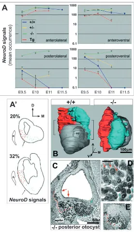
[image:5.612.52.329.69.546.2]expression over that in wild type (Fig. 1I,K; data not shown). The Tbx1–/–otocyst has a uniform distribution of Ngn1/NeuroD Fig. 5. Ectopic neurogenesis correlates with an expansion of VIIIth ganglion rudiment form. (A) Mean occurrence of epithelial NeuroD signals (perinuclear rings) by stage and otocyst region for each of the phenotypes examined, obtained by analyses of serial 7 µm thick transverse sections. Between four and eight right otocysts per genotype per stage were analyzed. Data were normalized to account for differences with wild type in average number of sections when this exceeded 5% (normalization factors: E9.5 Tg, 1.08; E9.5 Tbx1–/–, 1.09; E10 Tbx1–/–, 1.22; E11 Tbx1–/–, 1.53; E11
Tbx1+/–, 1.09). Transgenic data are for Tg+/+, except at E10 (Tg+/–). Values are plotted on a log scale, and non-overlapping error bars represent significant differences with P<0.003. At later stages these regions are distinguished as shown in A′. (A′) Tracings of E11 wild-type sections at 20 and 32% otocyst (with anterior pole set to zero) showing spatial distributions of NeuroD-positive lateral (red in B) and ventral (cyan in B) cells. a, anterior cardinal vein. Lateral and ventral regions at stages E9.5 and E10 are distinguished by line VL in Fig. S2, http://dev.biologists.org/supplemental/. (B) 3-D
expression (laterally and ventrally) along its entire AP length, and no AP midline border forms (Fig. 2J,K,L, Fig. 3I, Fig. 4B, Fig. 5A). Extreme dorsal and medial borders of wild-type Ngn1/NeuroD expression, together with signal intensity gradients for these markers along the medial-lateral axis, are preserved in Tbx1–/– otocysts (Fig. S1F-J,P-T, http://
dev.biologists.org/supplemental/).
Tbx1+/– otocysts show a stereotyped pattern of transient
ectopic neurogenesis, first posteroventromedially and then anterodorsolaterally (Fig. 2G,H,I, Fig. 4A,B). These loci correspond to sites and times of concerted Tbx1 expression onset and bHLH gene downregulation described above, suggesting that neurogenic suppression is delayed in the heterozygous state. Ectopic neurogenesis in heterozygotes correlates with increased accumulations of delaminated NeuroD-positive cells compared to wild type (Fig. S1K-O, http://dev.biologists.org/supplemental/).
The more persistent and widespread ectopic neurogenesis observed in Tbx1 null homozygotes correlates with ectopic delamination (Fig. 5C,E) and duplication of VIIIth ganglion rudiment form about the otocyst AP midline (Fig. 2K,L, Fig. 5B). At E10, Tbx1–/– ganglion volume, identified by NeuroD
signal, is 1.83-fold that of wild type (1.46±0.367×10⁶
vs. 0.798±0.206×106 µm3; n=8 ganglia/class; P<0.001). By
E10.5, anti-islet1/2 (mAb4D5) immunochemistry reveals histodifferentiation of the wild-type ganglion into lateral and ventromedial subdivisions (Fig. S3A,B, http:// dev.biologists.org/supplemental/), and islet-positive nuclear size and packing density vary between subdivisions (Fig. 5D; Fig. S3C, http://dev.biologists.org/supplemental/). These features are present along the entire AP length of the E10.5 Tbx1–/– ganglion rudiment (Fig. 5C). Evidence of ganglion
rudiment splitting near the otocyst AP midline is observed by E11.5 (data not shown), and early labyrinth-stage (E13.5)
Tbx1–/–ears commonly show a secondary compound ganglion
apposed to and innervating the epithelial posterior pole (Fig. 8E,F,G; 4/8 ears auditory and vestibular neuronal cytology; remainder vestibular only). No posterior VIIIth ganglia analyzed possessed a central projection. Anterior to the epithelium, all E13.5 Tbx1–/– VIIIth ganglia analyzed (8/8)
showed auditory and vestibular neuronal cytology and partial retention of auditory and vestibular ganglion morphology; in addition, all anterior ganglia possessed both peripheral and central projections (Fig. 8A-D). Tbx1–/–posterior and anterior
VIIIth ganglia are necrotic beyond E14.5 and largely absent by E16.5, suggesting an additional requirement for Tbx1 in neuronal survival at late embryonic stages.
Tbx1 is a determinant of anterior-posterior otocyst patterning
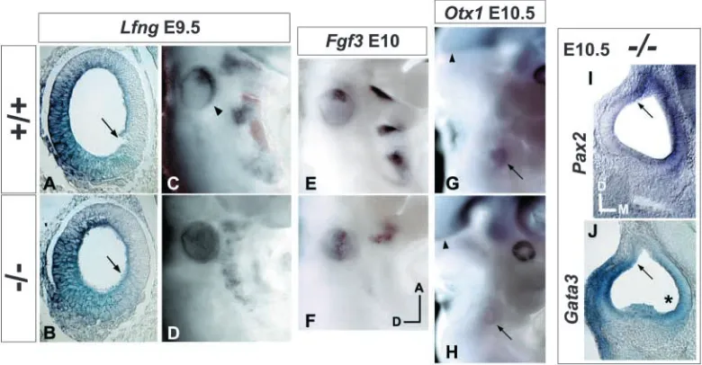
The foregoing results suggest that Tbx1 posteriorizes the otocyst by suppressing anterior identity. To test this hypothesis, the distributions of various regional markers were analyzed in Tbx1–/– embryos. Early otocyst Lfng expression
co-localizes with bHLH gene expression and is absent from the Tbx1-positive posterolateral region (Fig. 4A; Fig. 6C). In Tbx1–/–embryos between E9.5 and E10.5, Lfng is ectopically
expressed throughout the posterolateral otocyst (Fig. 3A,E; Fig. 6D). A medial-lateral intensity gradient of Lfng signal, similar to that present in the wild-type anterior otocyst (decreasing lateral to medial), is present along the entire AP length of the mutant otocyst (Fig. 6A,B). Dorsal and medial border positions of wild-type Lfng expression are also preserved in the mutant.
[image:6.612.108.498.75.278.2]et al., 1993). Fgf3 is selectively expressed in the anterolateral otocyst (McKay et al., 1996) and defines a portion of the common Lfng/bHLH gene domain (Fig. 6E). Fgf3 expression in E10-10.5 Tbx1–/–otocysts is expanded posterolaterally and
forms a stripe extending from anterior to posterior poles (Fig. 6F).
Pax2 and Gata3 expression was assayed to test for effects of Tbx1 mutation on medial-lateral otocyst patterning. Along this axis, their otocyst expression domains are reciprocal and partially overlapping (Lavoko-Kerali et al., 2002). Pax2 is expressed medially and is required for genesis of the cochlear duct (Nornes et al., 1990; Torres et al., 1996). Gata3 hybridization at E10.5 yields a strong lateral signal (Zheng et al., 2003), and the inner ear of Gata3–/– mice is severely
dysmorphic (Karis et al., 2001). No change was detected in expression patterning of either Pax2 or Gata3 in Tbx1–/–
otocysts between E9.5 and E10.5. Wild-type features of these genes’ expression, such as dorsal borders and ventral gradients are preserved in the mutant otocyst (Fig. 6I,J).
Otx1 is expressed in the posteroventrolateral otocyst by E10.25 and loss-of-function results in sensory organ absence (lateral vestibular canal/ampulla) or dysmorphogenesis (lateral crista, maculae, cochlea); no neurogenic phenotype pertaining to the ear has been reported (Fritzsch et al., 2001; Morsli et al., 1999; Acampora et al., 1996). E10.5 and E11 Tbx1–/– otocysts lack Otx1 expression (Fig. 6H) and show
ectopic neurogenesis posteroventrolaterally (Fig. 2L, Fig. 3I, Fig. 5A), which persists through to at least E11.5. Thus, Tbx1 loss-of-function causes anterior (neurogenic) transformation of a posterior otocyst region that contributes to sensory organ development.
Tbx1 is required for proper patterning and maintenance of Bmp4 expression
To further explore the effects of Tbx1 activity on sensory organ patterning, Bmp4 expression was analyzed in Tbx1–/– and
Tbx1+/–embryos. Local antagonism of BMP signaling in the
developing chick causes dysmorphogenesis of multiple inner ear sensory organs, with the most severe and frequent defects related to vestibular canal formation (Chang et al., 1999; Gerlach et al., 2000). In the E10.5 mouse otocyst, Bmp4 hybridization marks the presumptive anterior and lateral cristae (anterior stripe) and posterior crista (Morsli et al., 1998) (Fig. 3G). We have localized the origin of the anterior stripe at E10 to a juxtaposition of Tbx1 and Lfng/neural bHLH gene expression domains (Fig. 2B,E, Fig. 3B,C).
Bmp4 otocyst expression in E10 and E10.5 Tbx1–/–embryos
is mis-patterned. E10 mutant embryos show a diffuse anterodorsolateral signal in place of an anterior stripe, while an extreme dorsolateral band of signal terminating at the otocyst posterior pole is preserved (Fig. 3D). By E10.5, the wild-type anterior stripe and posterior focus form discrete signals (Fig. 3G), but Tbx1–/– otocysts of this stage show a
pattern similar to that of E10 mutants (Fig. 3H, Fig. 7D,E). The anterior stripe and posterior focus persist in wild-type otocysts through E11, however, Tbx1–/–embryos of this stage
showed little to no signal in the otocyst epithelium (Fig. 7F). Bmp4 otocyst signal in E10 and E10.5 Tbx1+/– embryos
(15/15) was indistinguishable from wild-type (data not shown). However, all E11 heterozygotes analyzed (four embryos) showed a diffuse anterodorsolateral signal
bilaterally in place of an anterior stripe (Fig. 7C), suggesting a requirement for proper Tbx1 gene dosage in patterning of Bmp4 otocyst expression.
Tbx1 loss-of-function blocks sensory organ morphogenesis
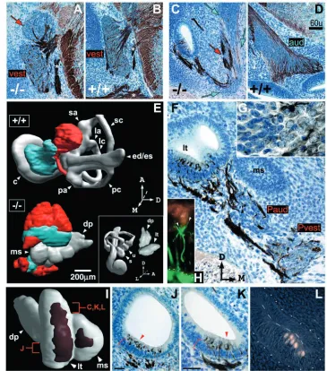
By E13.5, the six sensory organs of the membranous labyrinth are present in rudimentary form (Morsli et al., 1998). The cochlear duct is partially coiled, saccular and utricular outpouches are conspicuous, and, further dorsally, ampullary swellings are associated with each of the three semicircular canals (Fig. 8E and inset). The non-sensory endolymphatic duct projects dorsally from the medial wall of the labyrinth. At this stage the labyrinth is composed largely of simple or pseudostratified epithelium, however a patch of stratified epithelium that will differentiate into mechanosensory epithelium is associated with each of the developing sensory organs (Sher, 1971). Upper layer cells are innervated by neurites of the VIIIth ganglion (Sher, 1971) and express MATH1 (Chen et al., 2002; Shailam et al., 1999), a bHLH transcription factor that is necessary and sufficient for mechanosensory hair cell differentiation (Bermingham et al., 1999; Zheng and Gao, 2000; Kawamoto et al., 2003).
Identifiable vestibular and auditory sensory organs fail to develop in Tbx1–/– embryos (Fig. 8E,I). The E13.5 Tbx1–/–
[image:7.612.333.553.70.270.2]exhibits simple cuboidal epithelial morphology typical of the wild-type endolymphatic duct.
By E13.5, the mutant ventral epithelium, previously a site of neurogenesis, is stratified, innervated, and MATH1 positive (Fig. 8C,F,J,K,L). Neurites of the VIIIth ganglion envelop the base of upper layer MATH1-positive cells (Fig. 8H,J,K), and MATH1 immunoreactivity and innervation patterns are similar across the epithelium (Fig. 8K,L; data not shown). These patterns consist of two patches separated by the cleft between lateral tube and medial sac (Fig. 8I), thus segregation of an initially unitary neural/sensory-competent region may be related to morphogenetic movements of the late otocyst. Lateral patch structure is polarized along the AP axis with respect to the number and morphology of innervated, MATH1-positive cells (Fig. 8J,K,L). Architecture of the medial sac patch is similar to that of the posterior lateral tube.
Discussion
[image:8.612.197.562.72.486.2]The otocyst has been likened to the Drosophila imaginal disc (Fekete, 1996; Brigande et al., 2000). Both are transient embryonic structures derived by invagination of ectoderm and undergo extensive growth and morphogenesis during their maturation. Studies of imaginal disc patterning lead to a three-step model (selector gene action, boundary formation, boundary organizer function) for the transformation of positional information into differentiated structures (Garcia-Bellido et al., 1973; Lawrence and Struhl, 1996; Dahmann and Basler, 1999). Selector genes encoding transcription factors are regionally expressed in primordial epithelia and specify regional identity autonomously and non cell-autonomously. Selector genes mediate compartment boundary formation, a local process involving differential cell affinity and Notch pathway signaling (Milan and Cohen, 2003). For example, Fng, a Drosophila Notch signaling modulator, is Fig. 8. Tbx1–/–inner ear and VIIIth
ganglion morphology at E13.5. (A-D,F,G,J,K) Transverse sections through wild-type and Tbx1–/– ears/ganglia at E13.5, reacted with mAb2H3 (anti-neurofilament) and counter-stained with Cresyl Violet. Axes in F apply to all except G. Red arrows in A and C indicate regions of vestibular (vest) neuronal cytology and cyan arrows in C auditory (aud) neuronal cytology. The distinguishing cytological features (nuclear size and basophilia) from a mutant posterior ganglion are shown at high magnification in G, with vestibular neuronal cytology shown at right. White asterisks in A and B indicate a branch of the facial nerve. Section shown in F passes through a Tbx1–/–posterior compound ganglion and epithelial posterior pole. Pvest, presumptive vestibular ganglion; Paud, presumptive auditory ganglion. Brackets and arrowheads in J and K indicate cellular stratification. Scale bars: in D, 60 µm for A-D; 20 µm for J,K. (E) 3-D
reconstructions of serial sections reacted as described above. Presumptive vestibular (red) and auditory (cyan) ganglion tissue is shown in relation to the inner ear epithelium. c, cochlea; sa, superior ampulla; la, lateral ampulla; pa, posterior ampulla; sc, superior canal; lc, lateral canal; pc, posterior canal; ed/es, endolymphatic duct/sac; u, utricle; s, saccule; lt, lateral tube; ms, medial sac; dp, dorsal projection. (H) Double immunolabeling with mAb2H3 and anti-MATH1 shows neurites (in green, arrowheads) enveloping the base of a MATH1-positive cell. Dotted line indicates the basement membrane.
regulated by the selector gene apterous and is critical for boundary formation at the wing imaginal disc (Irvine and Weischaus, 1994; Kim et al., 1995). Boundaries, in turn, secrete long-range signaling molecules of the BMP or WNT families that serve as effectors of global growth and morphogenesis (Nellen et al., 1996; Zecca et al., 1996; Day and Lawrence, 2000).
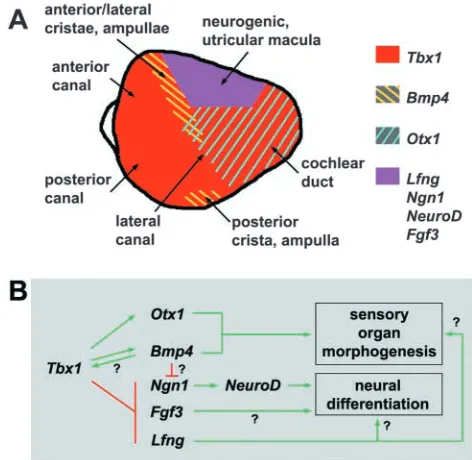
Tbx1 specifies regional identity and establishes a fate boundary in the otocyst
It is hypothesized that compartment boundaries exist along the three major axes of the otocyst (Fekete, 1996). At the lateral wall of the otocyst, a putative AP midline boundary is defined by anterior expression of NeuroD/Lfng/Fgf3 and posterior expression of Otx1 (Fekete and Wu, 2002). Otx1 exhibits particular attributes of an otocyst selector gene. Its regional expression encompasses primordia of the non-sensory cochlear duct, lateral canal and lateral crista, and it is required for proper morphogenesis of these end organs and the positioning/ segregation of the lateral crista and other proximate sensory epithelia (Morsli et al., 1999; Fritzsch et al., 2001). However, Otx1 otocyst expression is first detected at E10.25, which precludes a role for this gene in early otocyst patterning.
We find that Tbx1 expression is localized posterolaterally in the invaginating placode and defines an AP midline expression border prior to otocyst formation. Tbx1 expression is maintained in the posterolateral otocyst through the time of Otx1 expression onset. Apart from partial, transient overlap, Tbx1 and NeuroD expression domains are complementary throughout otocyst stages. TBX1 gain-of-function displaces the NeuroD domain border anteriorly. Tbx1 loss-of-function eliminates AP midline borders of NeuroD, Lfng and Fgf3 expression as early as E9.5 and causes ectopic posterolateral expression of all three genes. By contrast, Otx1 expression is not detected in Tbx1–/–otocysts, and the posterolateral otocyst
is a site of ectopic neural progenitor delamination between E9.5 and E11.5. At later stages, Otx1-dependent sensory organ structures are missing or indistinguishable by morphological criteria. These results suggest that Tbx1 patterns sensory organ and neural fate assignment in parallel, through local control of regional gene expression (Fig. 9B). Tbx1 appears to establish a midline boundary between an anterior region of combined neural and sensory epithelial competence and posterior tissue destined to form sensory organ structural components such as the lateral wall of the cochlear duct and lateral canal plate (Fig. 9A). Ectopic expression of Ngn1 in the Tbx1–/–posterolateral
otocyst further supports this hypothesis, as Ngn1 activates NeuroD expression and may mediate the selection of neural progenitors from otic epithelium through Notch pathway signaling (Ma et al., 1998).
Otocyst fate boundaries deviate from the major embryonic axes
Initially, the otic placode is symmetrical. Proceeding from a simple model of otocyst compartmentalization, selector gene loss could result in a complete duplication of form about a major axis, and such ‘enantiomorphic twin’ ear phenotypes have been observed following surgical rotations of the amphibian otic primordium (Harrison, 1936) or perturbation of Hedgehog signaling in zebrafish (Hammond et al., 2003). In Tbx1–/–embryos, gene expression patterning and early VIIIth
ganglion rudiment form are highly suggestive of anterior otocyst identity duplication in the posterior otocyst. We failed to find an effect of Tbx1 loss-of-function on a number of early otocyst medial-lateral patterning features, and Tbx1 may be linked specifically to AP patterning at early otocyst stages. However, the labyrinth-stage Tbx1–/–inner ear shows structural
variation along its AP axis, and one or more pathways probably mediate inner ear AP patterning in the absence of Tbx1. Transplantation studies of avian inner ear primordia provide evidence for a multi-step acquisition of axial polarity (Wu et al., 1998).
Incomplete symmetry of the Tbx1–/– ear about the AP
[image:9.612.323.559.72.302.2]midline may also result from complex and dynamic changes of Tbx1 expression border morphology during otocyst growth. At E10, the dorsal part of the AP midline border deviates anteriorly from the midline. By E11, it has shifted to a position perpendicular to the AP midline and forms a dorsal-ventral border. During these stages, a stripe of Bmp4 expression co-localizes with Tbx1 expression at its interface with the NeuroD/Lfng domain border (Fig. 9A). decapentaplegic (dpp), a Drosophila homolog of Bmp4, is expressed in a narrow stripe of cells adjacent to the imaginal wing disc AP compartment boundary and encodes a morphogen that controls growth and patterning of the disc (Nellen et al., 1996; Lecuit et al., 1996). The existence of a DPP morphogen gradient is implied by expression of its target genes, one of which is optomotor-blind, a T-box gene that controls wing disc regional identity (Podos and Ferguson, 1999). Interestingly, in the otocyst, we find high-intensity Tbx1 signal at all sites of Bmp4 expression.
In the mouse, Bmp4 expression marks sites of developing cristae (Morsli et al., 1998), however the effects of Bmp4 loss-of-function on inner ear development are unknown because of early embryonic lethality (Winnier et al., 1995). Perturbation of BMP signaling in the chick ear causes a range of sensory organ defects, and vestibular canal morphogenesis is most frequently and severely affected (Chang et al., 1999; Gerlach et al., 2000). Tbx1–/–ears lack vestibular canals and
it was recently reported that Tbx1 is required for otocyst Bmp4 expression (Vitelli et al., 2003). Contrary to this result, we find that Tbx1–/–otocysts express Bmp4 but do not form
an anterior stripe and epithelial expression is lost by E11. Tbx1+/– otocysts show normal patterning of Bmp4 through
E10.5 and a loss of the anterior stripe by E11. Thus, Bmp4 patterning appears sensitive to Tbx1 gene dose in a stage-specific manner. These results, together with the observation of high-intensity Tbx1 signal at all sites of Bmp4 expression, suggest that a complex regulatory interaction between Tbx1 and Bmp4 is localized to a fate boundary in the anterodorsolateral otocyst (Fig. 9A,B). Studies of comparative expression and epistatic relationships between Tbx1 and other regulators of Bmp4 patterning and vestibular canal morphogenesis, such as Hmx2 (Wang et al., 2001), Hmx3 (Wang et al., 1998) and Dlx5 (Merlo et al., 2002) may elucidate cooperative interactions among these genes.
Tbx1 suppresses neurogenesis at some prospective sensory epithelial territories
Evidence of a common progenitor for VIIIth ganglion neurons and mechanosensory cells has been obtained by clonal analyses in chick (Satoh and Fekete, 2003). Furthermore, expression overlap of Lfng and neural fate markers in both chick and mouse leads to the suggestion that neural progenitors and utricular and saccular maculae derive from a common anterior otocyst region (Fekete and Wu, 2002; Cole et al., 2000). In the wild-type anterior otocyst, we find overlapping Ngn1, NeuroD, and Lfng expression that is complementary to the Tbx1 domain (Fig. 9A). Neural bHLH gene expression persists in this region through E11.5, the latest stage assayed for these markers. Tbx1 loss-of-function has little to no effect on neurogenic activity in this region and does not preclude the subsequent development of anteroventral sensory epithelium. Thus, Tbx1-independent pathways probably control neural and sensory epithelial fate assignment at this otocyst region.
The Lfng-positive posteroventral otocyst is the presumptive anlage of the organ of Corti (Fekete and Wu, 2002) and initially, this region is Tbx1-negative. We show that transient wild-type expression of Ngn1 and NeuroD, together with delamination, precedes the local onset of Tbx1 expression in this region (see Fig. 4A). Regression of posteroventral neurogenesis is delayed in Tbx1 heterozygotes, and neurogenesis persists in this region through E11.5 in Tbx1–/–
otocysts. Conversely, TBX1 gain of function effectively eliminates posteroventral neurogenesis. Interestingly, Tbx1 heterozygotes at E11 show delayed regression of neurogenesis at the anterodorsolateral otocyst (see Fig. 2I) and loss of a definitive Bmp4 anterior stripe. These phenotypes are observed toward the end of a period (E9.75-E11) during which Tbx1 expression expands into the anterodorsolateral otocyst. Together these results suggest that at some otocyst regions, Tbx1 regulates the developmental timing by which neural and
sensory epithelial competent states are expressed. Functionally, this differs from the effect of Tbx1 activity at the posterolateral otocyst, where neural competence is fully suppressed at all times and sensory organ structural epithelium is formed.
Conclusion
In this study we have characterized the differential effects of Tbx1 mutation on neurogenesis and inner ear sensory organ development. Tbx1 specifies regional identity in the otocyst and is required for the positioning of a fate boundary. Our data support the hypothesis of a relationship between neural and sensory epithelial competence in the otocyst. Furthermore, absence of Tbx1 causes expression of neural competence in a portion of the otocyst associated with formation of sensory organ structural epithelia. Taken together, our results suggest that Tbx1 regulates otocyst gene expression locally but affects inner ear growth and morphogenesis in a global manner. Tbx1 may therefore function as an otocyst selector gene in its control of neurogenesis and sensory organ development. Studies aimed at dissecting the contributions of epithelial and mesenchymal Tbx1 activity to various aspects of inner ear development using tissue-specific gene inactivation strategies are currently in progress.
In Xenopus gastrula ectoderm, BMP signaling suppresses neural competence and induces epidermal fate (Weinstein and Hemmati-Brivanlou, 1999). In the mouse otocyst, the specific effects of Bmp4 on neural and sensory organ development and its range of action are unknown. Nevertheless, co-localization of Bmp4 expression and robust ectopic neurogenesis at the Tbx1–/– otocyst posterior pole through E10.5 provides a
preliminary clue that Tbx1-mediated neural suppression may proceed independently of Bmp4 activity. Finally, the homeobox genes Eya1 and Six1 (Xu et al., 1999; Zheng et al., 2003) may function reciprocally to Tbx1 with respect to neurogenesis, as both are expressed at regions of neural progenitor determination and are required for VIIIth ganglion rudiment formation.
We thank Paula Cohen, Andres Collazo, Andrew Groves, Jean Hebert, Zaven Kaprielian, Doris Wu and two anonymous reviewers for their invaluable commentary. We also thank Jane E. Johnson for a gift of anti-MATH1 antibody, Adam Hartley for suggestions on image analysis, and Haftan Eckhold for assistance with statistical analysis. This work was supported in part by grants from the March of Dimes Foundation #1-FY02-193, American Heart Association Established Investigator Grant, and NIH #1RO1 DC05186-01.
References
Acampora, D., Mazan, S., Avvantaggiato, V., Barone, P., Tuorto, F., Lallemand, Y., Brulet, P. and Simeone, A. (1996). Epilepsy and brain
abnormalities in mice lacking the Otx1 gene. Nat. Genet. 14, 218-222.
Adam, J., Myat, A., le Roux, I., Eddison, M., Henrique, D., Ish-Horowicz, D. and Lewis, J. (1998). Cell fate choices and the expression of Notch,
Delta, and Serrate homologues in the chick inner ear: parallels with
Drosophila sense-organ development. Development 125, 4645-4654.
Adkins, W. Y. and Gussen, R. (1974). Temporal bone findings in the third
and fourth pharyngeal pouch (DiGeorge) syndrome. Arch. Otolaryngol. 100, 206-208.
Bamshad, M., Lin, R. C., Law, D. J., Watkins, W. C., Krakowiak, P. A., Moore, M. E., Franceschini, P., Lala, R., Holmes, L. B., Gebuhr, T. C. et al. (1997). Mutation in human TBX3 alter limb, apocrine and genital
development in ulnar-mammary syndrome. Nat. Genet. 16, 311-315.
al. (1997). Mutations in human TBX5 cause limb and cardiac malformation
in Holt-Oram syndrome. Nat. Genet. 15, 30-35.
Bermingham, N. A., Hassan, B. A., Price, S. D., Vollrath, M. A., Nissim, B.-A., Eatock, R. A., Bellen, H. J., Lysakowski, A. and Zoghbi, H. Y.
(1999). Math1: An essential gene for the generation of inner ear hair cells.
Science 284, 1837-1841.
Black, F. O., Spanier, S. S. and Kohut, R. I. (1975). Aural abnormalities in
partial DiGeorge syndrome. Arch. Otolaryngol. 101, 129-134.
Bollag, R. J., Siegfried, Z., Cebra-Thomas, J., Garvey, N., Davidson, E. M. and Silver, L. M. (1994). An ancient family of embryonically expressed
mouse genes sharing a conserved protein motif with the T locus. Nat. Genet.
7, 383-389.
Braybrook, C., Doudney, K., Marcano, A. C., Arnason, A., Bjornsson, A., Patton, M. A., Goodfellow, P. J., Moore, G. E. and Stanier, P. (2001).
The T-box transcription factor gene TBX22 is mutated in X-linked cleft palate and ankyloglossia. Nat. Genet. 29, 179-183.
Brigande, J. V., Kiernan, A. E., Gao, X., Iten, L. E. and Fekete, D. M.
(2000). Molecular genetics of pattern formation in the inner ear: Do compartment boundaries play a role? Proc. Natl. Acad. Sci. USA 97, 11700-11706.
Chang, W., Nunes, F., de Jesus-Escobar, J. M., Harland, R. M. and Wu, D. K. (1999). Ectopic noggin blocks sensory and nonsensory organ
morphogenesis in the chicken inner ear. Dev. Biol. 216, 369-381.
Chapman, D. L., Garvey, N., Hancock, S., Alexiou, M., Agulnik, S. I., Gibson-Brown, J. J., Cebra-Thomas, J., Bollag, R. J., Silver, L. M. and Papaioannou, V. E. (1996). Expression of the T-box family genes,
Tbx1-Tbx5, during early mouse development. Dev. Dyn. 206, 379-390.
Chapman, D. L. and Papaioannou, V. E. (1998). Three neural tubes in mouse
embryos with mutations in the T-box gene Tbx6. Nature 391, 695-697.
Chen, P., Johnson, J. E., Zoghbi, H. Y. and Segil, N. (2002). The role of
Math1 in inner ear development: uncoupling the establishment of the sensory primordium from hair cell fate determination. Development 129, 2495-2505.
Chesley, P. (1935). Development of the short-tailed mutation in the house
mouse. J. Exp. Zool. 29, 437-438.
Cole, L. K., le Roux, I., Nunes, F., Laufer, E., Lewis, J. and Wu, D. K.
(2000). Sensory organ generation in the chicken inner ear: contributions of Bone Morphogenetic Protein 4, Serrate1, and Lunatic Fringe. J. Comp.
Neurol. 424, 509-520.
D’Amico-Martel, A. and Noden, D. M. (1983). Contributions of placodal and
neural crest cells to avian cranial peripheral ganglia. Am. J. Anat. 166, 445-468.
Dahmann, C. and Basler, K. (1999). Compartment boundaries at the edge of
development. Trends Genet. 15, 320-326.
Day, S. J. and Lawrence, P. A. (2000). Measuring dimensions: the regulation
of size and shape. Development 127, 2977-2987.
Fekete, D. M. (1996). Cell fate specification in the inner ear. Curr. Opin.
Neurobiol. 6, 533-541.
Fekete, D. M. and Wu, D. K. (2002). Revisiting cell fate specification in the
inner ear. Curr. Opin. Neurobiol. 12, 35-42.
Fritzsch, B., Beisel, K. W., Jones, K., Farinas, I., Maklad, A., Lee, J. and Reichardt, L. F. (2002). Development and evolution of inner ear sensory
epithelia and their innervation. J. Neurobiol. 53, 143-156.
Fritzsch, B., Signore, M. and Simeone, A. (2001). Otx1 null mutant mice
show partial segregation of sensory epithelia comparable to lamprey ears.
Dev. Genes Evol. 211, 388-396.
Funke, B., Epstein, J. A., Kochilas, L. K., Lu, M. M., Pandita, R. K., Liao, J., Bauerndistel, R., Schuler, T., Schorle, H., Brown, M. C. et al. (2001).
Mice overexpressing genes from the 22q11 region deleted in velo-cardio-facial syndrome/DiGeorge syndrome have middle and inner ear defects.
Hum. Mol. Genet. 10, 2549-2556.
Garcia-Bellido, A., Ripoll, P. and Morata, G. (1973). Developmental
compartmentalization of the wing disk of Drosophila. Nature 245, 251-253.
George, K. M., Leonard, M. W., Roth, M. E., Lieuw, K. H., Kioussis, D., Grosveld, F. and Engel, J. D. (1994). Embryonic expression and cloning
of the murine GATA-3 gene. Development 120, 2673-2686.
Gerlach, L. M., Hutson, M. R., Germiller, J. A., Nguyen-Luu, D., Victor, J. C. and Barald, K. F. (2000). Addition of the BMP4 antagonist, noggin,
disrupts avian inner ear development. Development 127, 45-54.
Goldberg, M. E. and Hudspeth, A. J. (2000). The vestibular system. In
Principles of Neural Science (ed. E. R. Kandel, J. H. Schwartz and T. M.
Jessell), pp. 801-815. New York: McGraw-Hill.
Hadrys, T., Braun, T., Rinkwitz-Brandt, S., Arnold, H. H. and Bober, E.
(1998). Nkx5-1 controls semicircular canal formation in the mouse inner ear. Development 125, 33-39.
Hammond, K. L., Loynes, H. E., Folarin, A. A., Smith, J. and Whitfield, T. T. (2003). Hedgehog signaling is required for correct anteroposterior
patterning of the zebrafish otic vesicle. Development 130, 1403-1417.
Helms, A. W. and Johnson, J. E. (1998). Progenitors of dorsal commissural
interneurons are defined by MATH1 expression. Development 125, 919-928.
Harrison, R. G. (1936). Relations of symmetry in the developing ear of
Amblystoma punctatum. Proc. Natl. Acad. Sci. USA 22, 238-247.
Herrmann, B. G., Labeit, S., Pustka, A., King, T. R. and Lehrach, H.
(1990). Cloning of the T gene required in mesoderm formation in the mouse.
Nature 343, 617-622.
Hudspeth, A. J. (2000). Hearing. In Principles of Neural Science (ed. E. R.
Kandel, J. H. Schwartz and T. M. Jessell), pp. 801-815. New York: McGraw-Hill.
Irvine, K. D. and Wieschaus, E. (1994). fringe, a boundary-specific signaling
molecule, mediates interactions between dorsal and ventral cells during Drosophila wing development. Cell 79, 595-606.
Jerome, L. A. and Papaioannou, V. E. (2001). DiGeorge syndrome
phenotype in mice mutant for the T-box gene, Tbx1. Nat. Genet. 27, 286-291.
Karis, A., Pata, I., Hikke van Doorninck, J., Grosveld, F., de Zeeuw, C. I., de Caprona, D. and Fritzsch, B. (2001). Transcription factor GATA-3
alters pathway selection of olivocochlear neurons and affects morphogenesis of the inner ear. J. Comp. Neurol. 429, 615-630.
Kawamoto, K., Ishimoto, S.-I., Minoda, R., Brough, D. E. and Raphael, Y. (2003). Math1 gene transfer generates new cochlear hair cells in mature
guinea pigs in vivo. J. Neurosci. 23, 4395-4400.
Kiernan, A. E., Nunes, F., Wu, D. K. and Fekete, D. M. (1997). The
expression domain of two related homeobox genes defines a compartment in the chicken inner ear that may be involved in semicircular canal formation. Dev. Biol. 191, 215-229.
Kiernan, A. E., Steele, K. P. and Fekete, D. M. (2002). Development of the
mouse inner ear. In Mouse development (ed. J. Rossant and P. Tam), pp. 539-566. San Diego: Academic Press.
Kim, J., Irvine, K. D. and Carroll, S. B. (1995). Cell recognition, signal
induction, and symmetrical gene activation at the dorsal-ventral boundary of the developing Drosophila wing. Cell 82, 795-802.
Kim, W.-Y., Fritzsch, B., Serls, A., Bakel, L. A., Huang, E. J., Reichardt, L. F., Barth, D. S. and Lee, J. E. (2001). NeuroD-null mice are deaf due
to a severe loss of the inner ear sensory neurons during development.
Development 128, 417-426.
Kopp, A. and Duncan, I. (1997). Control of cell fate and polarity in the adult
abdominal segments of Drosophila by optomotor-blind. Development 124, 3715-3726.
Lavoko-Kerali, G., Rivolta, M. N. and Holley, M. (2002). Expression of the
transcription factors GATA3 and Pax2 during development of the mammalian inner ear. J. Comp. Neurol. 442, 378-391.
Lawrence, P. A. and Struhl, G. (1996). Morphogens, compartments, and
pattern: Lessons from Drosophila? Cell 85, 951-961.
Lecuit, T., Brook, W. J., Ng, M., Calleja, M., Sun, H. and Cohen, S. M.
(1996). Two distinct mechanisms for long-range patterning by Decapentaplegic in the Drosophila wing. Nature 381, 387-393.
Lee, J. E., Hollenberg, S. M., Snider, L., Turner, D. L., Lipnick, N. and Wientraub, H. (1995). Conversion of Xenopus ectoderm into neurons by
NeuroD, a basic helix-loop-helix protein. Science 268, 836-844.
Li, Q. Y., Newbury-Ecob, R. A., Terrett, J. A., Wilson, D. I., Curtis, A. R., Yi, C. H., Gebuhr, T., Bullen, P. J., Robson, S. C., Strachan, T. et al.
(1997). Holt-Oram syndrome is caused by mutation in TBX5, a member of the Brachyury (T) gene family. Nat. Genet. 15, 21-29.
Lindsay, E. A., Vitelli, F., Su, H., Morishima, M., Huynh, T., Pramparo, T., Jurecic, V., Ogunrinu, G., Sutherland, H. F., Scambler, P. J. et al.
(2001). Tbx1 haploinsufficiency in the DiGeorge syndrome region causes aortic arch defects in mice. Nature 410, 97-101.
Liu, M., Pereira, F. A., Price, S. D., Chu, M.-J., Shope, C., Himes, D., Eatock, R. A., Brownell, W. E., Lysakowski, A. and Tsai, M.-J. (2000).
Essential role of BETA2/NeuroD1 in development of the vestibular and auditory systems. Genes Dev. 14, 2839-2854.
Ma, Q., Anderson, D. J. and Fritzsch, B. (2000). Neurogenin1 null mutant
ears develop fewer, morphologically normal hair cells in smaller sensory epithelia devoid of innervation. JARO 1, 129-143.
Ma, Q., Chen, Z., del Barco Barantes, I., de la Ponpa, J. L. and Anderson, D. J. (1998). neurogenin1 is essential for the determination of neuronal
Mansour, S. L., Goddard, J. M. and Capecchi, M. R. (1993). Mice
homozygous for a targeted disruption of the proto-oncogene int-2 have developmental defects in the tail and inner ear. Development 117, 13-28.
McKay, I., Lewis, J. and Lumsden, A. (1996). The role of FGF-3 in early
inner ear development: an analysis in normal and kreisler mutant mice. Dev.
Biol. 174, 370-378.
Merlo, G. R., Paleari, L., Mantero, S., Zerega, B., Adamska, M., Rinkwitz, S., Bober, E. and Levi, G. (2002). The Dlx5 homeobox gene is essential
for vestibular morphogenesis in the mouse embryo through a BMP4-mediated pathway. Dev. Biol. 248, 157-169.
Merscher, S., Funke, B., Epstein, J. A., Heyer, J., Anne, P., Lu, M. M., Xavier, R., Demay, M. B., Russell, R. G., Factor, S. et al. (2001). TBX1
is responsible for cardiovascular defects in velo-cardio-facial/DiGeorge syndrome. Cell 104, 619-629.
Milan, M. and Cohen, S. M. (2003). A re-evaluation of the contributions of
Apterous and Notch to the dorsoventral lineage restriction boundary in the Drosophila wing. Development 130, 553-562.
Morsli, H., Choo, D., Ryan, A., Johnson, R. and Wu, D. K. (1998).
Development of the mouse inner ear and origin of its sensory organs. J.
Neurosci. 18, 3327-3335.
Morsli, H., Tuorto, F., Choo, D., Pia Postiglione, M., Simeone, A. and Wu, D. K. (1999). Otx1 and Otx2 activities are required for the normal
development of the mouse inner ear. Development 126, 2335-2343.
Nellen, D., Burke, R., Struhl, G. and Basler, K. (1996). Direct and
long-range action of a DPP morphogen gradient. Cell 85, 357-368.
Nornes, H. O., Dressler, G. R., Knapik, E. W., Deutsch, U. and Gruss, P.
(1990). Spatially and temporally restricted expression of Pax2 during murine neurogenesis. Development 109, 797-809.
Ohtani, I. and Schuknecht, H. F. (1984). Temporal bone pathology in
DiGeorge’s syndrome. Ann. Otol. Rhinol. Laryngol. 93, 220-224.
Pauley, S., Wright, T. J., Pirvola, U., Ornitz, D., Beisel, K. W. and Fritzsch, B. (2003). Expression and function of FGF10 in mammalian inner ear
development. Dev. Dyn. 227, 203-215.
Pirvola, U., Spencer-Dene, B., Xing-Qun, L., Kettunen, P., Thesleff, I., Fritzsch, B., Dickson, C. and Ylikoski, J. (2000). FGF/FGFR-2(IIIb)
signaling is essential for inner ear morphogenesis. J. Neurosci. 20, 6125-6134.
Pirvola, U., Ylikoski, J., Trokovic, R., Hebert, J., McConnell, S. and Partanen, J. (2002). FGFR1 is required for the development of the auditory
sensory epithelium. Neuron 35, 671-680.
Podos, S. D. and Ferguson, E. L. (1999). Morphogen gradients: new insights
from DPP. Trends Genet. 15, 396-402.
Riccomagno, M. M., Martinu, L., Mulheisen, M., Wu, D. K. and Epstein, D. J. (2002). Specification of the mammalian cochlea is dependent on Sonic
hedgehog. Genes Dev. 16, 2365-2378.
Rubel, E. W. and Fritzsch, B. (2002). Auditory system development:
primary auditory neurons and their targets. Annu. Rev. Neurosci. 25, 51-101.
Satoh, T. and Fekete, D. M. (2003). Mechanosensory epithelial cells and
ganglion cells are clonally related. Assoc. Res. Otolaryngol. 23 (Abstr. No. 1776).
Shailam, R., Lanford, P. J., Dolinsky, C. M., Norton, C. R., Gridley, T. and Kelly, M. W. (1999). Expression of proneural and neurogenic genes in the
embryonic mammalian vestibular system. J. Neurocytol. 28, 809-819.
Sher, A. E. (1971). The embryonic and postnatal development of the inner ear
of the mouse. Acta Otolaryngologica, Suppl. 285, 5-77.
Theiler, K. (1989). The House Mouse: Atlas of Embryonic Development. New
York: Springer.
Torres, M., Gomez-Pardo, E. and Gruss, P. (1996). Pax2 contributes to inner
ear patterning and optic nerve trajectory. Development 122, 3381-3391.
Vitelli, F., Viola, A., Morishima, M., Pramparo, T., Baldini, A. and Lindsay, E. A. (2003). TBX1 is required for inner ear morphogenesis. Hum.
Mol. Genet. 12, 2041-2048.
Wang, W., Chan, E. K., Baron, S., van de Water, T. R. and Lufkin, T.
(2001). Hmx2 homeobox gene control of murine vestibular morphogenesis.
Development 128, 5017-5029.
Wang, W., van de Water, T. R. and Lufkin, T. (1998). Inner ear and maternal
reproductive defects in mice lacking the Hmx3 homeobox gene.
Development 125, 621-634.
Weinstein, D. C. and Hemmati-Brivanlou, A. (1999). Neural induction.
Annu. Rev. Cell Dev. Biol 15, 411-433.
Wilkinson, D. G. (1992). In Situ Hybridization: A Practical Approach.
Oxford: IRL Press.
Wilkinson, D. G., Bhatt, S. and Herrmann, B. G. (1990). Expression pattern
of the mouse T gene required in mesoderm formation in the mouse. Nature
343, 617-622.
Winnier, G., Blessing, M., Labosky, P. A. and Hogan, B. L. M. (1995). Bone
morphogenetic protein-4 is required for mesoderm formation and patterning in the mouse. Genes Dev. 9, 2105-2116.
Wu, D. K., Nunes, F. D. and Choo, D. (1998). Axial specification for sensory
organs versus non-sensory structures of the chicken inner ear. Development
125, 11-20.
Xu, P.-X., Adams, J., Peters, H., Brown, M. C., Heaney, S. and Maas, R.
(1999). Eya1-deficient mice lack ears and kidneys and show abnormal apoptosis of organ primordia. Nat. Genet. 23, 113-117.
Yanagisawa, K. O., Fujimoto, H. and Urushihara, H. (1981). Effects of the
Brachyury (T) mutation on morphogenetic movement in the mouse embryo.
Dev. Biol. 87, 242-248.
Zecca, M., Basler, K. and Struhl, G. (1996). Direct and long-range action of
a wingless morphogen gradient. Cell 87, 833-844.
Zheng, J. L. and Gao, W.-Q. (2000). Overexpression of Math1 induces robust
production of extra hair cells in postnatal rat inner ears. Nat. Neurosci. 3, 580-586.
Zheng, W., Huang, L., Zhu-Bo, W., Silvius, D., Tang, B. and Xu, P.-X.
(2003). The role of Six1 in mammalian auditory system development.