0022-538X/92/063609-07$02.00/0
Copyright C 1992, American Society for Microbiology
Structural and
Functional Analysis of the Visna
Virus
Rev-Response Element
LAURENCES. TILEY1 AND BRYAN R. CULLEN'2,3*
HowardHughes Medical Institute,1 Section of Genetics,2andDepartmentofMicrobiology and
Immunology,3Duke University MedicalCenter, Durham, North Carolina 27710 Received 13 December 1991/Accepted 25 February 1992
The distantly related lentiviruses human immunodeficiency virustype1(HIV-1)andvisna viruseach encode
a posttranscriptional regulatory protein, termed Rev, that is critical for expression of the viral structural proteins. We genetically mapped the cis-acting target sequence for visna virus Rev, the visna virus
Rev-response elementorRRE-V, to acomplex 176-nucleotide RNA stem-loop structure that coincides with
sequences encoding the N terminus of the transmembrane component of envelope. The computer-predicted structureof the RRE-Vwasvalidated by in vitroanalysis of structure-specific RNase cleavage patterns. The visna virus Revproteinwasshowntointeractspecifically with the genetically defined RRE-V in vitro butwas
unableto bind the HIV-1 RRE. Similarly, HIV-1 Revwas also unable tobind the RRE-Vspecifically. We therefore conclude that the HIV-1 and visna virus Rev proteins, while functionally analogous, nevertheless display distinct RNA sequence specificities. These findings provide a biochemical explanation for the
observation that thesetwoviral regulatory proteins arefunctional only in the homologous viral system.
Lentiviruses are a family of complex retroviruses that
inducechronic,degenerative diseases in the infected host (7, 10). Phylogenetic analysis suggests that lentiviruses canbe
broadly divided into twosubgroups (27, 34). These arethe
relatively cohesive primate immunodeficiency viruses anda
somewhat more diverse group of nonprimate lentiviruses thatprimarily infect ungulates (27, 34). Theprototypeof the nonprimate lentiviruses is visna virus, first described asthe
etiologicagentofachronicdegenerative syndrome observed
in domestic sheep (10). Visna virus and other nonprimate lentiviruses such as caprine arthritis-encephalitis virus and
equine infectious anemia virus continue tobe economically significant livestock pathogens. However, the most signifi-cantlentivirus isclearly human immunodeficiency virustype 1(HIV-1), the etiologicagentofAIDS and theprototype of theprimate lentivirus subgroup (4, 7).
Although HIV-1 and visna virus display little primary sequencehomology(32), theydo shareacomplexpatternof viral gene expression that is facilitated by the action of
virally encoded regulatory proteins (4, 6, 8, 12, 15, 25, 31, 35, 39). In particular, both HIV-1 and visna virus have been shown to encode a nuclear protein, termed Rev, that is required for thecytoplasmic expressionof theunsplicedand singly spliced mRNAs that encode theviral structural pro-teins (8, 9, 15, 22, 31, 35, 39). In the case of HIV-1, this action requires the direct binding of multiple Rev protein molecules to a highly structured viral RNA target site, the Rev-response element or RRE (5, 11, 21, 24, 28, 40). We havepreviously provided geneticevidence for the existence ofan RRE in visna virus (35). Here, we precisely mapthe visna virusRRE(RRE-V)toa176-nucleotide(nt)
computer-predicted RNA stem-loop structure located in the viral envelopegene. We show that the RRE-Vindeedforms the
predicted RNA secondary structure and demonstrate that the RRE-V can serve as a highly specific binding site for
visna virus Revprotein (Rev-V).
* Correspondingauthor.
MATERIALS AND METHODS
Plasmid constructs. The eukaryotic expression plasmids pBC12/CMV, pcL, pL/Rev, pRev/L, pcRev, pgTat; the prokaryotic expression plasmid pGST-Rev; and the HIV-1 RRE RNA expression construct pGEM/RRE have been describedpreviously(21, 22, 24, 35, 36). The coding regions of Rev-Vand of theL/Rev and Rev/L chimericproteinswere
isolated by using the polymerase chain reaction (26). The primers used introducedaunique NcoI sitecoincident with the first AUG ofRev/L or the second AUG of Rev-V and L/Rev and also introduced EcoRI sites downstream of each translation termination codon. These sequences were
sub-stituted into theglutathione-S-transferase (GST) fusion
pro-teinexpressionvectorpGST-Rev (21) in place of the HIV-1
Revgeneaftercleavage withNcoIandEcoRI. The resultant
pGST-Rev-V, pGST-L/Rev, andpGST-Rev/L plasmids are
predictedto encode each of these Revproteins fusedto the Cterminus ofGST.
Visna virus RNA segments are named according totheir location within the visna virus proviral genomic sequence
reported by Sonigo et al. (32). pSK+/RRE-V(7970-8262)
contains the PstI(7970)-to-Dral (8262) fragment of the visna virusenvgenecloned between the PstI andEcoRV sites of pBluescript SK+ (Stratagene). pGEM/RRE-V(7923-8124)
and pGEM/RRE-V(8001-8202)were constructed by cloning
polymerase chain reaction-generated DNA fragments from the indicatedregionsofthe visna virusenvgenebetweenthe EcoRI and HindIll sitesofpGEM-3Zf(+) (Promega). These
same DNAfragmentswere alsocloned between the EcoRI
and HindIll sites of pBluescript SK+ and subsequently excised asBamHI-to-HindIII DNA fragments, thus acquir-ingaBamHIsitefromthepBluescript polylinker. Theywere
theninserted between theBglIIand HindIll sites ofpgTAT
to produce pgTAT/RRE-V(7923-8124) and pgTAT/RRE-V (8001-8202).
Purification of GST fusion proteins. HIV-1 Rev, Rev-V,
L/Rev, and Rev/L were expressed in Escherichia coli as
GST fusion proteinsaspreviously described (21, 30). After
adsorptionof the celllysatetoglutathione-Sepharose affinity columns, the recombinant proteins were either eluted as
3609
on November 9, 2019 by guest
http://jvi.asm.org/
intact fusion proteins bydisplacement with excess glutathi-one orreleasedbyproteolytic digestion with thrombin. The GST-Rev-V fusion protein possesses a unique thrombin cleavage site at thejunction between GST and Rev-V. The column matrixwasincubated with 0.1 U of thrombin (Boehr-inger Mannheim) in 10mMTris(pH8.2)-150mMNaCl-2.5 mM CaCl2 for 30 min at room temperature. The eluted proteins wereconcentrated, and their diluents were changed by three rounds ofcentrifugalultrafiltration in Centricon 10 microconcentrators (Amicon). Polyacrylamide geland West-ern blot (immunoblot) analyses suggested that these recom-binant protein preparations were between 25 and 50% pure (data not shown). The proteinswerestoredin10% glycerol-10 mM HEPES
(N-2-hydroxyethylpiperazine-N'-2-ethane-sulfonic acid) (pH 7.5)-50 mM NaCl-10 mM KCI-0.5 mM
EGTA[ethyleneglycol-bis(13-aminoethyl ether)-N,N,N',N'-tetraacetic
acid]-2
mM dithiothreitol at -70°C.Invitrotranscription and binding assays. RNAtranscripts were synthesized byusing standardmethodology and mate-rials from a commercially available kit (Promega). The combinations oftemplateplasmid, site oflinearization, and RNA polymerase used for the various transcripts were as
follows: pGEM/RRE-V(7923-8124), Hindlll, T7; pGEM/
RRE-V(8001-8202), HindlIl, T7; pSK+/RRE-V(7970-8262), HindlIl, T3;pGEM/RRE, XbaI,T7.The 7970to8091 RNA probe was generated from pSK+/RRE-V(7920-8262) by T3 transcription afterbeing linearizedat anNcoI site locatedat
8091 within the inserted visna virus sequence. Transcripts were radioactively labeled either by incorporation of
[ax-32P]UTP during the transcription reaction orby labeling
at the 5' or3' endsaspreviously described (37).
Gel shiftassaysweredoneaspreviouslydescribed(21,24, 37). Standard binding reactions (10 ,ul) contained 105 cpm (-1 ng) of RNA probe in 10 mM HEPES (pH 7.5)-0.5 mM EGTA-2 mM MgCl2-10% glycerol-25 mM NaCI-150 mM KCl-1 mM dithiothreitol-7.5 mg of bovine serum albumin
per ml-175 U of RNAGuard (Pharmacia) per ml-20 p,g of yeast tRNA per ml. Purified REV proteins and additional RNAcompetitorswere addedasindicated in the text.
Structure probing and footprinting. Reaction volumes of 100 ,ul identical in composition to those described for gel shift assays, except for the omission of RNAGuard, were subjectedtolimiteddigestion with either RNase T2
(GIBCO-BRL) (20 U/ml)orRNase
Vi
(Pharmacia) (0.04 U/ml) for 10 min at 37°C. The reactions were terminated byphenol-chloroform extraction and then ethanol precipitation. The digestion products were resolved on a 6% polyacrylamide sequencing gelcontaining 8M urea.Identification of specific
cleavage productswas facilitatedby comparison with RNA
sequencingladders generatedbyusing base-specific RNases supplied in a commercially available kit (Pharmacia). The influence of Rev-V protein on the structure-specific cleav-ageswasexaminedbypreincubating thetestRNAwith 100 ,ugofpurified Rev-V protein per ml for 10 min on ice prior to
the RNase digestion step.
RESULTS
The HIV-1 RREcoincides with a 234-nt RNA secondary structure that also encodes the N-terminal portion of the transmembrane protein component of the viral envelope
protein (22). Similarly, the sequences that encode the N terminus of the visna virus transmembrane protein are also predicted to form a highly significant RNA folding region (29, 35). We have previously demonstrated that this se-quence, extending from 7923 to8262 within the visna virus
LU> > >
LU>LS Lll LL U m LUi
4. 4 >+ + + + >+ +
-29 -18.4
-14.3
--- 6.2 1 2 3 4 5 6 7 8
FIG. 1. Identification of the RRE-V. COS cell cultures were transfected(2)with the indicator constructionpgTAT(22) contain-ingthe HIV-1 RRE(lanes 1 to3)orwith derivativesinwhichthe HIV-1RRE hadbeenreplaced withthevisnavirussequence7923to 8124(lanes4and 5)or8001to8202(lanes 6 and7).Cultures were cotransfected with the HIV-1 RevexpressionvectorpcRev (lane2),
theRev-VexpressionvectorpcL (lanes 3, 5, and 7),orthenegative control vectorpBC12/CMV (NEG) (lanes 1, 4, 6, and 8).At 72 h aftertransfection, cultureswerelabeledwith[35S]cysteineas previ-ously described (2) and subjected to immunoprecipitation with a 1:140dilution of a rabbitpolyclonal antiserum specificfortheHIV-1 Tatprotein (2, 22). Precipitated proteinswereresolvedon a discon-tinuous sodiumdodecyl sulfate-14% polyacrylamide gel and visual-izedby autoradiography.Revactivity isindicated bytheappearance of atruncated, -14-kDa form of theHIV-1 Tatprotein. Numberson rightshow sizes inkilodaltons.
genome, indeed contains a biologically active RRE (35).
However,computeranalysis indicates that this regioncanbe folded to give two highly stable, but mutually exclusive,
RNA secondary structures (data not shown). The first of these is a 176-nt sequence extending from 7933 to 8108 within the visna virus genome, while the second potential RNAstructure is202nt in length and extends from 8001 to
8202 (29, 35).
To distinguish which of these potential RNA structures
forms the actual RRE-V, we used a previously described
assayfor Rev functionbased on theexpressionvectorpgTat (22, 23). Thisconstructcontainsboth exons ofthe HIV-1Tat proteinseparatedbyanintronthat includes the HIV-1RRE. In the absence ofHIV-1 Rev, the pgTat constructexpresses
exclusivelyasplicedcytoplasmicTatmRNA thatencodesa
16-kDa form of Tat visualizable by immunoprecipitation (Fig. 1, lane 1). Inthepresence of HIV-1 Rev, an unspliced cytoplasmicTat mRNAencodingatruncated 14-kDa form of
Tat is alsoexpressed (Fig. 1, lane 2). As previously shown (36), Rev-V is not active in this HIV-1 RRE-based assay
(Fig. 1, lane 3). However, if the HIV-1 RRE is substituted with the RRE-V, then it is predicted that Rev-V should be
able to rescue the expression of the unspliced mRNA
encoding the 14-kDa form of Tat. A derivative of pgTat
containingthe visnavirussequence 7923 to 8124, but nota constructcontaining the 8001 to 8202 sequence, was indeed highly responsive to Rev-V (Fig. 1, lanes 4 to 7). We
therefore conclude that the RRE-V is contained between
7923 and 8124 in the visna virus genome.
Recombinant HIV-1 Revprotein canspecifically bind to, andmultimerize on, the HIV-1 RRE in vitro(5, 11, 17, 21, 24,28, 40). It haspreviously beendemonstrated thatHIV-1
No wm- in
on November 9, 2019 by guest
http://jvi.asm.org/
[image:2.612.386.504.83.252.2]Visna RRE
1 2 3 4 5
HIV RRE
6 7 8 9 10
Complexed Probe
Free Probe
0 0.1 0.4 1
GSTREV-V[I.g1
FIG. 2. RRE-Vspecifically binds multipleRev-V protein mole-cules invitro.A constantlevelofauniformly labeled202-ntprobe, derived from7923to8124inthe visna virusgenome, wasincubated (24) withincreasinglevels of the recombinant GST-Rev-V protein, as indicated. Binding of probe by the Rev-V fusion protein was detected as slowermigration through anativepolyacrylamide gel. Atleast two distinctprotein-RNAcomplexes are visualized by this procedure. The GSTprotein itself was unable to bind the RRE-V probe (datanotshown).
Revexpressed as a fusionprotein attached to the C terminus of GST retains the same in vitro RNA-bindingspecificityas nonfusion Rev (21, 37). Purified recombinant GST-Rev-V protein was therefore examined for its ability to bind the
geneticallydefined RRE-V 7923to8124 sequence. Asshown in Fig. 2, the GST-Rev-V protein indeed bound a radiola-beled RNAprobe derived from thisregion. Asfirst reported for HIV-1 Rev(21), increasinglevels ofGST-Rev-V resulted in the formation of first one and then multiple specific protein-RNAcomplexes that could be separated by electro-phoresis through a nondenaturing polyacrylamide gel. To further demonstrate the specificity of this protein-RNA interaction, we asked whether binding of GST-Rev-V to the radiolabeled 7923to8124 RNAprobe could be blocked by an
excess of selected unlabeled RNAs. As shown in Fig. 3,
1 2 3 4 5 6 7 8
Complexed
Probe
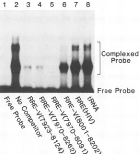
Free Probe
FIG. 3. Comlpetition analysis of the visna virus Rev-RRE inter-action. The indicated visna virus RNA sequences, as well as the entire HIV-1 RRE, were assessed for their ability to competitively inhibit binding of the labeled 7923 to 8124 RRE-V probe by the GST-Rev-V protein. A constant level (160 ng) of each unlabeled competitor RNA was preincubated with 400 ng of GST-Rev-V before addition of -1 ng of the labeled 7923 to 8124 RRE-Vprobe. The effect of this preincubation on the level of protein-RNA complex formation was then determined by gel retardation analysis. No Competitor indicates standard reaction conditions, which in-clude 200 ng of E. coli tRNA.
wk
Xf jOomplexed
^ Probe
-__ Free Probe
X k k
00P.'A P. '.A 00P,. A A pl
o~~~~~~~~~-FIG. 4. Sequence specificityof the HIV-1 Rev and Rev-V pro-teins. The L/Rev protein consists of the N-terminal domain of Rev-VfusedtotheC-terminalactivation domain of HIV-1 Rev (36). Similarly, the Rev/L protein contains the N-terminal domain of HIV-1 Rev fusedtothe C-terminal domain of Rev-V (36). Each of thesetwoRevchimeraswasexpressed as aGST fusionprotein, and their abilityto bind the RRE-V (lanes1 to 5) or the HIV-1 RRE (lanes6to 10)wascompared withthatofthe parental visna virus (GST-Rev-V) and HIV-1[GST-Rev(HIV)] proteins. Only proteins containingthe N-terminal basic domain of Rev-V boundtheRRE-V (lanes 2 and 3), while proteins containing the HIV-1 Rev basic domainspecificallybound only the HIV-1 RRE (lanes 9 and 10). This pattern therefore recapitulates the target specificity of these Revproteins previouslydetermined in vivo (36).
RNA segmentsderived fromvisna virus coordinates 7923 to 8124 (i.e., the probe sequence), 7970 to 8262, and, mini-mally, 7970 to 8091 all efficiently competed for Rev-V
binding. In contrast, tRNA as well as RNAs derived from visna virus coordinates 8001 to 8202 (i.e., containing the alternate RNAstructure)orcontaining the full-lengthHIV-1
RREwereunable tocompete effectively for Rev-V binding
(Fig. 3). Theconclusion that thespecific binding site(s) for Rev-Vonthe RRE-V is located between 7970 and 8091 in the visna virus genome was further supported by the direct demonstration of Rev-Vbinding to a radioactively labeled 7970to 8091probe (data notshown).
The HIV-1 Rev protein can be divided into at least two distinct functional domains (13, 20, 21, 28, 38, 40). Se-quencesadjacenttotheproteinNterminus thatinclude,but extendbeyond,ahighlybasic sequencearecritical both for
bindingto theHIV-1 RREandfor Revmultimerization (18, 20, 21, 28,40). Asecond essentialdomain,located adjacent
to the C terminus of HIV-1 Rev, is not involved in Rev
bindingor multimerization in vitro yet is essential for Rev function in vivo(13,20, 21, 23,28,40). Ithas therefore been proposed that this leucine-rich sequence forms the Rev activation domain (20, 23). Previously,wehave shown that chimeric proteins consisting of the N-terminal region of
HIV-1 Rev and theC-terminal region ofRev-V(Rev/L),or
viceversa(L/Rev),werefully activeontheRREcognatefor theproteinNterminus(36).This resultsuggestedthat HIV-1 Rev and Rev-V, despite very limited sequence homology,
were likely to share the same domain organization.
How-ever, these chimeric proteins, like theparental HIV-1 Rev
and Rev-V, were completely inactive on the noncognate
RRE (35, 36). To address the molecular basis for the sequencespecificityof HIV-1Rev, Rev-V, and thechimeric
1 2 3 4
VW WM
on November 9, 2019 by guest
http://jvi.asm.org/
[image:3.612.128.225.77.193.2] [image:3.612.330.533.78.245.2] [image:3.612.103.245.472.629.2]SINGLE-STRANDED (STRONG HIT)
* SINGLE-STRANDED (WEAK HIT)
+ DOUBLE-STRANDED (STRONG HIT) DOUBLE-STRANDED (WEAK HIT)
0 SINGLE AND DOUBLE-STRANDED HIT
ORD BASES PROTECTEDBY REV-V
'80
44
170 164
[image:4.612.71.561.86.359.2]202 (8124)
FIG. 5. Secondary structure of the RRE-V. The figure shows the entire 202-nt 7923 to 8124 sequence shown by both genetic and biochemical criteria tocontain the RRE-V(Fig. 1to3).Position 1equalscoordinate 7923 in the visna virusprovirussequencereported by
Sonigoetal.(32).Thestartof the visna virustransmembraneprotein(TMP)coincides withposition2(7924).Thecomputer-predictedRRE-V stem-loopstructure extends from 11(7923)to 186(8108)within thislargersequence. The minimal sequence showntobind Rev-V invitro extends from 7970(48)to8091(169)(Fig. 3)and is indicatedbyadarker line.Specific cleavagesitesforthesingle-strand-specificRNaseT2 and thedouble-strand-specificRNase Vlweredeterminedasdescribed in thelegendtoFig.6.The datapresentedhere representasummary ofcleavage data derived from threeindependent experiments.
Rev/L and L/Rev proteins, we purified each protein as a
GST-Rev fusion anddetermined whethertheycould specif-icallybindtothe HIV-1 RREorRRE-V.Asshown inFig. 4,
both proteins active on the RRE-V bound the RRE-V
efficientlybut failedtobind theHIV-1 RRE(Fig.4, lanes2,
3, 7, and8).Incontrast, thetwoproteinsactiveontheHIV-1 RREbound the HIV-1 RREefficiently but failedto interact with theRRE-V(Fig. 4, lanes 4, 5, 9, and 10). Wetherefore
conclude that theability of these Revproteinstofunction in onlyone of these twoviral systems is due to their distinct
RNAsequence specificities. These are, in turn, determined
bytheoriginof the basic domain of each protein.
The datapresented in Fig. 1 to 4 support the hypothesis that the RRE-V coincides with acomputer-predicted 176-nt RNAstem-loop structure locatedbetween 7933 and 8108 in
the viral genome (Fig. 5) (29, 35). As previously shown for
the somewhat longer HIV-1 RRE (17, 22), this structure is predicted to consistofa long presentation stemsurmounted bya set of shorter stem-loop structures that, in the HIV-1 RRE, contain primary sequence information critical for
HIV-1 Rev binding (1, 11, 17, 24, 37, 40). To assess the validity of the RNA secondary structure visualized in Fig. 5,
we radioactively labeled either the 5' or the 3' end of the 202-nt 7923 to 8124 RNA probe that contains the entire predicted RRE-V secondary structure (Fig. 5). These mole-cules were then subjected to limited digestion with the
single-strand-specificRNaseT2orthedouble-strand-specific RNase
Vi.
Sites of cleavage were identified afterelectro-phoresis through a denaturing polyacrylamide gel by refer-ence to sequence-specific RNase cleavage ladders run in
parallel (Fig. 6). These data, which are compiled in Fig. 5, strongly support the validity of the predicted RRE-V
sec-ondarystructure.
Asafirst step towardlocalizingthe sites of Rev-Vbinding
ontheRRE-V, we nextasked whether thebinding ofRev-V to the RRE-V would interfere with any of these specific
RNase cleavageevents (Fig. 6, lanes 9to 12and 21 to 24).
Nucleotidesprotected by Rev-Vbindingareindicated inFig.
6andarecompiledinFig.5.Theprotectednucleotideswere
foundtobe concentrated inseveralclusters near the apex of the RRE-V RNAstructure.This pattern is therefore similar
tothatreported for RNaseprotection of the HIV-1 RREby
Rev (17). The hypothesis that one or more of the shorter RNA stem-loops predicted for the RRE-V are critical for
Rev-Vbinding is further supported by the observation that the short 7970 (48) to 8091 (169) RRE-V sequence, which does not contain the RRE-V presentation stem, is both necessary and sufficient for specificRev-V binding in vitro
(Fig. 3).
DISCUSSION
AlthoughbothHIV-1 andvisna virus aremembersof the lentivirus family, they have diverged considerably during
evolution and now retain only limited primary sequence
identity (32). In addition, visna virus appears to have a
on November 9, 2019 by guest
http://jvi.asm.org/
5
13
r IoZ-
T2
Doo ~2 VIr ++om m m
F--)
F-}(I
_a
UN"b
a
:
qul-a.
~
- T2
Vl
no z
I ++ 0ommm
< <<:« 0-)(-)(+)(-)(+)
-0
*m. -.
So-..a.
_-A
122 -U121 zqCl16 Ul 15
G80-G68-
L
FF
$I
-C64L W~~~~_,G60
'6G59
*1- -} NC57
G54- -g54
F-
t~~~/-C48
L.. _ r:~G47
*
yU46
G44- C045
C43 A37 !-C36
G34- i G635
G26-G20
_,^3~
G99-ie
Gil12-
6125-
6139-6146
L.
6155- 4
6164-*
I.
6170-:e
GM15-* a
kILLj k t~ k
FIG. 6. RNasecleavage andprotectionpatternsof the RRE-V. The 202-nt 7923to8124 RRE-V probe shown in Fig.5wassynthesized
in vitroby usingT7 RNApolymerase (24). Subsequently, this RNA probewasuniquely end labeledatthe 5' end,using [-y-32P]ATPand T4
polynucleotidekinase (lanes1to12),oratthe 3' end, using [32P]pCP and T4 RNA ligase (lanes 13to24) (37). Aliquots of each RNAwere
incubated onice in the presence(+) orabsence (-) of Rev-V protein (lanes 8, 10, 12, 20, 22, and 24 and lanes 7, 9, 11, 19, 21, and 23,
respectively). The sampleswerethensubjectedtolimited digestion with either single-strand-specific RNase T2 (lanes 9, 10, 21, and22)or
double-strand-specific RNaseVi (lanes 11, 12, 23, and 24). Lanes 6, 7, 19, and 20 correspondtothe digestion controls inwhichnoRNase
wasadded. Alkalinehydrolysis (AH) lanesaswellaslanes showing cleavagepatternsinduced bysequence-specificRNases(lanes 1to5and
13to 17)were runinparalleltofacilitate identification of cleavage sites. RRE-V nucleotides thatwerespecificallyprotectedfromRNase
cleavage by bound Revareindicated.The RNasecleavage and protection dataderived from this and other experimentsarecompiled in Fig.5.
significantly lesscomplex genomic organizationthan HIV-1 and isnotbelievedto encodeequivalents of the HIV-1vpr,
vpu, and nefgenes (4, 29, 32). Furthermore, while both viruses encode transcriptional activators of their
homolo-gous longterminal repeat promoters, thesearefunctionally
distinct. HIV-1 Tat is ahighlypotenttrans activator that is
believedtoprimarilyfunctionbystimulating the elongation
of nascent proviral transcripts (reviewed in reference 3). Remarkably, thetargetsequenceforTathasbeen shownto
bean RNAstem-loop structure.Incontrast,the visna virus
S gene product only modestly enhances visna virus
tran-scription and appears to do so by directly or indirectly
interacting with AP-1 and AP-4 DNA target sites that are
locatedin theU3regionofthe virallongterminalrepeat(12).
G99--C64
r U1 15 C1 16 U121 A 122
U149
-N-U150
-G151
-/Cl56
_-A157
X -U158
XC0159 -C165 dw --C166
.l_.
on November 9, 2019 by guest
http://jvi.asm.org/
[image:5.574.111.447.64.535.2]Despite the considerable differences that exist between these two prototypic lentiviruses, genetic and biochemical analyses of the Rev-V trans activator reveal that this protein is remarkably similar in both function and mechanism of
action to the Rev trans activator of HIV-1. Previously, we had demonstrated that Rev-V, like HIV-1 Rev, activates the cytoplasmic expression of the incompletely spliced mRNAs that encode the viral structural proteins, including Gag and Env (35). Subsequently, we reported that Rev-V and HIV-1 Rev display the same functional organization, marked par-ticularly by an N-terminal basic domain that determines sequence specificity and by a conserved C-terminal leucine-rich motif that acts as the Rev activation domain (36). Here, we extend our previous genetic data for the existence of RRE-V by precisely mapping the RRE-V to a 176-nt RNA stem-loop structure that coincides with sequences encoding the N terminus of the transmembraneprotein component of the viral envelope. This locationis therefore identical tothat observed for the HIV-1 RRE(22). The secondary structure
adopted by the RRE-Vwasvalidated byanalysisof in vitro RNase cleavage patterns and appears generally similar to
that adopted by the somewhat larger
(234-nt)
HIV-1 RRE(22). Inparticular, both RREs consist ofahelicalregionof 30 to 40 bp surmounted by three or four shorter RNA
stem-loops. In the case of HIV-1, these apical stem-loop
struc-tures have been shown to contain primary sequence infor-mation that is critical forHIV-1 Revbinding (1, 11, 17, 24,
37, 40). Similarly, we demonstrated that Rev-V can also specificallybind to, andmultimerize on, the RRE-V.Invitro RNase protection data, as well as a preliminary mutational analysis of the RRE-V, indicate that the primary target for Rev-V bindingis alsolikelytobe located withinone or more
of the shorter stem-loops observed in the RRE-V.
The remarkably similarproperties of the Rev
proteins
of HIV-1 and visna virus areparticularly
surprising
given
the completely different mechanisms of actionreported
for the HIV-1 Tat and visna virus Stranscription
factors(3,
12).
ThisevolutionaryconservationsuggeststhatRevfunction is dependent on the specific interaction of Rev with a
highly
conserved cellular factor(s) involved in the
splicing and/or
transportofeukaryoticmRNA(9, 13,
18,
20,
23, 28, 36,
40).
This hypothesis is supported
by
the observation that HIV-1 Rev isfunctional inawidevariety
ofspecies,
including
cells ofavian and insectorigin(14,
23).
The extensivesimilarity
of the Rev proteins of theprototypic
lentiviruses HIV-1 and visnavirusreported heremay alsoimply
that the Revtransactivatorsrecently
reported
toexist in severalother nonpri-mate lentiviruses(16,
33)
arelikely
toretain many ofthese same functional characteristics.Although it is evident that HIV-1 Revand Rev-Vfunction by essentially identical
mechanisms,
it is also clear thatRev-Visunableto
functionally
substitutefor the HIV-1Rev protein and vice versa.Here,
we demonstrate that this results from theinability
of Rev-Vtobindthe HIV-1 RRE. HIV-1 Rev hasrecently
been showntospecifically
recognize
the primary sequence in and
adjacent
to a 5-nt RNAbulge
observed in the HIV-1 RRE
(1,
11,
37).
As this structured sequenceisnotconservedin theRRE-V,
it isnotsurprising
that HIV-1 Rev is also unable to bind to, and function
through, this visna virus element.It will
clearly
be of interestto determine the
precise
sequencerequirements
for Rev-Vbindingtodeterminewhether anyaspects ofthesequenceor
structure of the HIV-1 Rev
primary
binding
site have beenconserved during the
evolutionary
divergence
of these twodistinct viruses.Such
comparative
analyses
mayeventually
lead toanunderstandingof themechanismsthat underliethe
sequence
specificity
ofthese novelarginine-rich
RNA-bind-ing proteins
(19, 20)
andmight
also prove useful in thedevelopment
ofspecific
antagonists
of this critical viralregulatory
step.ACKNOWLEDGMENTS
We thankSharon Goodwinforsecretarialsupport.
This workwas supportedbythe Howard
Hughes
Medical Insti-tuteandbyPublic HealthServicegrantA128662from the National Institute ofAllergy
and Infectious Diseases.REFERENCES
1. Bartel,D.P.,M. L.Zapp,M. R.Green,andJ.W.Szostak.1991. HIV-1 Rev
regulation
involvesrecognition
ofnon-Watson-Crick basepairs
in viral RNA. Cell 67:529-536.2. Cullen,B. R. 1987. Use of
eukaryotic
expression
technology
in the functionalanalysis
of cloned genes. MethodsEnzymol.
152:684-704.3. Cullen,B. R.1990. The HIV-1 Tat
protein:
anRNAsequence-specific
processivity
factor? Cell63:655-657.4. Cullen,B. R.1991. Human
immunodeficiency
virusas aproto-typic
complex
retrovirus. J. Virol. 65:1053-1056.5. Daly,T. J., K. S. Cook, G. S.
Gray,
T. E. Maione, andJ. R. Rusche. 1989.Specific
binding
of HIV-1 recombinant Revprotein
totheRev-responsive
element in vitro. Nature(London)
342:816-819.
6. Davis,J.L.,S.Molineaux,andJ.E.Clements.1987. Visna virus exhibits a
complex
transcriptional
pattern: oneaspectof geneexpression
shared with theacquired
immunodeficiency
syn-dromeretrovirus. J.Virol. 61:1325-1331.7. Fauci,A.S. 1988. Thehuman
immunodeficiency
virus:infectiv-ity
andmechanisms ofpathogenesis.
Science239:617-622. 8. Feinberg,M.B.,R. F.Jarrett,A.Aldovini,R. C.Gallo,andF.Wong-Staal.
1986. HTLV-IIIexpression
andproduction
involvecomplex
regulation
at the levels ofsplicing
and translation of viral RNA. Cell 46:807-817.9. Felber,B.K.,M.
Hadzopoulou-Cladaras,
C.Cladaras,T.Cope-land,and G. N.Pavlakis. 1989. Rev
protein
ofhumanimmuno-deficiency
virustype 1affectsthestability
andtransportof the viral mRNA. Proc.Natl.Acad. Sci. USA 86:1495-1499. 10. Haase,A. T. 1986.Pathogenesis
of lentivirus infections.Nature(London)
322:130-136.11.
Heaphy,
S.,J.T.Finch,M.J.Gait,J.Karn,and M.Singh.
1991. Humanimmunodeficiency
virus type 1regulator
of virionexpression,
rev,formsnucleoprotein
filamentsafterbinding
toapurine-rich
"bubble" located within therev-responsive
region
of viral mRNAs. Proc. Natl.Acad. Sci. USA88:7366-7370. 12. Hess,J.L.,J.A. Small,andJ.E.Clements.1989.Sequences
inthe visna virus
long
terminalrepeatthatcontroltranscriptional
activity
andrespond
to viral trans-activation: involvement of AP-1 sites in basalactivity
and trans-activation. J. Virol. 63:3001-3015.13. Hope,T. J.,B.L.Bond,D.McDonald,N. P. Klein,and T.G. Parslow. 1991. Effector domains of human
immunodeficiency
virustype1 Revand humanT-cell leukemiavirustypeI Rexare
functionally interchangeable
and share an essentialpeptide
motif.J. Virol.65:6001-6007.
14.
Ivey-Hoyle,
M., and M. Rosenberg. 1990.Rev-dependent
expression
of humanimmunodeficiency
virus type 1gpl60
inDrosophila
melanogaster
cells. Mol. Cell.Biol. 10:6152-6159. 15. Kim, S., R.Byrn,
J.Groopman,
and D. Baltimore. 1989.Temporal
aspects ofDNA and RNAsynthesis during
humanimmunodeficiency
virusinfection: evidencefordifferential geneexpression.
J. Virol.63:3708-3713.16. Kiyomasu, T., T. Miyazawa,T. Furuya, R.Shibata, H. Sakai,
J.-I.Sakuragi,M.Fukasawa,N.Maki,A.Hasegawa,T.Mikami,
and A.Adachi. 1991. Identification of feline
immunodeficiency
virusrevgene
activity.
J. Virol.65:4539-4542.17. Kjems, J., M. Brown, D. D. Chang, and P. A. Sharp. 1991. Structural
analysis
of theinteractionbetween the humanimmu-nodeficiency
virus Revprotein
and the Rev response element. Proc. Natl. Acad. Sci. USA88:683-687.on November 9, 2019 by guest
http://jvi.asm.org/
18. Kjems, J., A. D. Frankel, and P. A. Sharp. 1991. Specific regulation ofmRNAsplicingin vitrobya peptide from HIV-1 Rev. Cell 67:169-178.
19. Lazinski, D., E. Grzadzielska, and A. Das. 1989. Sequence-specificrecognition of RNA hairpins bybacteriophage antiter-minatorsrequires aconservedarginine-rich motif. Cell 59:207-218.
20. Malim, M. H., S. Bohnlein, J. Hauber, and B. R. Cullen. 1989. Functionaldissection of the HIV-1 rev trans-activator--deriva-tion of a trans-dominant repressorofRevfunction.Cell 58:205-214.
21. Malim, M. H., and B. R. Cullen. 1991. HIV-1structuralgene expression requires the binding of multiple Rev monomers to the viral RRE:implications forHIV-1latency. Cell 65:241-248. 22. Malim, M. H., J. Hauber, S.-Y. Le, J. V. Maizel, and B. R. Cullen. 1989. The HIV-1 rev trans-activator acts through a structured target sequence to activate nuclear export of un-spliced viralmRNA. Nature (London)338:254-257.
23. Malim, M. H., D. F. McCarn, L. S. Tiley, and B. R. Cullen. 1991. Mutational definition of the human immunodeficiency virus type 1 Revactivation domain.J. Virol.65:4248-4254. 24. Malim, M. H., L. S. Tiley, D. F. McCarn, J. R. Rusche, J.
Hauber, and B. R.Cullen. 1990. HIV-1structural gene expres-sion requires binding of the Rev trans-activator to its RNA target sequence. Cell 60:675-683.
25. Muesing, M. A., D. H.Smith, C. D. Cabradilla, C. V. Benton, L. A.Lasky, and D.J.Capon.1985. Nucleicacid structure and expression of the human AIDS/lymphadenopathy retrovirus. Nature(London) 313:450-458.
26. Mullis, K. B., and F. A. Faloona. 1987. Specific synthesis of DNA in vitro via apolymerase-catalyzed chain reaction. Meth-ods Enzymol. 155:335-350.
27. Myers,G.,andG. N. Pavlakis. 1991. Evolutionary potentialof complexretroviruses,p. 1-37. In R. R. Wagnerand H. Fraen-kel-Conrat (ed.), Viruses. The Retroviridae, vol. 1. Plenum Press, New York.
28. Olsen, H. S., A. W. Cochrane, P. J. Dillon, C. M. Nalin, and C. A. Rosen.1990. Interaction ofthe humanimmunodeficiency virus type 1 Revproteinwithastructuredregion inenvmRNA is dependentonmultimer formation mediated through a basic stretch of amino acids. Genes Dev. 4:1357-1364.
29. Saltarelli,M.,G.Querat,D. A. M.Konings, R.Vigne, and J. E. Clements. 1990. Nucleotidesequenceandtranscriptional analy-sis of molecular clones of CAEV which generate infectious
virus. Virology179:347-364.
30. Smith, D. B., and K. S. Johnson. 1988.Single-step purificationof polypeptides expressed in Escherichia coli as fusions with glutathione S-transferase. Gene 67:31-40.
31. Sodroski, J., W. C. Goh, C. Rosen, A. Dayton, E. Terwilliger, and W. Haseltine. 1986. A second post-transcriptional trans-activator gene required for HTLV-IIIreplication.Nature (Lon-don)321:412-427.
32. Sonigo,P., M. Alizon, K. Staskus, D. Klatzmann, S. Cole, 0. Danos, E. Retzel, P. Tiollais, A. Haase, and S. Wain-Hobson. 1985. Nucleotide sequence of the visna lentivirus: relationship totheAIDS virus. Cell42:369-382.
33. Stephens, R. M., D. Derse, and N. R. Rice. 1990. Cloning and characterization ofcDNAs encodingequine infectious anemia virus Tat and putative Rev proteins. J.Virol. 64:3716-3725. 34. Talbott, R. L., E. E. Sparger, K. M. Lovelace, W. M. Fitch,
N.C. Pedersen, P. A. Luciw, and J. H. Elder. 1989. Nucleotide sequenceand genomicorganization offelineimmunodeficiency virus. Proc. Natl. Acad. Sci. USA 86:5743-5747.
35. Tiley, L. S., P. H. Brown, S.-Y. Le, J. V. Maizel, J. E. Clements, andB. R. Cullen. 1990. Visna virus encodesa post-transcrip-tional regulatorofviral structuralgene expression.Proc. Natl. Acad.Sci. USA87:7497-7501.
36. Tiley, L.S., M. H. Malim, and B. R. Cullen. 1991. Conserved functional organization of the human immunodeficiency virus type 1 andvisna virus Revproteins. J. Virol. 65:3877-3881. 37. Tiley, L. S., M. H.Malim, H. K.Tewary, P.G. Stockley, and
B. R.Cullen. 1992.Identificationof ahigh affinityRNA-binding site forthe humanimmunodeficiencyvirus type 1 Rev protein. Proc. Natl. Acad.Sci. USA89:758-762.
38. Venkatesh, L. K., and G. Chinnadurai. 1990. Mutants in a
conserved region near the carboxy-terminus of HIV-1 Rev identifyfunctionally important residues andexhibit a dominant negativephenotype.Virology178:327-330.
39. Vigne, R.,V.Barban, G.Querat, V. Mazarin, I. Gourdou, and N. Sauze. 1987. Transcription of visna virus during its lytic cycle: evidence of asequentialearly andlategene expression. Virology161:218-227.
40. Zapp, M. L., T. J. Hope, T. G.Parslow, and M. R. Green. 1991. Oligomerizationand RNAbindingdomains of the type 1 human immunodeficiency virus Rev protein: a dual function for an
arginine-rich binding motif. Proc. Natl. Acad. Sci. USA 88: 7734-7738.