I
COMPA
MODIFICA
IMPLANT
TH
ARATIVE
ATIONS O
SUPPORTE
TW
HE TAMILN
In
M
PROSTH
EVALUAT
OF “PEEK”
ED PROVI
WO DIFFER
AN
IN-Disserta
NADU Dr.
n partial fulfi
MASTER OF
B
HODONTIC
A
TION OF TH
ABUTMEN
SIONAL R
RENT LUTI
-VITRO ST
ation submit
M.G.R. ME
fillment for th
F DENTAL
BRANCH I
CS AND CR
APRIL 2011
HE EFFEC
NTS ON TH
RESTORAT
NG AGENT
TUDY
tted to
EDICAL UN
he Degree of
SURGERY
ROWN & BR
CERTIFICATE
This is to certify that the dissertation titled “
COMPARATIVE EVALUATION
OF THE EFFECT OF SURFACE MODIFICATIONS OF “PEEK”
ABUTMENTS ON THE RETENTION OF IMPLANT SUPPORTED
PROVISIONAL RESTORATIONS LUTED WITH TWO DIFFERENT
LUTING AGENTS”
is a bonafide record work done by
Dr. NIRMAL GEORGE
SAIBU
under our guidance and to our satisfaction during his post graduate study
period between 2008 – 2011.
This dissertation is submitted to
THE TAMILNADU DR. M.G.R.
MEDICAL UNIVERSITY
, in partial fulfillment for the degree of
MASTER OF
DENTAL SURGERY – PROSTHODONTICS AND CROWN & BRIDGE,
BRANCH I.
It has not been submitted (partial or full) for the award of any other
degree or diploma.
Guided by
Dr. N.S. Azhagarasan, M.D.S.
Dr.Manoj Rajan, M.D.S. D.N.B
Professor and Head of the Department, Professor,
Department of Prosthodontics
Department of Prosthodontics,
and Crown & Bridge,
and Crown & Bridge,
Ragas Dental College & Hospital
Ragas Dental College & Hospital
Chennai.
Chennai.
Dr. S. Ramachandran, M.D.S.
Principal,
ACKNOWLEDGEMENT
I thank THE LORD for all the grace bestowed upon me.
This dissertation is the result of work with immense support from many
people and it is a privilege now that I have the opportunity to express my heartfelt
gratitude to all of them.
I would be failing in my duty if I do not adequately express my deep sense of
gratitude and my sincere thanks to my Prof. Dr. N. S. Azhagarasan M.D.S.,
Head of the Department, Department of Prosthodontics, Ragas Dental college
and Hospital, Chennai, for his exceptional guidance, tremendous encouragement,
well-timed suggestions and heartfelt support throughout my postgraduate
programme which has never failed to derive the best out of me. I would like to
profoundly thank him for giving an ultimate sculpt to this study.
I wish to express my gratitude to Dr. S. Ramachandran M.D.S., Principal,
Ragas Dental College, Chennai, for his timely help and encouragement throughout
my post graduate course. I also thank him for permitting me to make use of the
amenities in the institution.
I would like to solemnly thank my Professor, Dr. K. Chitra Shankar M.D.S.,
for her untiring help, back up and constructive criticism extended to me throughout
the period of my postgraduate study.. I admire my madam for her patience and all
the care rendered to me when I needed it most.
I would also like to thank my Professor, Dr. K. Madhusudan M.D.S., a
constant source of support, encouragement and insightful guidance. He has been
the most able task master I have been privileged to work under, ever willing to
indulge me and always enthusiastic to readily share his limitless knowledge. He has
always pushed me towards my goals at all times and promoted my interests with
utmost love and concern, in him I have truly found the noblest of teachers.
I wish to convey my heartfelt gratitude towards my Professor, Dr. S
Jayakrishnakumar, M.D.S., for his valuable suggestions and help throughout my
postgraduate study. A lot would not have been learned by me if not for his gentle
yet firm manner of persuasion and purposeful diligence.
I would also like to thank Dr. Saket Miglani, M.D.S., my mentor and guide
throughout my postgraduate study. He has been the epitome of support and
encouragement all throughout and unfailingly given me the kindest of advice and
guidance. Unflinching in his belief of perseverance and practice he has been
instrumental in helping me complete my postgraduate study.
pursuit of knowledge have been my strength whenever I was in doubt and failed to
comprehend the intricacies of this work.
My sincere thanks to Dr. Manikandan, M.D.S., for all the help rendered
during my study.
I am grateful to Dr. Saravana Kumar, M.D.S., for sharing his knowledge
which helped me learn a lot in my field.
I would like to thank Dr. Vallabh Mahadevan, M.D.S., for being a
fantastic friend who always smiled while helping me to do better.
It would not be justifiable on my part if I do not acknowledge the help of my
fellow colleagues Dr Anil Kumar P, Dr Laju S, Dr Sangeetha S, Dr Sudarson K
and Dr Vidhya J, my seniors and juniors for their criticism and continued support
throughout my postgraduate course and also the non teaching staff in the
department
My sincere thanks to Dr. Syed Ammnula, Senior Technical officer, Central
Institute of Plastic Engineering and Technology, Guindy, Chennai for permitingt
me to do testing of the samples.
My sincere thanks to the Bio-statistician, Mr. Bhoopathi Kanguswamy,
National Institute of Epidemiology (Indian Council of Medical Research), Chennai
for his valuable guidance and statistical analysis of the data. .
CONTENTS
TITLE PAGE No.
1.
INTRODUCTION 1
2.
REVIEW OF LITERATURE 9
3.
MATERIALS AND METHODS 32
4.
RESULTS 42
5.
DISCUSSION 57
6.
CONCLUSION 64
7.
SUMMARY 69
LIST OF TABLES
Table No. Title Page No.
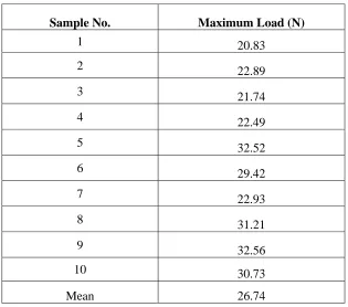
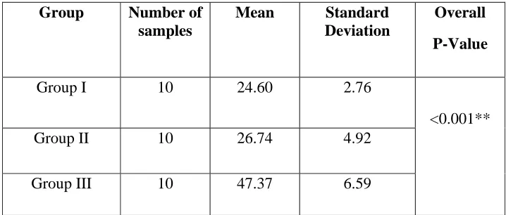
1.
Basic data of tensile bond strength for Group I
(PEEK abutments with retentive grooves luted with
non-eugenol zinc oxide cement) samples
46
2.
Basic data of tensile bond strength for Group II
(PEEK abutments milled with tungsten carbide bur and
air abraded with 110 µm aluminum oxide powder luted
with non-eugenol zinc oxide cement) samples
47
3.
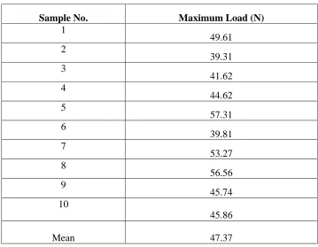
Basic data of tensile bond strength for Group III
(PEEK abutments milled with diamond abrasive luted
with non-eugenol zinc oxide cement) samples
48
4.
Comparison of mean and standard deviation of tensile
bond strength for Groups I, II and III by one-way
ANOVA for non-eugenol zinc oxide cement
49
5.
Comparison of mean tensile bond strength of Groups
I & II, Groups I & III and Groups II & III using
Tukey-HSD procedure (non-eugenol zinc oxide cement)
50
6.
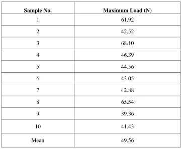
Basic data of tensile bond strength for Group IV
(PEEK abutments with retentive grooves luted with
polymeric implant cement) samples
51
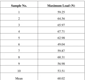
7.
Basic data of tensile bond strength for Group V (PEEK
abutments milled with tungsten carbide bur and air
abraded with aluminum oxide powder 110µm luted
with polymeric implant cement) samples
8.
Basic data of tensile bond strength for Group VI
(PEEK abutments milled with diamond abrasive luted
with polymeric implant cement ) samples
53
9.
Comparison of mean and standard deviation of tensile
bond strength for Groups IV, V and VI by one-way
ANOVA for polymeric implant cement
54
10.
Comparison of mean tensile bond strength of the pairs
Groups IV & V, Groups IV & VI and Groups V & VI
using Tukey-HSD procedure (polymeric implant
cement)
55
11.
Comparison of mean tensile bond strength Groups I &
IV, Groups II & V and Groups III & VI using
Tukey-HSD procedure (between polymeric implant cement
and non-eugenol zinc oxide cement)
56
ANNEXURE
LIST OF FIGURES
Fig no. Title
Fig.1a
:
Torque ratchet
Fig.1b
:
Ratchet hex driver
Fig.1c
:
Hand hex driver
Fig.1d
:
PEEK implant abutment
Fig.1e
:
Abutment screw
Fig.1f
:
Implant analog
Fig.1g
:
Implant cover screw
Fig.2
:
Polyvinyl siloxane putty and light body impression material
Fig.3
:
Silicone mold for resin block
Fig.4a
:
Dental surveyor
Fig.4b
:
Positioning of the implant analog in silicone mold
Fig.5
:
Clear auto polymerizing acrylic resin
Fig.6a
:
Fastening the PEEK implant abutment to implant analog with
hand
hex
driver
Fig.6b
:
Fastening the PEEK implant abutment to implant
analog with torque ratchet and ratchet hex driver
Fig.7a
:
Dental surveyor with micro motor
Fig.7b
:
Tungsten carbide bur
Fig.7c
:
Diamond abrasive
Fig.8a
:
PEEK implant abutment with retentive grooves (Groups I and IV)
Fig.8b
:
PEEK implant abutment milled with tungsten carbide bur and
air abraded implant (Groups II and V)
Fig.8c
:
PEEK implant abutment milled with diamond abrasive
[image:9.612.111.519.169.679.2]Fig.9a
:
Sandblaster
Fig.9b
:
Aluminum oxide power (110µm)
Fig.10
:
Scanning electron microscope (SEM)
Fig.11
:
Scanning electron microscope with specimen chamber
Fig.12
:
Inlay wax
Fig.13a
:
Inlay wax pattern
Fig.13b
:
Index over wax pattern
Fig.14
:
Sectioned index for acrylic coping
Fig 15a
:
Tooth coloured auto-polymerizing resin
Fig 15b
:
Fabricated provisional restoration
Fig.16a
:
Non eugenol zinc oxide cement
Fig.16b
:
Equal parts of non-eugenol zinc oxide cement
Fig.16c
:
Mixing procedure for non-eugenol zinc oxide cement
Fig.16d
:
Luting procedure for acrylic copying with non-eugenol zinc
oxide
cement
Fig.17a
:
Polymeric implant cement
Fig.17b
:
Automixing tips for polymeric implant cement
Fig.18
:
Luting procedure for acrylic coping with polymeric implant
cement
Fig.19
:
Two 1kg weight stones
Fig.20
:
Custom-made clear autopolymerizing acrylic resin table
Fig.21
:
Cementation of acrylic coping to implant abutment
Fig.22
:
Artificial saliva
Fig.23
:
Aging of test samples in artificial saliva
Fig.24
:
Custom-made hook embedded in autopolymerizing acrylic
block
Fig.25
:
Universal testing machine
Fig.27a
:
Debonded coping (Group I)
Fig.27b
:
Debonded coping (Group II)
Fig.27c
:
Debonded coping (Group III)
Fig.28a
:
Debonded coping (Group IV)
Fig.28b
:
Debonded coping (Group V)
LIST OF SEM PHOTOMICROGRAPHS
Fig. No.
Title
Fig. 29:
Fig. 30:
Fig. 31:
SEM photomicrograph of Groups I and IV (PEEK abutments with
retentive grooves) sample
SEM photomicrograph of Groups II and V (PEEK abutments milled with
tungsten carbide bur and air abraded with 110µm aluminum oxide)
sample
SEM photomicrograph of Groups III and VI (PEEK abutments milled
LIST OF GRAPHS
Graph No. Title
1.
2.
3.
4.
5.
6.
Basic data of tensile bond strength of Group I
(PEEK abutments with retentive grooves and luted with
non-eugenol zinc oxide cement) samples
Basic data of tensile bond strength of Group II
(PEEK abutments milled with tungsten carbide bur, air abraded
with 110µm aluminum oxide powder) and luted with
non-eugenol zinc oxide cement) samples
Basic data of tensile bond strength of Group III
(PEEK abutments milled with diamond abrasive and luted with
non-eugenol zinc oxide cement) samples
Comparison of mean tensile bond strength of Groups I, II & III
Basic data of tensile bond strength of Group IV
(PEEK abutments with retentive grooves and luted with
polymeric implant cement) samples
Basic data of tensile bond strength of Group V
(PEEK abutments milled with tungsten carbide bur, air abraded
7.
8.
9.
Basic data of tensile bond strength of Group VI
(PEEK abutments milled with diamond abrasive, and luted with
polymeric implant cement) samples
Comparison of mean tensile bond strength of Groups IV,V & VI
Comparison of mean tensile bond strength of Groups I & IV,
INTRODUCTION
The use of osseointegrated dental implants is recognized as a
predictable and successful treatment method for functional restoration of the
fully or partially edentulous patients.17
Restoration over implants can be screw retained, cement retained or a
combination of both. The factors that influence the different methods of
fixation of prostheses include passivity of the framework, retention, occlusion,
esthetics and retrievability of prosthesis. Screw retention in implant-supported
prostheses was developed in response to the need for retrievability of the
prosthesis. The restorative screw was designed to provide abutment -
restorative joint integrity. But it has the disadvantages of compromise in
esthetics due to the visibility of screw access hole. An analysis of occlusal
table width and screw hole size reveals that screw holes can occupy 50% or
more than the width of the occlusal table. Also it can affect the development
of ideal occlusal contacts. 1,20,22,24,39,52
The advantage of cement retained restoration include enhanced
ability to develop esthetics and occlusion, easier access to restorations of
posterior teeth and reduced complexity of clinical and laboratory procedures.
But it has disadvantages of difficulty in retrievability of prosthesis, reduced
in peri-implant sulcus and reduced retention in areas of limited inter-ridge
space.5,14
Several factors influence the amount of retention in
cement-retained restorations, whether they exist on natural teeth or implant
abutments. These factors are taper or parallelism, surface area and height,
surface finish or roughness, type of restorative material and types of
cement.22
The cements used in fixed prosthodontics are either definitive or
provisional. The choice of cement is one of the most important factors
controlling the amount of retention attained. Selection of cement that is too
retentive could lead to damage to implant, implant abutment, abutment screw
and the prosthesis if an aggressive removal technique is used. The selection of
cement that is not retentive enough could be a potential source of failure of
retention of the restoration. 3,10,22,52, 60
Studies have demonstrated that cements such as resin composite, zinc
phosphate, zinc polycarboxylate, ionomer, and resin modified
glass-ionomer are used on implant abutment to increase retention, provide good
marginal seal and to significantly enhance the cement failure loads of the
prostheses. but retrievability is more difficult when definitive cements are
used. Retrievability is highly desirable for cleaning and it facilitates evaluation
The literature reports suggested the use of provisional cements such as
reinforced zinc-oxide eugenol, non-eugenol zinc-oxide and calcium hydroxide
for ease of retrievability. But the problems encountered are inadequate
retention to resist functional force, cement washout, mobility of restoration.
Also gap between the prepared finish line and the margin of restoration causes
bacterial collection and may jeopardize the health of the soft tissue and the
implant or tissue interface.5,22, 29,40,48
Resin based cements used for luting definitive restoration on implant
abutment are well documented. Literature report indicates that polyurethane
and resin cements have comparative tensile bond strength on implant abutment
in comparison with other oxide eugenol, non-eugenol oxide,
zinc-phosphate and radiopaque reinforced glass ionomer cements.The polyurethane
and resin based provisional luting agents are typically stable intraorally. Thus
it is anticipated that the cement gap and the tensile bond strength of these
luting agents would change less over time.So the newer polymer based resin
cements could satisfy the requirements of adequate retention to resist
functional force, marginal seal and also retrievability of the
superstructure.2, 11,22,48
The factors that influence retention of cement retained restorations
are the same as those for natural teeth. Abutment with nearly parallel axial
was increased. The change in the angle of convergence affects the retention.
10,27,39.
There are literature reports that an increase in surface roughness of
prepared teeth will result in an increase in retention of cemented prosthesis,
due to mechanical interlocking of the cementing medium with the roughened
tooth surface. The surface modification on implant abutment can increase the
retention of the cement-retained implant-supported prosthesis. Surface
roughness of the implant abutment and luting agents are factors that can be
controlled by the clinician.In the literature, authors have advocated different
implant abutment surface modifications namely air-borne particle abrasion,
abrasion with diamond rotary cutting instrument,retentive groovesto enhance
the retention of cement-retained implant-supported cast copings. 2,29
Implant abutments are available as either smooth surface or with
retentive grooves. In most clinical situations to achieve parallelism, it is
necessary to mill the implant abutment resulting in the loss of retentive
grooves on the abutments. In clinical situations like implant in posterior
region, the implant abutment height is less and milling of abutment causes
further decreases in retention of the implant crown. In order to achieve
retention of the restorations, the surface modifications of the implant
abutments are advocated.4,7,29,40,41
surface modification of titanium abutment has been shown to influence
retention of implant restoration. Since predictable results are achieved with
immediate loading of implants, there is an increasing need for use of
provisional abutments. Development of a provisional prosthesis that will be
stable and esthetic should begin with the initial examination and should
involve all members of the implant team. To address these concerns, dentists
began modifying final abutments and using them to achieve acceptable labial
or buccal contours and support for the papillary tissues. Hence the PEEK
abutment cost effective modality that can easily be modified to support a
transitional prosthesis at the time of implant placement. Provisional
abutments are available with titanium, polymer ( plastic) or gold
abutment. Since 1980 a newer thermoplastic polymer poly (acryl ether)
ketone (PEEK) have been increasingly employed as biomaterials for
trauma, orthopedics and spinal implants. PEEK is a semicrystalline
thermoplastic with good mechanical properties.9,22,30,42
In dentistry, PEEK material is used as a plastic temporary abutment for
implants in the fabrication of temporary crowns. This biocompatible material
features a natural tooth colour appearance. PEEK can be also used as healing
abutments. The material has low surface energy and resistance to surface
modification by chemical treatments. It can be easily shaped with dental burs
is lack of research on the effect of surface modifications of PEEK abutments
on retention of provisional restoration. 49,50,56
In view of the above, the present in vitro study was conducted with the
aim of comparatively evaluating the effect of surface modifications of PEEK
abutments on the retention of implant-supported provisional restorations luted
with two different luting agents. The luting cements employed in this study
were, non-eugenol zinc oxide cement and polymeric implant cement. Also
glued to this aim were the following objectives:
1. To evaluate the effect of the retentive grooves present on PEEK implant
abutment on the tensile bond strength of non-eugenol zinc oxide cement
used for luting the provisional restorations. (Group I)
2. To evaluate the effect of milling with tungsten carbide bur and air abrasion
of PEEK implant abutments on the tensile bond strength of non-eugenol
zinc oxide cement used for luting the provisional restorations. (Group II)
3. To evaluate the effect of milling with diamond abrasive of PEEK implant
abutments on the tensile bond strength of non-eugenol zinc oxide cement
used for luting the provisional restorations. (Group III)
4. To comparatively evaluate the effect of the three different surface
modifications of PEEK implant abutments on the tensile bond strength of
5. To evaluate the effect of the retentive grooves present on PEEK implant
abutments on the tensile bond strength of polymeric implant cement used
for luting the provisional restorations. (Group IV)
6. To evaluate the effect of milling with tungsten carbide bur and air abrasion
of PEEK implant abutments on the tensile bond strength of polymeric
implant cement used for luting the provisional restorations. (Group V)
7. To evaluate the effect of milling with diamond abrasive of PEEK implant
abutments on the tensile bond strength of polymeric implant cement used
for luting the provisional restorations. (Group VI)
8. To comparatively evaluate the effect of the three different surface
modification of PEEK implant abutments on the tensile bond strength of
polymeric implant cement . ( Between Groups IV, V & VI)
9. To compare the effect of retentive grooves present on PEEK abutments on
the tensile bond strength of non-eugenol zinc oxide cement with the tensile
bond strength of polymeric implant cement (Between Groups I & IV)
10.To compare the effect of milling with tungsten carbide bur and air abrasion
on PEEK abutments on the tensile bond strength of non-eugenol zinc
oxide cement with the tensile bond strength of polymeric implant cement
(Between Groups II, & V)
11.To compare the effect of milling with diamond abrasive on PEEK implant
abutments on the tensile bond strength of non-eugenol cement with tensile
bond strength of polymeric implant cement (Between Groups III, & VI)
12.To compare the mean tensile bond strength of Groups I, II and III
non-eugenol zinc oxide for three different surface modification with that of
poly implant cement Groups IV,V & VI.
13.To evaluate qualitatively the surface of the PEEK abutments of Groups I
and IV samples (retentive grooves) using a scanning electron microscope
(SEM )
14.To evaluate qualitatively the surface of the PEEK abutments of Groups II
and V samples (milling with tungsten carbide bur and air abrasion with
110 µm) using a scanning electron microscope (SEM )
15.To evaluate qualitatively the surface of the PEEK abutments of Groups III
and VI samples (milling with diamond abrasive) using a scanning electron
microscope (SEM )
16.To correlate the qualitative and quantitative analysis of the study.
REVIEW OF LITERATURE
Schneider RL. (1987)51studied to evaluate the retention of gold castings
with the use of four dental cements such as zinc phosphate, polycarboxylate,
glass ionomer and zinc silicophosphate to various dental implants manufactured
in different materials and varying head designs. A significant difference was
found among all four cements tested. Glass ionomer cement was the most
retentive, followed by zinc phosphate, zinc silicophosphate, and
polycarboxylate, respectively.
Felton DA et al. (1987)16 Studied on the effect of surface roughness of
crown preparation using carbide and diamonds burs on retention of cemented
castings. Each crown was cemented with zinc phosphate cement with a 25kg
compressive force that was held for 10 minutes. All crowns were removed
parallel to the axis of draw by using Instron Universal testing machine with a
crosshead speed of 0.02cm/min until failure occurred. Conclusions drawn from
this study were: (1) Teeth prepared for full crowns by using diamond burs will
have 31% greater retention than preparations made with carbide burs. (2) If the
dentist wishes to use the more efficient carbide burs, alternative retentive
features should be considered in the preparation design.
Franchina N L et al. (1991)18 studied semicrystalline amorphous PEEK
by testing the resistance to solvents that were likely to be used for surface
modification reactions. The amorphous film is stable to methanol, ethanol at
group in semicrystalline PEEK is a versatile reactive handle for the surface
modification of PEEK.
Mash LK et al. (1991)35 Studied on leakage of various types of luting
agents. Freshly extracted molar teeth were prepared for complete cast gold
crowns cemented with zinc phosphate cement, polycarboxylate cement, glass
ionomer cement, a resin luting agent, or zinc oxide-eugenol temporary cement.
The specimens were tested at 1-, 6-, and 12-month intervals with radioactive
45Ca. The specimens were sectioned, auto-radiographs were made, and the
marginal leakage was evaluated on a scale of 0 to 3. The results showed that zinc
phosphate, polycarboxylate, and glass ionomer cements are equally suited for
permanent cementation of restorations. The resin luting agent showed high initial
leakage, indicating that it is not as desirable for permanent cementation
purposes. The zinc oxide-eugenol cement showed increased leakage with time
but is well suited for its indicated purpose, temporary cementation.
Breeding LC et al. (1992)5 compared the retentive strengths of castings
cemented to machined titanium implant abutments and to a human premolar with
three provisional luting agents. And also compared the retentive strengths of cast
noble metal implant abutments cemented into titanium fixtures with three
permanent luting agents both dry and after storage in 0.9% physiologic saline for
30 days at 37° C. Author concluded that no significant differences were noted in
retentive values between the cemented castings on the titanium abutments and
the natural tooth. The Temp Bond zinc oxide-eugenol luting agent exhibited a
Life calcium hydroxide luting agents. Ketac Cem glass-ionomer cemented
abutments that were stored in saline exhibited a significantly higher mean
retentive strength than abutments cemented with either Core Paste or Resiment
resin luting agents.
Dixon DL et al. (1992)11 studied to determine the amount of die space
necessary to reduce seating discrepancies of castings cemented onto implant
abutments and to determine the effect that this space created for the luting-agent
has on crown retention. Noble metal castings were made with 0.000 inch, 0.001
inch, 0.002 inch, and 0.003 inch spacing for pre-manufactured titanium implant
abutments. The castings were cemented onto the abutment with three permanent
luting agents using Core Paste, Resin cement, and Zinc Phosphate. Seating
discrepancies of each casting/abutment combination were measured, and the
castings were pulled from the abutments by use of tensile force. The results of
this study concluded that: (1) Spacing did not reduce .retentive values for any of
the specimen groups. The resin luting agent groups exhibited consistently higher
retentive strength than the zinc phosphate specimens. (2) Zinc phosphate and
Resiment luting agents exhibited seating discrepancy values below 25 µm with
0.001 inch luting agent spacing. Core Paste cemented specimens required 0.003
inch spacing to show values below 25µm.
Lorey RE et al. (1993)32 studied on the potential for bonding titanium
restorations by cementing with various adhesives: metal to metal, metal to
enamel, and comparing with a known procedure of bonding nickel-chromium.
Panavia. These were compared with nickel-chromium cones sandblasted and
bonded to nickel-chromium with Panavia. The author concluded that titanium
was most effectively bonded with All-Bond 2 and Panavia, with Panavia
samples significantly better than Panavia to nickel-chromium samples.
Kallus T et al. (1994)26 investigated the possible occurrence of loose
gold and abutment screws retaining full-arch osseointegrated prostheses which
had been in use for at least 5 years. Author found that Gold screw loosening
were related to framework misfit and was considered to be operator dependent to
some extent and he recommended that full-arch fixed prostheses be retightened
after 5 years.
GaRey DJ et al. (1994)19 compared the effects of thermocycling,
load-cycling, and human blood contamination on the retentive strength of five
different cements for luting posts to root-form implants. This study using an
Instron machine indicated that thermocycling did not significantly reduce
retentive strength of the test cements, but that cyclical compressive loading did.
However, the decrease is small and may not be clinically apparent. The
combination of thermocycling, cyclical load-stressing, and blood contamination
substantially reduced the retentive strengths for all of the cements. This suggests
that blood adversely affects the retentive strength of the cements tested more
than other variables.
Singer A et al. (1996)53 conducted a 6 month to 3-year follow-up study
implants were placed (86 in maxilla and 139 in mandible) for 92
implant-supported Fixed Partial Dentures, including single tooth restorations, which were
cemented using Temp-Bond or IRM. On 6-month to 3-year follow-up study, the
complications encountered were cement washout, porcelain fracture, loose
central screw and implant failure. The most common failure was cement
washout (9.8%), porcelain fracture (2.2%), loose central screw (2.2%) and
failure of one implant. This study suggests an alternative method to
screw-retained prostheses; the method presented may lower the reported complications.
Hebel KS et al. (1997)22 discussed how the choice to use screw-retained
or cement-retained implants dramatically influences the occlusion and esthetics
and concluded that occlusion and esthetics should not be arbitrarily discarded
through the use of screws to achieve retrievability. With dramatically increased
survival rates for dental implants, the once centrally important issue of
retrievability takes on less significance. The proper handling of cement-retained
implant prostheses provides for retrievability without compromising the
occlusion, esthetics, and stress distribution to the prosthetic components and
bone-implant interface. The impact of offset loading on the bone-implant
interface is not well understood and further research is required in this area.
Ayad MF et al. (1997)2 studied to determine the relationship between
surface characteristics of teeth prepared for complete cast crowns and retention
of respective cemented restorations. Three luting cements selected for this study:
zinc phosphate cement (Fleck's), glass ionomer cement (Ketac-Cem), and
carbide burs improved retention of complete cast crowns cemented with zinc
phosphate cement by 46% to 55% compared with tooth preparations completed
with diamond stones or finishing burs (2) The rotary instrument used for tooth
preparation did not have a significant difference on retentive strength of either
glass ionomer cement or Panavia-EX resinous cement (3) Panavia-EX resinous
cement provided greater tensile resistance to dislodgment of the casting and
more strength regardless of type of instrumentation selected to finish the tooth
preparations.
Preiskel HW et al. (1998)46 studied on the outcome of 73 telescopic
implant-supported fixed prostheses. Fifty-four prostheses were entirely
cement-retained, and 19 incorporated a screw-clamping unit. Author concluded that
cement-retained implant-supported telescopic prostheses provided a versatile and
reliable method of treatment. However, cement-retained telescopic prostheses
involving a distal cantilevered extension required the greatest postoperative
maintenance.
Ramp MH et al. (1999)48 compared the tensile bond strengths of 6
provisional luting agents such as Temp Bond, Provilink, Prototype, IRM,
Neo-Temp with releasing agent, and Neo-Neo-Temp used with cemented superstructures
and 1 implant system. Ten castings were fabricated and randomly paired with
abutment specimens. Author concluded that: (1) Temp Bond and Provilink
luting agents exhibited the lowest mean tensile bond strengths. (2) The prototype
cement was significantly stronger in tension than Temp Bond and Provilink
Neo-Temp luting agent exhibited tensile bond strength more than 3 times that of
Temp Bond luting agent. (4) Neo-Temp luting agent exhibited the greatest
tensile bond strengths of the luting agents tested.
Hofstede TM et al. (1999)23 has describes an alternative technique for
the fabrication of a complete-arch, cement-retained, metal-acrylic resin implant–
supported fixed partial denture. The prosthesis provides an esthetic and
inexpensive alternative to the traditional PFM implant-supported fixed partial
denture. The cemented framework over milled abutments provides excellent
prostheses retention, resistance, and stability. The cemented design ensures
optimum esthetics, good occlusal contacts, and a passive fit. The use of a base
metal alloy, with its high modulus of elasticity, enables the framework to be
designed with smaller dimensions. Waxing the framework on the refractory cast
eliminates any distortion caused by removal and handling of the wax pattern, and
acrylic resin denture teeth processed with a heat-polymerized acrylic resin
allows for easy and predictable repairs.
Keith SE et al. (1999)28 studied on the marginal discrepancy of the
implant-to-prosthetic-crown interface on nonsubmerged dental implants restored
with either a cemented or a screw-retained approach. Author concluded that: (1)
the mean marginal discrepancy of screw-retained metal-ceramic crowns on
implant abutments is significantly smaller than that of cemented metal-ceramic
crowns (2) the mean marginal discrepancy of metal-ceramic crowns cemented
on implant abutments with glass-ionomer is significantly smaller than those
Covey DA et al. (2000)10 studied on Effects of abutment size and luting
cement type on the uniaxial retention force of implant-supported crowns. Test
specimens consisted of standard and wide CeraOne (Nobel Biocare) titanium
abutments and matching CeraOne gold cylinders. Type III gold cast onto the
cylinders formed an attachment mechanism for testing. Three sizes of implant
abutments such as standard, wide, and experimental were evaluated. Two types
of cement were evaluated: a zinc phosphate permanent cement and a zinc oxide
eugenol provisional cement. Author has concluded that: Permanent luting
cement (zinc phosphate) produced uniaxial retention forces 2.5 to 4.7 times
greater than provisional cement (zinc oxide eugenol). The increase in surface
area provided by the Wide CeraOne abutment did not result in improvement in
retention strengths. Abutment height and height to width ratio were positively
related to retention strength, whereas an abutment’s total surface area and width
were not.
Michalakis KX et al. (2000)38 studied to evaluate the retentive strengths
of 4 provisional luting agents used to cement restorations supported by 2 or 4
implants 24 hours after cementation. The provisional luting agents used for this
study were: (1) ImProv, (2) Nogenol, (3) Temp Bond, and (4) Temp Bond NE.
Author has concluded that: (1) Twenty-four hours postcementation, ImProv
cement exhibited higher retentive strength values than Temp Bond NE, Temp
Bond, and Nogenol for both the 2-unit and the 4-unit implant-supported FPDs.
(2) Nogenol provisional luting agent seems to be more appropriate for the
Valbao FPB et al. (2001)59 describes a simple, quick, and economical
technique that makes use of a light-polymerized resin system to facilitate the
retention and removal of cement-retained implant prostheses. The technique
described offers restorative versatility, reversibility, and security because the
hexagon gold UCLA selection allows realignment of the restoration. Moreover,
the use of 2 burs with different diameters to prepare the access holes in the
abutment and the use of provisional cement and light-polymerized resin facilitate
removal of the restoration. An ultrasonic device or other prosthetic removal
device can be used, without harming the abutment or the coping, to help remove
the prosthesis once the light-polymerized resin has been removed. One
disadvantage of this technique is that it may not be applicable when there is
limited interocclusal distance.
Squier RS et al. (2001)54 compared retentiveness of dental cements used
with metallic implant components. The cements used for this study were zinc
phosphate, resin composite, glass ionomer, resin-reinforced glass ionomer, and
zinc oxide–non-eugenol. Author has concluded that: (1) Resin cement
demonstrated the highest mean retentive strengths. (2) Glass-ionomer and zinc
oxide–non-eugenol cements exhibited the lowest mean retentive strengths. (3)
Zinc phosphate and resin-reinforced glass ionomer showed intermediate mean
retentive strengths. (4) Use of an anodized abutment surface does not appear to
affect retentive strength. (5) Resin and resin-modified glass-ionomer cements
failed cohesively, leaving residual cement on the abutment and the implant
Randi AP et al. (2001)49 compared the dimensional accuracy or the fit of
retrievable cement-retained implant-supported prostheses using a bis-GMA
composite luting cement to a traditional wax and cast, screw-retained frame
work. And evaluate the retentive strength of a retrievable cement-retained,
implant-supported FPD. The control group consisted of 10 frameworks
fabricated with traditional wax and casting techniques directly on the gold
cylinders. Frameworks were analyzed for distortion in the z-axis and compared
with retrievable cement-retained group using scanning electron microscopy and
a single screw test. Results demonstrated that the retrievable cement-retained
group had a decreased gap distance and improved angular distortion compared
the control group. Retentive strength measurements for the cement-retained
group with a direct pull-out test revealed a mean pull-out force of 65.7 kg. This
retentive test supports a simplified technique of clinically luting
implant-supported frameworks with adequate retentive strength.
Dumbrigue HB et al. (2002)14 describe a technique to minimize the
amount of excess cement used to lute implant restorations with the use of ITI
solid abutments. The luting cement used only occlusal half the intaglio of the
restoration. This amount will provide sufficient flow to the axial walls cervically
and reduce the amount of excess cement along the restorative margin. The
disadvantage of this technique is that incomplete sealing of the restorative
margin with the luting agent may result. The resultant microgap between the
implant fixture and restoration may harbor subgingival microorganisms with the
In another technique the intaglio of the implant restoration fills with
luting agent, and seats it extraorally on the practice abutment or implant analog.
Wipe excess cement with a gloved finger or cotton tip applicator; immediately
remove the crown from the analog, and cement it intraorally. Removal of the
restoration from the analog should be in line with the long axis of the analog to
avoid the elimination of too much cement from the axial walls of the restoration.
The advantage of this technique is that a more complete flow of cement to the
axial walls and restorative margins of the implant restoration is achieved.
However, a practice abutment or abutment analog needs to be used with a luting
agent that has a longer working time.
Doerr J (2002)12 describes an accurate method for locating the implant
abutment access chamber and abutment retaining screw to facilitate the removal
of a cemented implant restoration. The described technique is a time-saving and
accurate way to retrieve a cemented implant restoration without destroying the
restoration or abutment but disadvantages are need to retain the original implant-
and abutment-level casts used to fabricate the original restorations and the
access hole made in the restoration must be filled and the necessary material may
be dissimilar to that used to fabricate the original restoration.
Okamoto M et al. (2002)45 describe a technique for removing a
cemented superstructure from an implant abutment. A cylindrical guide hole on
the lingual surface of the abutment is prepared and an access hole on the lingual
side of the superstructure. To remove the superstructure from the abutment,
removing driver to generate a shear force to raise the superstructure. The shear
force will cause the temporary cement layer to fracture and enable removal of
the superstructure from the abutment. This technique is easy and reliable.
Mansour A et al. (2002)34 compared the retention of metal copings
fabricated to fit on the one-groove, one flat-sided solid titanium abutment using
six different cements such as eugenol-free zinc oxide (Temp Bond NE),
zinc-oxide eugenol (IRM), zinc phosphate (Hy-Bond), resin-modified glass ionomer
(Protec Cem), zinc polycarboxylate (Durelon) and 10-methacryloyloxydecyl
dihydrogen phosphate resin (Panavia 21). Author concluded that the results do
not suggest that one cement type is better than another, but they do provide a
ranking order of the cements in their ability to retain the castings. This ranking is
somehow different than that obtained when the same cements are used on natural
teeth. The material and surface characteristics of the implant abutment are likely
responsible for this difference. Cement retention values obtained from studies
that use teeth as abutments may be misleading when used in cement-retained
implant-supported crowns.
Ergin S et al. (2002)15 Compared the retentive properties of five
different luting cements such as zinc phosphate (Phosphate cement), glass
ionomer (Meron), resin-modified glass ionomer (Principle), resin-modified glass
ionomer (Fuji Plus) and resin (Avanto) cements on base and noble metal
copings. Author concluded that Fuji Plus and Avanto showed significantly
higher retentive strength for nobel metal alloys and Phosphate, Principle and
Bernal G et al. (2003)3 Compared the effect of 20 degrees and 30
degrees of abutment taper, total occlusal convergence (TOC), the
occlusocervical dimension of 4 mm (S) and 8 mm (L), and cement such as zinc
phosphate cement (Fleck's cement), zinc oxide eugenol cement(Temp-Bond),
zinc oxide eugenol cement plus Vaseline, and acrylic/urethane cement (IMProv)
on the tensile resistance to dislodgement of cement-retained, implant-supported
restorations. Author concluded that preparations with 20 degrees of TOC and 8
mm of occIusocervical dimension had significantly higher mean retentive values
for all of the cements tested. Significant differences in mean tensile strength
were observed, with the highest tensile resistance seen with IMProv, followed by
Fleck's cement, and the lowest tensile resistance seen with Temp-Bond plus
Vaseline.
Michalakis KX et al. (2003)39 reviewed on Cement-Retained versus
Screw-Retained implant restorations. The advantages, disadvantages, and
limitations have been discussed on both types of restorations. Several factors are
essential to the long-term success of any implant were reviewed with regards to
the both method of fixation. These factors include (1) ease of fabrication and
cost, (2) passivity of the framework, (3) retention, (4) occlusion, (5) esthetics, (6)
delivery, and (7) retrievability
Retrievability is advantageous for reservicing, replacement, or salvaging
of the restorations and implants necessitated by (1) the need for periodic
replacement of prosthodontic components; (2) loosening or fracture of the
after loss of an implant; and (5) surgical reintervention. The main disadvantage
of cemented prostheses is the difficulty of their retrievability. Although retrieval
is needed less often because of the dramatically increased survival rates for
dental implants, the need for future removal of FPDs should not be overlooked.
For this reason, provisional luting agents are widely used for the cementation of
cement-retained restorations. From various laboratory researches it was
concluded that there is a statistically significant difference in the tensile strengths
of provisional cements. Clinicians are encouraged to use the least retentive
cements so that prostheses can be retrieved if necessary.
Zidan O et al (2003)61 The retention of complete crowns prepared with
three different tapers and luted with four different cements. One hundred twenty
sound human molar teeth were assigned randomly to 1 of 12 groups. The groups
represented the 4 cements: zinc phosphate (Fleck’s), a conventional glass
ionomer (Ketac-Cem); 2 adhesive resin cements (C&B Metabond and Panavia);
and 3 tapers of 6-degrees, 12-degrees, and 24-degrees within each cement.
Crowns were cast with a high noble alloy. The 6-degree taper was considered the
control within each cement group. Retention was measured (MPa) by separating
the metal crowns from the prepared teeth under tension on a universal testing
machine. Analysis of variance was used to test the main effects on the retentive
strength of full crowns, namely cements, tapers, and failure modes. Author
concluded that within the limitations of this study, the retentive values of the
adhesive resins at 24-degree taper were 20% higher than the retentive values of
yielded retention values that were double the values of zinc phosphate or
conventional glass ionomer cement.
Rajan M et al. (2004)47 describe a method to fabricate a retrievable,
cement- and screw-retained implant crown for a single molar. A complete arch
closed tray impression using the indirect transfer coping assembly made. The
implant analog screwed to the transfer coping and cast is poured using Type IV
stone. The friction fit abutment is attached to the implant analog. The abutment
is prepared for a metal-ceramic crown. The hexagonal screwdriver is placed in
position to maintain the screw access channel. Wax pattern is made for the
implant crown coping and casting is done and verified intraorally. With keeping
the occlusal screw access channel open ceramic is added incrementally. Using
the crown as a repositioning device, the abutment to the implant is screwed and
the hexagonal screwdriver is passing through the open screw access channel.
The abutment screw is tightened and torque. The crown with a definitive cement,
such as zinc phosphate, glass ionomer, or resin cement is cemented. The excess
cement is removed through the access opening with an explorer and closes the
screw channel with gutta-percha. Author suggests that, this is a simple, practical,
and effective technique for fabricating a retrievable cemented implant restoration
has been described. The technique facilitated predictable prosthesis retrieval and
allowed for removal of excess cement.
Tomson P. L. M. et al (2004)57 reported a patient who developed
peri-implant bone loss around 2 maxillary endosseous root-form peri-implants after
occurred around 1 of the implants due to retained excess cement. Reparative
treatment consisted of a guided bone regeneration technique. Following a
9-month period of submerged healing, the implants were re-exposed and restored
to complete function.
Maydan. L et al (2004)36 evaluated the effect of the addition of
circumferential grooves to the abutments on the retention of cemented crowns in
an implant system using two types of cements. 52 implant abutments (MIS,
Israel) were divided into four groups: no grooves, one groove, two grooves and
three grooves. Other than the number of grooves, all abutments were identical.
13 NiCr identical casts were prepared to fit all 52 abutments. The casts were
cemented to each group of abutments using a temporary cement (Tempbond NE,
Peterborough, UK) and a permanent cement (Harvard Cement, Harvard Dental,
Germany). The casts were separated from the abutments using an Instron testing
machine (crosshead speed of 0.5 cm/min) and the maximum retentive forces
were recorded. The data were subjected to one way-ANOVA and Tukey tests.
The mean retentive forces for Tempbond NE cement were 168±32 N for no
grooves,192±33 N for one groove, 208±23 N for two grooves and 246±17 N for
three grooves. The mean retentive forces for Harvard cement were 343±60 N for
no grooves, 579±30 N for one groove and 540±38 N for two grooves (results for
three grooves have not been obtained yet). For Tempbond cement groups,
retention values were increased in accordance to the number of grooves
(p<0.001). For Harvard cement, addition of one and two grooves increased
circumferential grooves to implant abutments increases the retention of crowns
cemented with Tempbond as well as Harvard cements. These grooves can be
crucial for the retention of crowns cemented to short abutments of implant
systems.
Bresciano M. et al. (2005)6 studied to evaluate the retention of four
cements such as zinc-phosphate, zinc oxide-eugenol, polyurethane resin with and
without vaseline cemented on Procera titanium abutments of 5, 7, and 9 mm of
height, and of 0 degrees , 4 degrees , and 8 degrees of convergence angle. Author
concluded that the most retentive cement was zinc-phosphate, followed by
polyurethane, polyurethane plus vaseline, and zinc oxide-eugenol.
Maeyama H. et al (2005)33 compared the retentive strength of metal
copings on prefabricated abutments with five different luting cements such as
zinc oxide-eugenol-free temporary, zinc phosphate, glass ionomer,
resin-reinforced glass ionomer, and composite resin cements. Author concluded that
the retentive strength of metal copings on implant abutments is somewhat
different from those of conventional cemented restorations on natural teeth.
These differences may be influenced by differences in surface roughness and the
height of the abutment.
Kim Y et al. (2006)29 compared the retention of provisional
autopolymerizing acrylic resin implant-supported single restorations with
combinations of different implant abutment surface conditions such as abraded
with 50-mm aluminum oxide and roughened with a medium-roughness diamond
TempBond NE, Life, and Zone. Author concluded that Surface modification of
an implant abutment by airborne-particle abrasion or diamond rotary cutting
instrument did not improve retention of a provisional acrylic crown when Life or
Zone was used as the luting agent. Airborne-particle abrasion may be an
effective method to increase retention of a provisional acrylic crown when
TempBond NE is used.
Kaar D et al. (2006)25 evaluates the effect of fatigue damage on the
force required to remove a restoration in a cement-retained implant system using
with three types of luting cements (ImProv, UltraTemp, and TempBond). Author
concluded that TempBond luting agent as the material of choice for provisional
cementation because it allows easier removal of the prosthesis and maintains
enough retention to prevent loosening of the restoration. ImProv had the highest
retentive value before and after the two cycles, and TempBond had the lowest.
UltraTemp had the highest percentage of retentive value lost. TempBond had no
significant loss under loading even though initially it was the weakest.
Chee et al. (2006)8 discussed the clinical perspective and the advantages
and disadvantages of both screw-retained and cement-retained implant prosthesis
under the heading of aesthetics, retrievability, retention, implant placement,
passivity, provisionals, occlusion, immediate loading, impression procedures,
long term treatment planning. Author concluded that although there is no clear
advantage of one type retention over the other, it is the clinical preference to use
screw retention as primary mode when restoring implants.
Michalakis K et al. (2007)40 compared the effects of thermal cycling
and surface roughness of metal implant abutments and the intaglio surface of the
copings on the retentive properties of 4 provisional luting agents commonly used
in the cementation of implant-retained fixed partial dentures (FPDs). Within the
limitations of this in vitro study it was concluded that: Thermal cycling had a
detrimental effect on the retentive properties of all cements tested. Air abrasion
significantly increased the cement failure loads of the provisional luting agents
used in the study and seems to be an effective way of increasing the retention of
implant-retained FPDs. Nogenol exhibited the lowest mean retentive values after
thermal cycling and after air abrasion for both the 2- and 4-unit FPD models.
Improv exhibited the highest mean retentive strength for both the 2- and 4-unit
FPDs after both the thermal cycling and air abrasion treatments.
Kurtz S M et al. (2007)30 studied about the PEEK employed as
biomaterials for trauma, orthopedic and spinal implants also discussed about the
structure, mechanical properties and chemical properties and chemical resistance
to PEEK biomaterials. This review concluded that PEEK had greatest clinical
impact in the field of spine implant design, total joint replacement and fracture
fixation implants. Radiolucency and inertness of PEEK biomaterials must be a
platform to further develop bioactive materials with the blending of
hydroxyapetite and tricalcium phosphate into sintered PEEK
Lawson CN et al. (2007)31 measured the retention and flexural strength
of base metal alloy castings to dentin provided by 8 provisional cement. This
based cement. Pemier implant cement, SensiTemp, TNE were the resin based
cement and GC Temp, TempBond NE, TempoSIL, TempoCem, and Zone were
zinc-oxide non-eugenol based cement. Author concluded that all three resin
based cement were highest crown retention strength and flexural strength
comparatively oxide non-eugenol based cement
Sheets JL et al. (2008)52 compared the retentive nature of common
dental cements that have been adapted for use in the implant abutment
cement-retained crown technique. Ten regular diameter implant analogs were embedded
in stainless steel disks. Unmodified CRC abutments were attached and torqued
to 30 Ncm. Test crowns were waxed and cast with base metal alloy. Castings
were fitted, cleaned with aluminum oxide, and steam cleaned prior to application
of the cement. The cements used were: (1) Temp Bond, (2) UltraTemp, regular,
(3) UltraTemp firm, (4) ImProv with petroleum jelly coating of crown, (5)
ImProv without petroleum jelly, (6) Premier Implant with KY Jelly coating of
abutment, (7) Premier Implant without KY jelly, (8) TR-2, (9) Fleck's, (10)
Ketac Cem Aplicap, and (11) Fuji Plus Capsule. Within the limitations of this in
vitro study, it is not suggested that any one cement variety is better than another
at retaining cement-retained crowns (CRCs) to implant abutments or that a
threshold value must be accomplished to ensure retention. The ranking of
cements presented is meant to be a discretionary guide for the clinician in
deciding the amount of desired retention between castings and implant
Ichikawa T et al. (2008)24 describe two methods for improving the
retrievability of cement-retained implant superstructures. One method involves
incorporating a removal screw in the superstructure and the second method uses
a small dimple on the abutment, accessed through a vent in the superstructure.
The technique uses a large removal screw in the same dimension as the gold
screw positioned parallel to the long axis of the abutment. A small removal
screw positioned at an oblique angle, thereby allowing the removal screw access
to nonesthetic and nonocclusal contact areas. The rotating lever system provides
the convenience of prosthesis retrievability as well as venting for removal of
excess provisional cement.
Urdaneta RA et al. (2008)58 studied on the integrated abutment crown
(IAC) technique for the fabrication of single-tooth implant-supported crowns
where the abutment and the crown are one unit. The abutment-crown complex is
connected to the implant with a locking taper. This technique does not use
cement to retain the crown or screws to retain the abutment. Author concluded
that the screw less and cementless implant restorations showed a survival rate of
98.7%, excellent marginal adaptation with a cementless interface, color stability,
and a reduced number of prosthetic components. Plaque accumulation was
observed around the crown material. The surface texture had higher roughness.
IACs located between a tooth and implants were 2.65 times more likely to have
postinsertion complications and IACs with incorrect anatomic form
(overcontoured) were 3.26 times more likely to have postinsertion
Mehl C. et al. (2008)37 studied on the retrievability of cemented implant
crowns using two different removal devices using five cements such as
eugenol-free zinc oxide (Freegenol), zinc phosphate (Harvard), glass ionomer (Ketac
Cem), polycarboxylate (Durelon) and so-called self-adhesive resin (RelyX
Unicem) cement. Author concluded that zinc phosphate and glass ionomer
cement might be suitable for a so-called 'semipermanent' (=retrievable)
cementation, while polycarboxylate seems to provide the most durable
cementation.
Dudley JE. (2008)13 studied on the influence of compressive cyclic
loading on the physical retention of cast crown copings cemented to Straumann
synOcta implant abutments. Author concluded that, with a resin, glass ionomer
and temporary cement was significantly affected by cement type but not
compressive cyclic loading. Resin cement is the cement of choice for the
definitive non-retrievable cementation of cast crown copings to Straumann
synOcta implant abutments out of the three cements tested.
Okada H et al. (2009)44 studied on development of a new temporary
luting agent consisting of PEMA and eugenol-residue ratio and bond strength of
luting cements such as for abutment materials and assess their clinical
applicability. The residue ratio of PE 1.0 on the abutment material after
temporary restoration removal was lower than those of comparable temporary
luting agents (polycarboxylate cement type, zinc oxide-eugenol cement type),
and no residue was recognized for PE 1.6. On bond strength, those of the
dentin surface after the removal of trial agents tended to be the same or increase
in comparison to commercial temporary luting agents. Author concluded that the
trial agents were suitable for clinical use
Schmidlin P R et al. (2010)50 studied on the effectof a composite resin
cement and an adhesive composite system to a non treated PEEK surface, acid
etching surface with sulfuric acid, sand blasting with aluminium oxide powder
110µm, silica coating using the rocatec system and polished sand blasted CP
titanium. Shear bond strength was measured in a shear testing machine and
failure was assessed. No bond Conclusion drawn from this study was no bond
MATERIALS AND METHODS
This in vitro study was conducted for the comparative evaluation of the
effect of surface modifications of PEEK implant abutments on the retention of
implant-supported restoration with two different cements (polymeric implant
cement and non-eugenol zinc oxide cement).
Materials used for this study:
• Torque ratchet (UniTi, Equinox medical technologies, Holland)( Fig. 1a)
• Ratchet hex driver (UniTi, Equinox medical technologies, Holland)
(Fig. 1b)
• Hand hex driver (UniTi, Equinox medical technologies, Holland) (Fig. 1c)
• PEEK implant abutment (Bio temp, UniTi, Equinox medical technologies,
Holland ) (Fig. 1d)
• Implant analog (UniTi, Equinox medical technologies, Holland )(Fig. 1f)
• Implant cover screw (UniTi, Equinox medical technologies, Holland)
(Fig.1g)
• Polyvinyl siloxane putty and light body impression material (Aquasil,
Dentsply, USA)(Fig. 2)
• Auto polymerizing acrylic resin (DPI, India) (Fig. 5)
• Tooth coloured autopolymerizing acrylic resin( DPI, India) (Fig.15a)
• Tungsten carbide milling bur (0110.023HP Edenta, Switzerland) (Fig. 7b)
• Diamond abrasive (5218/037,Edenta, Switzerland) (Fig. 7c)
• Non-eugenol zinc oxide cement( Rely X Temp NE, 3M ESPE, USA)
(Fig.16a)
• Polymeric implant cement with automixing tips (Implacem, Equinox
Medical Technologies B. V, Holland) (Fig. 17a)
• 1kg weight stones (Giri Brothers, Chennai, India) (Fig. 19)
• Artificial saliva(Wet mouth, ICPA health, India) (Fig. 22)
Equipments used for this study:
1. Dental surveyor (Bego, Germany) (Fig. 4a)
2. Micromotor with handpiece for milling (Sprint, Heraeus Kulzer
Dental, Germany) (Fig.7a)
3. Sandblaster (Delta, India) (Fig. 9a)
4. Scanning electron microscope (Hitachi d3400, Japan ) (Fig. 10)
5. Universal testing machine (Lloyd instruments,Farnham,U.K.)(Fig. 25)
Scanning electron microscope
The scanning electron microscope (SEM) (Fig. 10) is a type of electron
microscope that images the sample surface by scanning it with a high-energy
beam of electrons in a scan pattern.
The types of signals produced by an SEM include secondary electrons
and back scattered electrons (BSE) and transmitted electrons.
Metal objects require little special preparation for SEM except for
cleaning and mounting on a specimen stub. Nonconductive samples usually
gold, deposited on the sample either by low vacuum sputter coating or by high
vacuum evaporation. Coating prevents the accumulation of static electric
charge on the specimen during electron irradiation.
For PEEK, Carbon double-sided conductive tapes are used for
electrical conductivity and offer a clean background.
Universal testing machine:
To obtain the tensile bond strength of the specimens, universal
mechanical testing machine (Lloyd instruments, Farnham, U.K.) (Fig. 25) was
used. This machine rests on a table top. It consists of a lower chamber, upper
chamber, a display board to display the amount of force needed and a
computer. The upper member houses the hydraulic pressure machine. It also
has the fixture to hold the custom-made hook embedded to the clear
autopolymerizing resin block, (Fig. 26) on which a stylus can be attached for
determining the fracture bond strength. The lower portion has a bench vice test
specimen fixture to hold the test specimens. The whole unit is attached to a
METHODOLOGY
The following methodology was adapted for preparation and for testing the
samples.
1. Preparation of silicone mold
2. Positioning of the implant analog in the mold
3. Stabilizing the implant analog with auto polymerizing acrylic resin
4. Fastening the implant abutment to implant analog
5. Surface modification of test abutment samples
6. Scanning electron microscopy of surface topography of abutments
7. Fabrication of provisional acrylic copings
8. Cementation of acrylic copings to implant abutments
9. Aging of test samples with cemented copings
10.Testing the samples for tensile bond strength
11.Data tabulation and statistical analysis
1. Preparation of silicone mold:
Silicone mold is necessary for the fabrication of the acrylic blocks of
standard dimension for the positioning and stabilizing the implant analog
(Fig.1f). This mold is prepared from the custom fabricated acrylic block of 25
x 25 x 15 mmdimensions. The silicone mold was prepared with polyvinyl
siloxane with an internal mold space of 25 x 25 x 15 mm.(Fig.3)
2. Positioning of the implant analog in the mold:
The silicone mold (Fig.3) was positioned on the surveying table of a
cover screw (UniTi, Equinox medical technologies, Holland) (Fig.1g) was
attached to the implant analog with a hand hex driver (UniTi, Equinox medical
technologies, Holland).(Fig.1c) With the help of a straight mandrel an implant
analog (Fig.1f)with cover screw was attached to long axis of the surveying
arm of the dental surveyor. The surveying arm was adjusted to position the
implant analog in the centre of the silicone mold such that the platform for the
implant abutment was 1mm above the surface of silicone mold (Fig.4b)
3. Stabilizing the implant analog with auto polymerizing acrylic resin:
After positioning the implant analog,(Fig.4b) the space around the
analog was filled with clear auto polymerizing acrylic resin (DPI,
India)(Fig.5). The silicone mold(Fig.3) was completely filled such that the
platform of the implant abutment was 1mm above the surface of the resin
block. The resin was allowed to polymerize and the resin block removed from
the silicone mold. 60 clear acrylic blocks were made, each stabilizing one
implant analog.
4. Fastening the implant abutment to implant analog:
A PEEK implant abutment (Bio temp, UniTi, Equinox medical
technologies, Holland) (Fig.1d) was placed on the implant analog (Fig.1f) and
the abutment screw was first tightened with hand hex driver (UniTi, Equinox
medical technologies, Holland) (Fig.1c) (Fig.6a) and followed by tightening to
25 Ncm of torque with a ratchet hex driver (UniTi, Equinox medical