Neuro-Oncology
Neuro-Oncology 18(12), 1622–1633, 2016doi:10.1093/neuonc/now117 Advance Access date 13 June 2016
10.1093/neuonc/now117
Reactive astrocytes potentiate tumor aggressiveness in a murine
glioma resection and recurrence model
Onyinyechukwu Okolie, Juli R. Bago, Ralf S. Schmid, David M. Irvin, Ryan E. Bash, C. Ryan Miller
†,
and Shawn D. Hingtgen
†Division of Molecular Pharmaceutics, UNC Eshelman School of Pharmacy, University of North Carolina at Chapel Hill, Chapel Hill, North Carolina (O.O., J.R.B., S.D.H.); Division of Neuropathology, Department of Pathology and Laboratory Medicine, Department of Neurology, and Neuroscience Center, School of Medicine, University of North Carolina at Chapel Hill, Chapel Hill, North Carolina (C.R.M.); Lineberger Comprehensive Cancer Center, University of North Carolina at Chapel Hill, Chapel Hill, North Carolina (R.S.S., D.M.I., R.E.B., C.R.M., S.D.H.); Biomedical Research Imaging Center, University of North Carolina at Chapel Hill, Chapel Hill, North Carolina (S.D.H.)
Corresponding Authors:C. Ryan Miller, MD, PhD, University of North Carolina School of Medicine, 6109B Neurosciences Research Building, Chapel Hill, NC 27599 ([email protected]); Shawn D. Hingtgen, PhD, University of North Carolina Eshelman School of Pharmacy, 4212 Marsico Hall, 125 Mason Farm Road, Chapel Hill, NC 27599 ([email protected]).
†These authors contributed equally to this work.
Background.Surgical resection is a universal component of glioma therapy. Little is known about the postoperative microenviron-ment due to limited preclinical models. Thus, we sought to develop a glioma resection and recurrence model in syngeneic immune-competent mice to understand how surgical resection influences tumor biology and the local microenvironment. Methods.We genetically engineered cells from a murine glioma mouse model to express fluorescent and bioluminescent report-ers. Established allografts were resected using image-guided microsurgery. Postoperative tumor recurrence was monitored by serial imaging, and the peritumoral microenvironment was characterized by histopathology and immunohistochemistry. Cocul-ture techniques were used to explore how astrocyte injury influences tumor aggressiveness in vitro. Transcriptome and secretome alterations in injured astrocytes was examined by RNA-seq and Luminex.
Results.We found that image-guided resection achieved.90% reduction in tumor volume but failed to prevent both local and distant tumor recurrence. Immunostaining for glial fibrillary acidic protein and nestin showed that resection-induced injury led to temporal and spatial alterations in reactive astrocytes within the peritumoral microenvironment. In vitro, we found that astrocyte injury induced transcriptome and secretome alterations and promoted tumor proliferation, as well as migration.
Conclusions.This study demonstrates a unique syngeneic model of glioma resection and recurrence in immune-competent mice. Furthermore, this model provided insights into the pattern of postsurgical tumor recurrence and changes in the peritumoral microen-vironment, as well as the impact of injured astrocytes on glioma growth and invasion. A better understanding of the postsurgical tumor microenvironment will allow development of targeted anticancer agents that improve surgery-mediated effects on tumor biology.
Keywords:glioblastoma, glioma, migration, reactive astrocytosis, recurrence.
Glioblastoma (GBM) is the most prevalent and aggressive pri-mary brain tumor and portends a poor prognosis.1,2The current clinical treatment regimen involves surgical resection followed by chemo- and radiotherapy.3–5However, these treatments fail to prevent tumor recurrence, and median survival remains only 12 – 15 months.3,6As surgical resection is central to clinical care, a clear understanding of the postsurgical environment and the role that surgery-induced changes play in mediating tumor response to treatment is vital. It is known that resection
mediates pathological changes to residual tumor cells that in-clude increased proliferation as well as enhanced invasion.7,8 Additionally, surgery-related changes to the tumor microenvi-ronment have been implicated in reduced effectiveness of local intracavity chemotherapy.9Temporal analysis of clinical tissue samples would facilitate the study of these phenomena, yet the rates of surgery-related mortality are low for brain can-cer patients, and few clinical tissue samples are available for analysis.10This makes the use of animal models a necessity,
Received 16 November 2015; accepted 4 May 2016
#The Author(s) 2016. Published by Oxford University Press on behalf of the Society for Neuro-Oncology. All rights reserved. For permissions, please e-mail: [email protected].
Neuro-Oncology
Neuro-Oncology
2016;
0
, 1–12, doi:10.1093/neuonc/now117
but the mainstay of preclinical GBM models remains solid orthotopic human xenografts that fail to incorporate surgical tumor resection.11Thus, preclinical models of GBM resection/ recurrence are needed to understand the postoperative tumor environment and how it may contribute to tumor pro-gression, regrowth, or treatment failure.
Previously, we created an image-guided surgical model of glioma resection and recurrence in mice.8,9In this model, sur-gical resection of intracranial human GBM xenografts is guided by intraoperative fluorescence imaging. Pre- and postoperative multimodality imaging revealed that this approach allowed re-moval of�92% of the primary mass, followed by rapid recur-rence of residual GBM cells that displayed a faster growth rate and altered blood-vessel density compared with nonresected tumors. Although beneficial, this model fails to address several key aspects of clinical GBM resection. First, the use of human xenografts in immune-depleted animals has left the role of the immune system entirely unexplored. Immune cells within the tumor microenvironment are known to release soluble factors that regulate GBM growth, migration, and angiogenesis.12 – 14 After treatment, tumor cells are thought to hijack components of the host immune system and utilize these cells to mediate re-sistance to chemotherapy and apoptosis, increasing malignant tumor progression.15,16Secondly, our initial studies employed U87 and other established GBM cell lines. While the genetics of these extensively cultured cells vastly differ from their surgical counterparts, these lines also fail to recapitulate many histolog-ical hallmarks of GBM, such as the infiltrative growth pattern ob-served in human patients.11,17
We have recently described a new genetically engineered murine model of GBM in which key signaling pathways were tar-geted specifically to astrocytes.18 – 20These tumor cells, which we call TRP, were transformed from cultured astrocytes by in-ducing aberrations in 3 of the most commonly mutated GBM networks by inactivating the retinoblastoma protein family of G1/S cell cycle proteins (T), activating the Kirsten rat sarcoma viral oncogene homolog (R), and inactivating the phosphatase and tensin homolog tumor suppressor gene (P). Injection of TRP astrocytes into syngeneic, immune-competent hosts generates GBM that exhibits infiltrative growth patterns and recapitulates aspects of the clinical disease. Engineered TRP allograft models accurately recapitulate the full range of histological traits seen in clinical GBM, including elevated mitotic activity, microvascu-lar proliferation, and necrosis. Inoculation of TRP within an immune-competent host enables both the innate and acquired immune systems present to interact with the tumor microenvi-ronment, features that cannot be recapitulated in immune-depleted human xenograft models. Together, these features suggest that image-guided microsurgery of TRP allografts could be used to develop more accurate models of GBM.
In this study, we utilized TRP astrocytes to create a novel model of GBM resection/recurrence in immune-competent hosts and sought to characterize the key pathological features of the postoperative surgical cavity. We engineered the TRP model system with optical reporters to allow for microsurgical visualization, real-time monitoring of tumor growth, and as-sessment of the biological consequences of tumor resection. We used bioluminescence imaging and histology to monitor the pattern of postsurgical tumor recurrence, with a specific focus on local tumor growth within the resection cavity, as
well as distant growth by invasive tumor cells. Finally, we used immunohistochemistry and in vitro coculture techniques to in-vestigate dynamic changes in the reactive astrocyte component of the tumor microenvironment. We found that surgery induces a phenotypic switch in reactive astrocytes into a more primitive cell type. Using a model that mimics the in vivo astrocyte re-sponse to surgical trauma and astrocytes isolated from the in vivo resection model, we found that injury to astrocytes pro-motes tumor proliferation and migration in vitro. Additionally, through transcriptome and secretome analysis, we revealed thatCxcl5may play a key role in this effect. This suggests that traumatic brain injury induced by tumor resection alters the local microenvironment and that injured astrocytes may impact the aggressiveness of GBM cells adjacent to the surgical cavity.
Materials and Methods
Cell Culture
TRP, GL261, U251, and U87 cells were cultured at 378C and 5% CO2in complete medium consisting of Dulbecco’s modified Eagle’s medium (Gibco) with 10% fetal bovine serum (Sigma) and penicillin/streptomycin (100mg/mL; Sigma), as described.20
Fluorescent-Guided Microsurgical Resection
TRP–mCherry-FLuc (mcF) allografts were formed by intracranial tumor inoculation in C57BL/6 mice and established for 14 days. Cranial windows were created to aid visualization of fluorescent tumor and allow for tumor removal. TRP-mcF allografts were surgically excised under fluorescence microscopy using a com-bination of aspiration and microsurgical dissection. Biolumines-cent imaging (BLI) was used pre-resection, immediately post-resection, and at serial time points postsurgery to deter-mine extent of resection and growth of recurrent tumors. Post-surgical mouse brains harvested at various time points were characterized using histology and immunohistochemistry to observe patterns of tumor recurrence and determine patholog-ical consequences of resection. Animal studies were approved by the University of North Carolina Institutional Animal Care and Use Committee.
Cell Growth and Migration In vitro
Glioma cells (TRP-mcF, GL261, U87, U251), immortalized astro-cytes, and astrocytes derived from the in vivo resection model were seeded separately into adjacent wells located 0.5 mm apart in 2-chamber culture inserts (Ibidi), placed in microwell dish-es (MatTek) or 24-well platdish-es, and incubated overnight in complete media. Culture inserts were removed and astrocytes were either scratch injured or left uninjured. Cells were imaged over 24 h for migration. To assess proliferation, 5-ethynyl-2′-deoxyuridine
(EdU) was pulsed into culture medium at 24 h for 2 h. ImageJ was used to perform analysis.
To assess proliferation of TRP-mcF cells by bioluminescence, immortalized astrocytes were grown in 24-well plates, and cells were either scratched or left untreated. Twenty-four hours later, the scratch astrocyte conditioned medium (SACM) or control astrocyte conditioned medium (ACM) was collected. Cell proliferation was measured by BLI using an IVIS Kinetic (PerkinElmer).
Reactive astrocytes potentiate tumor aggressiveness in a murine
glioma resection and recurrence model
Onyinyechukwu Okolie, Juli R. Bago, Ralf S. Schmid, David M. Irvin, Ryan E. Bash, C. Ryan Miller
†,
and Shawn D. Hingtgen
†Division of Molecular Pharmaceutics, UNC Eshelman School of Pharmacy, University of North Carolina at Chapel Hill, Chapel Hill, North Carolina (O.O., J.R.B., S.D.H.); Division of Neuropathology, Department of Pathology and Laboratory Medicine, Department of Neurology, and Neuroscience Center, School of Medicine, University of North Carolina at Chapel Hill, Chapel Hill, North Carolina (C.R.M.); Lineberger Comprehensive Cancer Center, University of North Carolina at Chapel Hill, Chapel Hill, North Carolina (R.S.S., D.M.I., R.E.B., C.R.M., S.D.H.); Biomedical Research Imaging Center, University of North Carolina at Chapel Hill, Chapel Hill, North Carolina (S.D.H.)
Corresponding Authors:C. Ryan Miller, MD, PhD, University of North Carolina School of Medicine, 6109B Neurosciences Research Building, Chapel Hill, NC 27599 ([email protected]); Shawn D. Hingtgen, PhD, University of North Carolina Eshelman School of Pharmacy, 4212 Marsico Hall, 125 Mason Farm Road, Chapel Hill, NC 27599 ([email protected]).
†These authors contributed equally to this work.
Background.Surgical resection is a universal component of glioma therapy. Little is known about the postoperative microenviron-ment due to limited preclinical models. Thus, we sought to develop a glioma resection and recurrence model in syngeneic immune-competent mice to understand how surgical resection influences tumor biology and the local microenvironment. Methods.We genetically engineered cells from a murine glioma mouse model to express fluorescent and bioluminescent report-ers. Established allografts were resected using image-guided microsurgery. Postoperative tumor recurrence was monitored by serial imaging, and the peritumoral microenvironment was characterized by histopathology and immunohistochemistry. Cocul-ture techniques were used to explore how astrocyte injury influences tumor aggressiveness in vitro. Transcriptome and secretome alterations in injured astrocytes was examined by RNA-seq and Luminex.
Results.We found that image-guided resection achieved.90% reduction in tumor volume but failed to prevent both local and distant tumor recurrence. Immunostaining for glial fibrillary acidic protein and nestin showed that resection-induced injury led to temporal and spatial alterations in reactive astrocytes within the peritumoral microenvironment. In vitro, we found that astrocyte injury induced transcriptome and secretome alterations and promoted tumor proliferation, as well as migration.
Conclusions.This study demonstrates a unique syngeneic model of glioma resection and recurrence in immune-competent mice. Furthermore, this model provided insights into the pattern of postsurgical tumor recurrence and changes in the peritumoral microen-vironment, as well as the impact of injured astrocytes on glioma growth and invasion. A better understanding of the postsurgical tumor microenvironment will allow development of targeted anticancer agents that improve surgery-mediated effects on tumor biology.
Keywords:glioblastoma, glioma, migration, reactive astrocytosis, recurrence.
Glioblastoma (GBM) is the most prevalent and aggressive pri-mary brain tumor and portends a poor prognosis.1,2The current clinical treatment regimen involves surgical resection followed by chemo- and radiotherapy.3 – 5However, these treatments fail to prevent tumor recurrence, and median survival remains only 12 – 15 months.3,6As surgical resection is central to clinical care, a clear understanding of the postsurgical environment and the role that surgery-induced changes play in mediating tumor response to treatment is vital. It is known that resection
mediates pathological changes to residual tumor cells that in-clude increased proliferation as well as enhanced invasion.7,8 Additionally, surgery-related changes to the tumor microenvi-ronment have been implicated in reduced effectiveness of local intracavity chemotherapy.9Temporal analysis of clinical tissue samples would facilitate the study of these phenomena, yet the rates of surgery-related mortality are low for brain can-cer patients, and few clinical tissue samples are available for analysis.10This makes the use of animal models a necessity,
Received 16 November 2015; accepted 4 May 2016
#The Author(s) 2016. Published by Oxford University Press on behalf of the Society for Neuro-Oncology. All rights reserved. For permissions, please e-mail: [email protected].
Neuro-Oncology
2016;
0
, 1–12, doi:10.1093/neuonc/now117
but the mainstay of preclinical GBM models remains solid orthotopic human xenografts that fail to incorporate surgical tumor resection.11Thus, preclinical models of GBM resection/ recurrence are needed to understand the postoperative tumor environment and how it may contribute to tumor pro-gression, regrowth, or treatment failure.
Previously, we created an image-guided surgical model of glioma resection and recurrence in mice.8,9In this model, sur-gical resection of intracranial human GBM xenografts is guided by intraoperative fluorescence imaging. Pre- and postoperative multimodality imaging revealed that this approach allowed re-moval of�92% of the primary mass, followed by rapid recur-rence of residual GBM cells that displayed a faster growth rate and altered blood-vessel density compared with nonresected tumors. Although beneficial, this model fails to address several key aspects of clinical GBM resection. First, the use of human xenografts in immune-depleted animals has left the role of the immune system entirely unexplored. Immune cells within the tumor microenvironment are known to release soluble factors that regulate GBM growth, migration, and angiogenesis.12–14 After treatment, tumor cells are thought to hijack components of the host immune system and utilize these cells to mediate re-sistance to chemotherapy and apoptosis, increasing malignant tumor progression.15,16Secondly, our initial studies employed U87 and other established GBM cell lines. While the genetics of these extensively cultured cells vastly differ from their surgical counterparts, these lines also fail to recapitulate many histolog-ical hallmarks of GBM, such as the infiltrative growth pattern ob-served in human patients.11,17
We have recently described a new genetically engineered murine model of GBM in which key signaling pathways were tar-geted specifically to astrocytes.18–20These tumor cells, which we call TRP, were transformed from cultured astrocytes by in-ducing aberrations in 3 of the most commonly mutated GBM networks by inactivating the retinoblastoma protein family of G1/S cell cycle proteins (T), activating the Kirsten rat sarcoma viral oncogene homolog (R), and inactivating the phosphatase and tensin homolog tumor suppressor gene (P). Injection of TRP astrocytes into syngeneic, immune-competent hosts generates GBM that exhibits infiltrative growth patterns and recapitulates aspects of the clinical disease. Engineered TRP allograft models accurately recapitulate the full range of histological traits seen in clinical GBM, including elevated mitotic activity, microvascu-lar proliferation, and necrosis. Inoculation of TRP within an immune-competent host enables both the innate and acquired immune systems present to interact with the tumor microenvi-ronment, features that cannot be recapitulated in immune-depleted human xenograft models. Together, these features suggest that image-guided microsurgery of TRP allografts could be used to develop more accurate models of GBM.
In this study, we utilized TRP astrocytes to create a novel model of GBM resection/recurrence in immune-competent hosts and sought to characterize the key pathological features of the postoperative surgical cavity. We engineered the TRP model system with optical reporters to allow for microsurgical visualization, real-time monitoring of tumor growth, and as-sessment of the biological consequences of tumor resection. We used bioluminescence imaging and histology to monitor the pattern of postsurgical tumor recurrence, with a specific focus on local tumor growth within the resection cavity, as
well as distant growth by invasive tumor cells. Finally, we used immunohistochemistry and in vitro coculture techniques to in-vestigate dynamic changes in the reactive astrocyte component of the tumor microenvironment. We found that surgery induces a phenotypic switch in reactive astrocytes into a more primitive cell type. Using a model that mimics the in vivo astrocyte re-sponse to surgical trauma and astrocytes isolated from the in vivo resection model, we found that injury to astrocytes pro-motes tumor proliferation and migration in vitro. Additionally, through transcriptome and secretome analysis, we revealed thatCxcl5may play a key role in this effect. This suggests that traumatic brain injury induced by tumor resection alters the local microenvironment and that injured astrocytes may impact the aggressiveness of GBM cells adjacent to the surgical cavity.
Materials and Methods
Cell Culture
TRP, GL261, U251, and U87 cells were cultured at 378C and 5% CO2in complete medium consisting of Dulbecco’s modified Eagle’s medium (Gibco) with 10% fetal bovine serum (Sigma) and penicillin/streptomycin (100mg/mL; Sigma), as described.20
Fluorescent-Guided Microsurgical Resection
TRP–mCherry-FLuc (mcF) allografts were formed by intracranial tumor inoculation in C57BL/6 mice and established for 14 days. Cranial windows were created to aid visualization of fluorescent tumor and allow for tumor removal. TRP-mcF allografts were surgically excised under fluorescence microscopy using a com-bination of aspiration and microsurgical dissection. Biolumines-cent imaging (BLI) was used pre-resection, immediately post-resection, and at serial time points postsurgery to deter-mine extent of resection and growth of recurrent tumors. Post-surgical mouse brains harvested at various time points were characterized using histology and immunohistochemistry to observe patterns of tumor recurrence and determine patholog-ical consequences of resection. Animal studies were approved by the University of North Carolina Institutional Animal Care and Use Committee.
Cell Growth and Migration In vitro
Glioma cells (TRP-mcF, GL261, U87, U251), immortalized astro-cytes, and astrocytes derived from the in vivo resection model were seeded separately into adjacent wells located 0.5 mm apart in 2-chamber culture inserts (Ibidi), placed in microwell dish-es (MatTek) or 24-well platdish-es, and incubated overnight in complete media. Culture inserts were removed and astrocytes were either scratch injured or left uninjured. Cells were imaged over 24 h for migration. To assess proliferation, 5-ethynyl-2′-deoxyuridine
(EdU) was pulsed into culture medium at 24 h for 2 h. ImageJ was used to perform analysis.
Fluorescence Immunocytochemistry In vitro
To investigate the impact of astrocyte injury on nestin and glial fibrillary acidic protein (GFAP) expression in vitro, immortalized astrocytes were seeded in 24-well plates. Cells were formalin fixed and incubated with GFAP or nestin primary antibodies and dye-conjugated secondary antibodies as previously described.21
Bioluminescent Imaging and Survival Analysis
Pre-resection tumor volumes were monitored by BLI using an IVIS Kinetic. Surgery was performed on day 14 and tumor re-currence was monitored. Mice were monitored for neurological symptoms and sacrificed upon their development. Survival analysis was performed as previously described.8
Histopathology and Immunohistochemistry
Allograft mice were resected at day 14 after cell injection and randomized for sacrifice at days 1, 3, 5, and 7 post-resection. Formalin-fixed, paraffin-embedded brain sections were stained with hematoxylin and eosin (H&E), and Iba-1 immunohisto-chemistry was performed as previously described.22
RNA-Seq Analyses
Total RNA was extracted from injured or uninjured astrocyte cell pellets using the Qiagen RNeasy kit, followed by library prepa-ration using a Stranded mRNA-seq Kit (Kapa Biosystems) ac-cording to the manufacturer’s instructions. High-throughput sequencing with 75-bp single-end reads was performed on a NextSeq500 using the Illumina High Output Kit and analyzed as previously described.23,24
Luminex Analysis
Protein levels in the supernatant of injured or uninjured astro-cytes were determined using a mouse magnetic Luminex screening assay (R&D Systems) according to the manufactur-er’s protocol. Each sample was prepared in a 24-well plate as described above. Medium from each well was collected and used for protein analysis.
Supplementary Materials
Additional materials and methods are detailed in the Supple-ment.Supplementary figures and videoscan also be found online.
Results
Fluorescence-Guided Microsurgery Effectively Reduces
Orthotopic TRP Allograft Burden but Fails to Prevent Rapid
Tumor Recurrence
We infected murine TRP cells with a recombinant lentiviral vec-tor encoding mcF (TRP-mcF) to facilitate high-resolution fluo-rescence imaging–guided microsurgical tumor resection and noninvasive BLI to quantify tumor volumes (Fig.1A–D). Fluo-rescence imaging confirmed mCherry expression (Fig.1B and
D). TRP-mcF cells showed a small but statistically significant decrease in growth compared with uninfected TRP cells (Fig.1E). Dilution assays revealed that the luciferase activity of TRP-mcF correlated directly with cell number in vitro (Fig.1F). We have previously shown that TRP allografts develop the histopathological hallmarks of human gliomas18and progress to GBM 15–20 days after orthotopic cell injection.20Similar to the parental line, serial BLI showed that engineered TRP-mcF allografts developed into rapidly expanding tumors over 24 days in the parenchyma of C57BL/6 mice (Fig.1G). Fluorescence and histological analysis of postmortem tissue sections con-firmed the presence of large TRP-mcF tumors 14 days after in-jection (Fig. 1H and I). These tumors contained abundant microvascular proliferation and elevated mitotic activity (Fig.1J – M) but generally lacked necrosis at this time point. These results demonstrate that TRP glioma cells engineered to express mCherry and luciferase develop the histopathologi-cal hallmarks of human GBM.
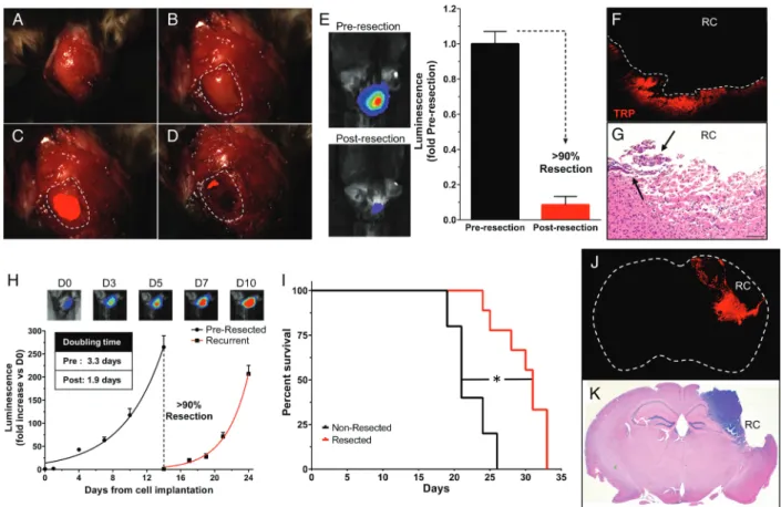
Our previous microsurgical resection model using U87 cells failed to recapitulate important hallmarks of clinical GBM, including a full immune system and brain invasion.8 We have previously shown that TRP-mcF allografts diffusely invade the brain parenchyma when established in syngeneic, immunocompetent mice.20We therefore examined whether fluorescence-guided microsurgery could be used to resect TRP-mcF allografts that accurately reflect the brain-invasive phenotype of human GBM in immune-competent C57 hosts. Fourteen days after intracortical injection of TRP-mcF cells, mCherry fluorescence was evident in the surgical field and was used to guide microsurgical dissection (Fig.2A– D). Pre-and postoperative BLI showed that resection achieved.90% reduction in tumor burden (Fig.2E). Histological analysis con-firmed these findings, as most mice examined displayed only small residual deposits of tumor remaining near the resection cavity on postoperative day 1 (Fig.2F and G).
To examine the therapeutic benefit of resection, we moni-tored TRP-mcF growth before and after surgery using BLI. Inter-estingly, recurrent TRP-mcF tumors grew more rapidly than their pre-resection counterparts (Fig.2H). Similar to clinical findings, resected mice survived significantly longer than non-resected mice but eventually succumbed from their tumor (Fig.2I).10,25,26Fluorescence imaging and histological examina-tion of symptomatic, terminally aged mice confirmed the pres-ence of recurrent mCherry+tumors around the resection cavity (Fig.2J and K). Taken together, these results demonstrate that fluorescence-guided microsurgery can be used to create a TRP allograft resection and recurrence model in a syngeneic immune-competent host where surgery extends the life of mice, but tumor recurrence eventually leads to death.
We next characterized the peritumoral microenvironment by performing histological analysis of tumor brains 1–7 days after resection to monitor patterns of postsurgical recurrence. Histology confirmed near-total tumor resection 1–3 days after microsurgery (Fig.3A–H). However, despite absence of tumor at the resection cavity, tumor invasion of the deep ipsilateral hemisphere led to distant recurrence (Fig.3I). Recurrent, mitot-ically active (Fig.3J) tumors exhibited extensive migration along blood vessels (Fig.3K) and diffusely invaded brain paren-chyma (Fig.3L). Five days after tumor microsurgery, locally re-current tumor became apparent near the resection cavity
(Fig. 3M) and was accompanied by reactive changes in the section bed, including hemosiderin-laden macrophages and re-active astrocytes (Fig. 3N – P). After 7 days, locally recurrent tumors became large, highly mitotically active, and necrotic (Fig. 3Q – T) and diffusely invaded the adjacent brain. Local tumor recurrence was accompanied by increased reactive changes in the resection bed, including infiltration of foamy and hemosiderin-laden macrophages (Fig. 3U–X). These results suggest that fluorescence-aided microsurgery in the syngeneic TRP-mcF allograft model system recapitulates aspects of clini-cal GBM, including invasion and distant recurrence, a process that occurs concomitantly with reactive, surgery-induced changes in the resection cavity.
Microsurgery Induces Dynamic Changes in the Reactive
Astrocyte Component of the Peritumoral
Microenvironment Surrounding TRP-mcF Allografts
Reactive astrocytes are the main cellular component of glial scars that develop in response to a variety of pathological stim-uli in the brain. During tumorigenesis, these cells play a key role in tumor progression and invasion,13,27 – 30where they develophypertrophic cytoplasmic processes and upregulate expression of cytoskeletal proteins such as GFAP. Indeed, hypertrophic GFAP+reactive astrocytes accumulated near the invasive edge of TRP-mcF allografts and became progressively dense as these tumors grew (Fig. 4A and B).
In addition to tumorigenesis, reactive astrocytes are a signif-icant component of the glial scars that develop in response to blunt force brain trauma.31 – 35In murine models, stab wound injury induces proliferation of GFAP+reactive astrocytes that also express stem cell markers, such as the intermediate fila-ment nestin.34,36 – 38Moreover, these cells have been shown to acquire the phenotypic properties of stem cells when cul-tured ex vivo, including unlimited self-renewal and multilineage differentiation.36,37To determine whether the reactive astro-cytes that accompanied TRP-mcF allograft growth might un-dergo a similar change in phenotype, we performed nestin immunostaining on established tumors. In contrast to their abundant GFAP immunoreactivity, reactive astrocytes around the invasive edge of TRP-mcF allografts did not express nestin, suggesting they lacked stemlike properties (Fig. 4C).
To examine the effects of blunt force trauma induced by mi-crosurgical resection on reactive astrocytes in the peritumoral Fig. 1. TRP cells engineered to express mCherry and luciferase proliferate in vitro and form GBM allografts in vivo.Cultured TRP glioma cells transduced with lentiviral vectors encoding TRP-mcF express mCherry and luciferase in vitro as determined by (A and C) white light and (B and D) fluorescence imaging. TRP-mcF cells (doubling time 1.1 days) showed a small but statistically significant decrease in growth compared with uninfected TRP cells (doubling time 0.9 days,P¼.01) (E). TRP-mcF cell number showed linear correlation with Fluc activity (R2¼.998,P,
Fluorescence Immunocytochemistry In vitro
To investigate the impact of astrocyte injury on nestin and glial fibrillary acidic protein (GFAP) expression in vitro, immortalized astrocytes were seeded in 24-well plates. Cells were formalin fixed and incubated with GFAP or nestin primary antibodies and dye-conjugated secondary antibodies as previously described.21
Bioluminescent Imaging and Survival Analysis
Pre-resection tumor volumes were monitored by BLI using an IVIS Kinetic. Surgery was performed on day 14 and tumor re-currence was monitored. Mice were monitored for neurological symptoms and sacrificed upon their development. Survival analysis was performed as previously described.8
Histopathology and Immunohistochemistry
Allograft mice were resected at day 14 after cell injection and randomized for sacrifice at days 1, 3, 5, and 7 post-resection. Formalin-fixed, paraffin-embedded brain sections were stained with hematoxylin and eosin (H&E), and Iba-1 immunohisto-chemistry was performed as previously described.22
RNA-Seq Analyses
Total RNA was extracted from injured or uninjured astrocyte cell pellets using the Qiagen RNeasy kit, followed by library prepa-ration using a Stranded mRNA-seq Kit (Kapa Biosystems) ac-cording to the manufacturer’s instructions. High-throughput sequencing with 75-bp single-end reads was performed on a NextSeq500 using the Illumina High Output Kit and analyzed as previously described.23,24
Luminex Analysis
Protein levels in the supernatant of injured or uninjured astro-cytes were determined using a mouse magnetic Luminex screening assay (R&D Systems) according to the manufactur-er’s protocol. Each sample was prepared in a 24-well plate as described above. Medium from each well was collected and used for protein analysis.
Supplementary Materials
Additional materials and methods are detailed in the Supple-ment. Supplementary figures and videos can also be found online.
Results
Fluorescence-Guided Microsurgery Effectively Reduces
Orthotopic TRP Allograft Burden but Fails to Prevent Rapid
Tumor Recurrence
We infected murine TRP cells with a recombinant lentiviral vec-tor encoding mcF (TRP-mcF) to facilitate high-resolution fluo-rescence imaging–guided microsurgical tumor resection and noninvasive BLI to quantify tumor volumes (Fig. 1A–D). Fluo-rescence imaging confirmed mCherry expression (Fig. 1B and
D). TRP-mcF cells showed a small but statistically significant decrease in growth compared with uninfected TRP cells (Fig. 1E). Dilution assays revealed that the luciferase activity of TRP-mcF correlated directly with cell number in vitro (Fig. 1F). We have previously shown that TRP allografts develop the histopathological hallmarks of human gliomas18and progress to GBM 15–20 days after orthotopic cell injection.20Similar to the parental line, serial BLI showed that engineered TRP-mcF allografts developed into rapidly expanding tumors over 24 days in the parenchyma of C57BL/6 mice (Fig. 1G). Fluorescence and histological analysis of postmortem tissue sections con-firmed the presence of large TRP-mcF tumors 14 days after in-jection (Fig. 1H and I). These tumors contained abundant microvascular proliferation and elevated mitotic activity (Fig. 1J – M) but generally lacked necrosis at this time point. These results demonstrate that TRP glioma cells engineered to express mCherry and luciferase develop the histopathologi-cal hallmarks of human GBM.
Our previous microsurgical resection model using U87 cells failed to recapitulate important hallmarks of clinical GBM, including a full immune system and brain invasion.8 We have previously shown that TRP-mcF allografts diffusely invade the brain parenchyma when established in syngeneic, immunocompetent mice.20We therefore examined whether fluorescence-guided microsurgery could be used to resect TRP-mcF allografts that accurately reflect the brain-invasive phenotype of human GBM in immune-competent C57 hosts. Fourteen days after intracortical injection of TRP-mcF cells, mCherry fluorescence was evident in the surgical field and was used to guide microsurgical dissection (Fig. 2A– D). Pre-and postoperative BLI showed that resection achieved.90% reduction in tumor burden (Fig. 2E). Histological analysis con-firmed these findings, as most mice examined displayed only small residual deposits of tumor remaining near the resection cavity on postoperative day 1 (Fig. 2F and G).
To examine the therapeutic benefit of resection, we moni-tored TRP-mcF growth before and after surgery using BLI. Inter-estingly, recurrent TRP-mcF tumors grew more rapidly than their pre-resection counterparts (Fig. 2H). Similar to clinical findings, resected mice survived significantly longer than non-resected mice but eventually succumbed from their tumor (Fig. 2I).10,25,26Fluorescence imaging and histological examina-tion of symptomatic, terminally aged mice confirmed the pres-ence of recurrent mCherry+tumors around the resection cavity (Fig. 2J and K). Taken together, these results demonstrate that fluorescence-guided microsurgery can be used to create a TRP allograft resection and recurrence model in a syngeneic immune-competent host where surgery extends the life of mice, but tumor recurrence eventually leads to death.
We next characterized the peritumoral microenvironment by performing histological analysis of tumor brains 1–7 days after resection to monitor patterns of postsurgical recurrence. Histology confirmed near-total tumor resection 1–3 days after microsurgery (Fig. 3A–H). However, despite absence of tumor at the resection cavity, tumor invasion of the deep ipsilateral hemisphere led to distant recurrence (Fig. 3I). Recurrent, mitot-ically active (Fig. 3J) tumors exhibited extensive migration along blood vessels (Fig. 3K) and diffusely invaded brain paren-chyma (Fig. 3L). Five days after tumor microsurgery, locally re-current tumor became apparent near the resection cavity
(Fig.3M) and was accompanied by reactive changes in the section bed, including hemosiderin-laden macrophages and re-active astrocytes (Fig.3N – P). After 7 days, locally recurrent tumors became large, highly mitotically active, and necrotic (Fig.3Q – T) and diffusely invaded the adjacent brain. Local tumor recurrence was accompanied by increased reactive changes in the resection bed, including infiltration of foamy and hemosiderin-laden macrophages (Fig.3U–X). These results suggest that fluorescence-aided microsurgery in the syngeneic TRP-mcF allograft model system recapitulates aspects of clini-cal GBM, including invasion and distant recurrence, a process that occurs concomitantly with reactive, surgery-induced changes in the resection cavity.
Microsurgery Induces Dynamic Changes in the Reactive
Astrocyte Component of the Peritumoral
Microenvironment Surrounding TRP-mcF Allografts
Reactive astrocytes are the main cellular component of glial scars that develop in response to a variety of pathological stim-uli in the brain. During tumorigenesis,these cells play a key role in tumor progression and invasion,13,27–30where they develophypertrophic cytoplasmic processes and upregulate expression of cytoskeletal proteins such as GFAP. Indeed, hypertrophic GFAP+reactive astrocytes accumulated near the invasive edge of TRP-mcF allografts and became progressively dense as these tumors grew (Fig.4A and B).
In addition to tumorigenesis, reactive astrocytes are a signif-icant component of the glialscars that develop in response to blunt force brain trauma.31–35In murine models, stab wound injury induces proliferation of GFAP+reactive astrocytes that also expressstemcell markers, such as the intermediate fila-ment nestin.34,36–38Moreover, these cells have been shown to acquire the phenotypic properties of stem cells when cul-tured ex vivo, including unlimited self-renewal and multilineage differentiation.36,37To determine whether the reactive astro-cytes that accompanied TRP-mcF allograft growth might un-dergo a similar change in phenotype, we performed nestin immunostaining on established tumors. In contrast to their abundant GFAP immunoreactivity, reactive astrocytes around the invasive edge of TRP-mcF allografts did not express nestin, suggesting they lacked stemlike properties (Fig.4C).
To examine the effects of blunt force trauma induced by mi-crosurgical resection on reactive astrocytes in the peritumoral Fig. 1. TRP cells engineered to express mCherry and luciferase proliferate in vitro and form GBM allografts in vivo.Cultured TRP glioma cells transduced with lentiviral vectors encoding TRP-mcF express mCherry and luciferase in vitro as determined by (A and C) white light and (B and D) fluorescence imaging. TRP-mcF cells (doubling time 1.1 days) showed a small but statistically significant decrease in growth compared with uninfected TRP cells (doubling time 0.9 days,P¼.01) (E). TRP-mcF cell number showed linear correlation with Fluc activity (R2¼.998,P,
microenvironment, mouse brains harvested 1–7 days after re-section were also immunostained for GFAP and nestin. A dra-matic decrease in the number of GFAP+reactive astrocytes was evident over this time course (Fig.4D – H). Few of these cells expressed nestin at postoperative day 1 (Fig.4I). However, reactive astrocytes that coexpressed both GFAP and nestin (Fig.4N–Q) increased 16-fold by day 3 and gradually decreased thereafter (Fig.4J – M). These results suggest that the blunt force trauma induced by resection alters the reactive astrocytes present in the peritumoral microenvironment and may induce a stem cell–like phenotype in these cells.36,37
Because recurrent tumors grew faster than their pre-resection counterparts (Fig.2H) and diffusely infiltrated the brain (Fig.3), we next used cell culture to explore whether as-trocyte injury influences tumor biology in vitro. Scratch injury of cultured astrocytes is an established in vitro model that mimics the blunt force trauma induced by surgical resection. Cultured astrocytes injured by scratching have been shown to undergo a phenotypic switch to a stemcell–like state accompanied by in-creased nestin expression.39–42We confirmed that scratch
injury of immortalized astrocytes induces a 2.5-fold increase in nestin expression within 24 h (Fig.5A and B). Next, we exam-ined the effects of astrocyte injury on TRP-mcF cell migration and proliferation using a coculture system (Fig.5C). In contrast to the effects of uninjured astrocytes, scratch injury of immor-talized astrocytes induced a 2.5-fold increase in migration of cocultured TRP-mcF cells over 24 h (Fig.5D–F). Astrocytes iso-lated from the in vivo resection model (Supplementary Fig. S1) increased TRP-mcF cell migration compared with uninjured as-trocytes but showed no difference relative to injured asas-trocytes (Fig.5E). In vitro injury to astrocytes derived directly from the in vivo model did not increase TRP-mcF migration further. Single cell analysis (Fig.5F) showed the TRP-mcF cells cultured with in-jured astrocytes migrated faster (Fig.5G), farther ( Supplemen-tary Fig. S2A), and more directly toward injured astrocytes (Supplementary Fig. S2B) than those cultured with uninjured astrocytes. These effects were specific to astrocyte injury, as scratch wounding of TRP-mcF cells themselves had no effect on their migration, in both the presence (Supplementary Fig. S3A) and absence of injured astrocytes (Supplementary
Fig. 2. Fluorescence-guided microsurgical resection reduces volumes of orthotopic TRP-mcF allografts that redevelop to induce death. A scalp incision (A) was made 14 days after injection of TRP-mcF cells. A craniotomy was performed (B) to visualize the underlying mCherry+tumors (C). Fluorescence-guided microsurgery significantly reduced tumor burden as determined by intraoperative fluorescence imaging (D). Postoperative BLI showed a 91% reduction in mean tumor burden (N¼6 mice per group,P,.0001) (E). Representative BLIs pre- and postresection are shown. Representative mCherry fluorescence (F) and H&E images (G) of brain sections taken 1 day postresection showed residual tumor (arrows) within the resection cavity (RC). Recurrent TRP-mcF allografts (H) grew faster than their pre-resection counterparts (data from Fig.1G), with doubling times of 1.9 vs 3.3 days, respectively (P¼.0003). Microsurgical resection extended survival, as resected mice survived significantly longer than nonresected mice (31 vs 21 days, log-rankP¼.001) (I). Fluorescence (J) and H&E images (K) of brain sections taken 7 days postresection show recurrent tumor near the RC. Original magnifications: 15x (J and K) and 100x (F and G).
Fig. S3B). Revealing the broad applicability of this effect, astro-cyte injury increased migration in established GBM mouse (GL261) and human (U251 and U87) cell lines in vitro (Fig. 5H). To examine the effects of astrocyte injury on TRP-mcF prolif-eration, we cocultured these cells with uninjured and injured astrocytes and monitored their proliferation by labeling S-phase cells with EdU. Compared with uninjured astrocytes, scratch injury of astrocytes induced significantly more TRP-mcF
microenvironment, mouse brains harvested 1–7 days after re-section were also immunostained for GFAP and nestin. A dra-matic decrease in the number of GFAP+reactive astrocytes was evident over this time course (Fig. 4D – H). Few of these cells expressed nestin at postoperative day 1 (Fig. 4I). However, reactive astrocytes that coexpressed both GFAP and nestin (Fig. 4N–Q) increased 16-fold by day 3 and gradually decreased thereafter (Fig. 4J – M). These results suggest that the blunt force trauma induced by resection alters the reactive astrocytes present in the peritumoral microenvironment and may induce a stem cell–like phenotype in these cells.36,37
Because recurrent tumors grew faster than their pre-resection counterparts (Fig. 2H) and diffusely infiltrated the brain (Fig. 3), we next used cell culture to explore whether as-trocyte injury influences tumor biology in vitro. Scratch injury of cultured astrocytes is an established in vitro model that mimics the blunt force trauma induced by surgical resection. Cultured astrocytes injured by scratching have been shown to undergo a phenotypic switch to a stem cell–like state accompanied by in-creased nestin expression.39 – 42We confirmed that scratch
injury of immortalized astrocytes induces a 2.5-fold increase in nestin expression within 24 h (Fig. 5A and B). Next, we exam-ined the effects of astrocyte injury on TRP-mcF cell migration and proliferation using a coculture system (Fig. 5C). In contrast to the effects of uninjured astrocytes, scratch injury of immor-talized astrocytes induced a 2.5-fold increase in migration of cocultured TRP-mcF cells over 24 h (Fig. 5D–F). Astrocytes iso-lated from the in vivo resection model (Supplementary Fig. S1) increased TRP-mcF cell migration compared with uninjured as-trocytes but showed no difference relative to injured asas-trocytes (Fig. 5E). In vitro injury to astrocytes derived directly from the in vivo model did not increase TRP-mcF migration further. Single cell analysis (Fig. 5F) showed the TRP-mcF cells cultured with in-jured astrocytes migrated faster (Fig. 5G), farther (Supplemen-tary Fig. S2A), and more directly toward injured astrocytes (Supplementary Fig. S2B) than those cultured with uninjured astrocytes. These effects were specific to astrocyte injury, as scratch wounding of TRP-mcF cells themselves had no effect on their migration, in both the presence (Supplementary Fig. S3A) and absence of injured astrocytes (Supplementary Fig. 2. Fluorescence-guided microsurgical resection reduces volumes of orthotopic TRP-mcF allografts that redevelop to induce death. A scalp incision (A) was made 14 days after injection of TRP-mcF cells. A craniotomy was performed (B) to visualize the underlying mCherry+tumors (C). Fluorescence-guided microsurgery significantly reduced tumor burden as determined by intraoperative fluorescence imaging (D). Postoperative BLI showed a 91% reduction in mean tumor burden (N¼6 mice per group,P,.0001) (E). Representative BLIs pre- and postresection are shown. Representative mCherry fluorescence (F) and H&E images (G) of brain sections taken 1 day postresection showed residual tumor (arrows) within the resection cavity (RC). Recurrent TRP-mcF allografts (H) grew faster than their pre-resection counterparts (data from Fig. 1G), with doubling times of 1.9 vs 3.3 days, respectively (P¼.0003). Microsurgical resection extended survival, as resected mice survived significantly longer than nonresected mice (31 vs 21 days, log-rankP¼.001) (I). Fluorescence (J) and H&E images (K) of brain sections taken 7 days postresection show recurrent tumor near the RC. Original magnifications: 15x (J and K) and 100x (F and G).
Fig. S3B). Revealing the broad applicability of this effect, astro-cyte injury increased migration in established GBM mouse (GL261) and human (U251 and U87) cell lines in vitro (Fig.5H). To examine the effects of astrocyte injury on TRP-mcF prolif-eration, we cocultured these cells with uninjured and injured astrocytes and monitored their proliferation by labeling S-phase cells with EdU. Compared with uninjured astrocytes, scratch injury of astrocytes induced significantly more TRP-mcF
astrocyte component of the peritumoral microenvironment that may promote tumor migration and proliferation.
To identify the factors that contribute to the observed tumor proliferation and migration induced by astrocyte injury, we per-formed RNA-seq analysis on cultured astrocytes before and after scratch injury. Differential gene expression analysis (Fig.6A) and hierarchical clustering (Fig.6B andSupplementary Fig. S5) revealed significant alterations to the astrocyte tran-scriptome after scratch injury. We found that injured astrocytes exhibited upregulation of genes involved in cytokine production and response (Fig.6C). Based on RNA-seq analysis, we identi-fied 5 candidates that were upregulated and known to encode secreted proteins (Cxcl5,Il33,Tnfsf12,Tnfsf13, andVegfa). Four
of these candidates (Cxcl5,Il33,Tnfsf12, andVegfa) were ex-amined using Luminex assays, which showed that onlyCxcl5 was upregulated in the supernatants of injured astrocytes (1.42-fold,P¼.0074; Fig.6D). These data suggest thatCxcl5 may contribute to increased tumor proliferation and migration following astrocyte injury.
Discussion
Surgical resection followed by radio- and chemotherapy is the standard of care for GBM patients. Nonetheless, these treatments ultimately fail because they are unable to reach the invasive can-cer cells that evade surgical resection and lie outside radiation
Fig. 4. Microsurgical resection induces dynamic changes in reactive astrocytes surrounding TRP-mcF allografts. Reactive astrocytes were abundant in the peritumoral brain parenchyma in mCherry+(red) TRP-mcF allografts established for 7 days (A), and their density increased further in tumors established for 14 days (B). Tumor cells, but not reactive astrocytes, expressed nestin (C). Over the course of 7 days following microsurgical tumor resection, a gradual decrease in the density of reactive astrocytes in the peritumoral microenvironment is highlighted by GFAP immunostaining (D– H). However, over this time course, reactive astrocytes gain expression of the stem cell marker nestin and their density peaks 3 days postresection (I–M). Double immunostaining (N–P, merge, Q) of brains 3 days postresection shows that reactive astrocytes express both GFAP (green, O) and nestin (red, P). Nuclei are stained with Hoechst (blue, I–L, N, and Q). All pairwise comparisons in panels H and M were significant except days 5 and 7 in (H) (P,.0001). Original magnifications: 100x. Scale bars¼100mm.
fields.10,25,26As a result, local and regional therapies that penetrate the brain have proven effective, specifically controlled-release poly-mers. Others, such as convection-enhanced drug delivery and cell-based therapies, hold further promise when used as an adjunct to surgical resection. However, their promise has yet to be clinically realized due to the current lack of accurate preclinical models in which to test these therapies. To address this gap, we developed a syngeneic, immunocompetent mouse model of GBM that mim-ics the clinical scenario of surgical resection and accurately recapit-ulates the invasive nature of the human disease.18,20
Surgical resection remains one of the most widely used, yet least studied components of GBM therapy. Many of the patho-physiological events following tumor resection remain unknown, emphasizing the need for a clinically relevant preclinical model. Previous models have used human U87 xenografts that fail to recapitulate important GBM features, such as brain invasion.8,9,11 In this current study, we developed a resection model that uses glioma cells that mimic clinical hallmarks of human GBM in syn-geneic immune-competent hosts and exhibits one of the most important GBM features, brain invasion. This biologically faithful
Fig. 5. Astrocyte injury promotes TRP migration and proliferation in vitro. Compared with cultured, uninjured astrocytes (A), scratch wounding induces significant increases in nestin (green) expression at 24 h (2.5-fold increase, P,.0001) (B). Astrocytes and TRP were seeded approximately 500mm apart and groups were constructed with and without scratch injury of each cell population (C). Time-lapse images were captured every 20 min for 24 h and used to construct movies that revealed the migration of TPRs in real time. Injuring astrocytes by scratch induced significantly increased migration of TRP-mcF tumor cells at 24 h (P,.0001) (D and E). Astrocytes isolated from the in vivo injury model (ExAC uninjured) increased the migration of TRP cells (P¼.0002) and injury in vitro (ExAC injured) did not alter migration further (P¼.3) (E). Single cell imaging analysis showed that injury of cocultured astrocytes (F) increased the velocity (P,.0001) (G) of individual TRP-mcF tumor cells. Astrocyte injury induced increased migration in established GL261 (P,.0001), U251 (P,.0001), and U87 (P,.0001) GBM cell lines (H). Compared with uninjured astrocytes, scratch injury of immortalized astrocytes increased proliferation of cocultured TRP-mcF tumor cells 32% as measured by EdU incorporation at 24 h (P,.0001) (I). TRP-mcF cell proliferation was significantly increased when cultured in scratch injured-astrocyte conditioned media (SACM) compared with uninjured astrocyte conditioned media (ACM) as measured by BLI (doubling time: 1.07 vs 1.19 days,P¼.02) (J). Scratch injury of astrocytes significantly increased proliferation of GL261 (P¼.0006), U251 (P,
astrocyte component of the peritumoral microenvironment that may promote tumor migration and proliferation.
To identify the factors that contribute to the observed tumor proliferation and migration induced by astrocyte injury, we per-formed RNA-seq analysis on cultured astrocytes before and after scratch injury. Differential gene expression analysis (Fig. 6A) and hierarchical clustering (Fig. 6B and Supplementary Fig. S5) revealed significant alterations to the astrocyte tran-scriptome after scratch injury. We found that injured astrocytes exhibited upregulation of genes involved in cytokine production and response (Fig. 6C). Based on RNA-seq analysis, we identi-fied 5 candidates that were upregulated and known to encode secreted proteins (Cxcl5,Il33,Tnfsf12,Tnfsf13, andVegfa). Four
of these candidates (Cxcl5,Il33,Tnfsf12, andVegfa) were ex-amined using Luminex assays, which showed that onlyCxcl5 was upregulated in the supernatants of injured astrocytes (1.42-fold,P¼.0074; Fig. 6D). These data suggest thatCxcl5 may contribute to increased tumor proliferation and migration following astrocyte injury.
Discussion
Surgical resection followed by radio- and chemotherapy is the standard of care for GBM patients. Nonetheless, these treatments ultimately fail because they are unable to reach the invasive can-cer cells that evade surgical resection and lie outside radiation
Fig. 4. Microsurgical resection induces dynamic changes in reactive astrocytes surrounding TRP-mcF allografts. Reactive astrocytes were abundant in the peritumoral brain parenchyma in mCherry+(red) TRP-mcF allografts established for 7 days (A), and their density increased further in tumors established for 14 days (B). Tumor cells, but not reactive astrocytes, expressed nestin (C). Over the course of 7 days following microsurgical tumor resection, a gradual decrease in the density of reactive astrocytes in the peritumoral microenvironment is highlighted by GFAP immunostaining (D– H). However, over this time course, reactive astrocytes gain expression of the stem cell marker nestin and their density peaks 3 days postresection (I–M). Double immunostaining (N–P, merge, Q) of brains 3 days postresection shows that reactive astrocytes express both GFAP (green, O) and nestin (red, P). Nuclei are stained with Hoechst (blue, I–L, N, and Q). All pairwise comparisons in panels H and M were significant except days 5 and 7 in (H) (P,.0001). Original magnifications: 100x. Scale bars¼100mm.
fields.10,25,26As a result, local and regional therapies that penetrate the brain have proven effective, specifically controlled-release poly-mers. Others, such as convection-enhanced drug delivery and cell-based therapies, hold further promise when used as an adjunct to surgical resection. However, their promise has yet to be clinically realized due to the current lack of accurate preclinical models in which to test these therapies. To address this gap, we developed a syngeneic, immunocompetent mouse model of GBM that mim-ics the clinical scenario of surgical resection and accurately recapit-ulates the invasive nature of the human disease.18,20
Surgical resection remains one of the most widely used, yet least studied components of GBM therapy. Many of the patho-physiological events following tumor resection remain unknown, emphasizing the need for a clinically relevant preclinical model. Previous models have used human U87 xenografts that fail to recapitulate important GBM features, such as brain invasion.8,9,11 In this current study, we developed a resection model that uses glioma cells that mimic clinical hallmarks of human GBM in syn-geneic immune-competent hosts and exhibits one of the most important GBM features, brain invasion. This biologically faithful
Fig. 5. Astrocyte injury promotes TRP migration and proliferation in vitro. Compared with cultured, uninjured astrocytes (A), scratch wounding induces significant increases in nestin (green) expression at 24 h (2.5-fold increase, P,.0001) (B). Astrocytes and TRP were seeded approximately 500mm apart and groups were constructed with and without scratch injury of each cell population (C). Time-lapse images were captured every 20 min for 24 h and used to construct movies that revealed the migration of TPRs in real time. Injuring astrocytes by scratch induced significantly increased migration of TRP-mcF tumor cells at 24 h (P,.0001) (D and E). Astrocytes isolated from the in vivo injury model (ExAC uninjured) increased the migration of TRP cells (P¼.0002) and injury in vitro (ExAC injured) did not alter migration further (P¼.3) (E). Single cell imaging analysis showed that injury of cocultured astrocytes (F) increased the velocity (P,.0001) (G) of individual TRP-mcF tumor cells. Astrocyte injury induced increased migration in established GL261 (P,.0001), U251 (P,.0001), and U87 (P,.0001) GBM cell lines (H). Compared with uninjured astrocytes, scratch injury of immortalized astrocytes increased proliferation of cocultured TRP-mcF tumor cells 32% as measured by EdU incorporation at 24 h (P,.0001) (I). TRP-mcF cell proliferation was significantly increased when cultured in scratch injured-astrocyte conditioned media (SACM) compared with uninjured astrocyte conditioned media (ACM) as measured by BLI (doubling time: 1.07 vs 1.19 days,P¼.02) (J). Scratch injury of astrocytes significantly increased proliferation of GL261 (P¼.0006), U251 (P,
mouse model allowed us to uncover potential mechanisms of tumor progression following tumor resection. Thus, this resec-tion model can help provide a better understanding of the path-ophysiological consequences of surgical resection.
Reactive astrocytes are recruited to the local microenviron-ment in response to an assortment of pathological stimuli, such as tumor growth13,36,43 and blunt force trau-ma.32,33,38,44–46The pattern of reactive astrocytosis after blunt force trauma in the CNS has been heavily studied. After cortical stab wound injury, astrocytes react by hypertro-phy and upregulation of nestin and GFAP,and subsequently dedifferentiate into multipotent cells.34,36,37Since glioma re-section models are rare, it was unclear how reactive astrocytes in the peritumoral microenvironment would react following trau-matic brain injury induced by surgical tumor resection. We found that reactive astrocytes express nestin following resection, sug-gesting that these astrocytes dedifferentiate in response to sur-gical tumor resection. One potential weakness of this study is that we used marker expression, instead of ex vivo functional analysis, to suggest astrocyte dedifferentiation. However, in a similar context, reports have already shown that traumatic brain injury causes astrocytes to acquire stem cell features, in-cluding neurosphere formation, self-renewal, and multilineage potential.36,37
As a prevalent component of the glioma microenvironment, reactive astrocytes play a major role in tumor progression and
invasion.12,28,29,47,48Since our data suggest that these astro-cytes dedifferentiate in response to surgical resection, we fur-ther extended these findings by investigating how this response may influence tumor biology, specifically tumor growth and migration. Using an in vitromodel that mimics the in vivo astrocyte response to injury,39–42,49we found that the astrocyte response promotes tumor proliferation and mi-gration, suggesting that reactive astrocytes may potentiate tumor aggressiveness after resection. Moreover, we found that recurrent tumors are highly invasive and exhibited signifi-cantly faster growth compared with their nonresected counter-parts. Since in vitro tumor growth showed a marginal increase compared with the in vivo response, this suggests that reactive astrocytes may be only a contributing factor in promotion of the increased proliferation of recurrent tumors. Although tumor growth and proliferation are more straightforward to monitor, it is difficult to assess in vivo tumor invasion due to mass effects from the malignant tumor. Therefore, surgical tumor resection provided a unique opportunity to study tumor cell invasion. While it is challenging to compare the inva-sion of pre- versus postoperative tumors, we found that recur-rent tumors extensively invaded the brain. Moreover, surgical intervention alters the reactive astrocyte composition within the tumor microenvironment and may have deleterious conse-quences on patient survival. While glioma resection remains the standard of care in qualified patients, this report highlights
Fig. 6. Astrocyte injury induces transcriptome and secretome changes. Differential expression analysis (A) and hierarchical clustering showed that scratch injury of astrocytes (S1–S4) induced significant transcriptome alterations (2093 genes,q,0.001) compared with uninjured (C1–C4) astrocytes (B). Ontology analysis of upregulated genes revealed enrichment in genes associated with cytokine production and response (C). Compared with uninjured astrocytes, astrocyte injury increased secretion ofCxcl5(P¼.0074) (D) as measured by Luminex analysis.
the role of reactive astrocytes in inducing a more favorable mi-croenvironment for tumor regrowth and recurrence. This sug-gests that our model may provide the means to further understand how the postsurgical microenvironment influences tumor properties and allows for the development of drugs to combat resection-mediated effects. Our data suggest that Cxcl5contributes to increased tumor invasion and proliferation after GBM resection, but further experimentation is needed to fully characterize and identify the factors responsible for in vivo response. We are currently exploring these molecular mechanisms.
Microglia/macrophages are likely to play an important role in the postsurgical microenvironment as tissue scavengers and may potentiate the aggressiveness of recurrent tumors.13,50,51 These cells have been implicated in glioma pathogenesis and may promote a tumor-permissive microenvironment.52 – 54We found that recurrent tumors exhibited a significant increase in Iba-1+cells in the tumor compared with pre-resection (Supple-mentary Fig. S6). This suggests that microglia/macrophages may play a role in the increased tumor migration and/or prolif-eration observed after resection.
Surgery alters the tumor microenvironment, which is largely composed of reactive astrocytes. The microenvironment in es-tablished nonresected tumors contains a high tumor-to–reac-tive astrocyte ratio, while, after tumor resection, recurrent tumors harbor a much lower proportion. Though surgery ma-nipulates the microenvironment, it remains unclear how this af-fects local treatment strategies such as stem cell therapies. Engineered stem cells extensively home to glioma cells and have shown therapeutic efficacy55,56—though, in a similar con-text, implanting stem cells within a CNS lesion has been shown to reduce stem cell survival, alter differentiation, and decrease therapeutic efficacy.9,57However, the molecular mechanism and signals associated with this response remain unclear. To better develop stem cell therapies, we are currently investigat-ing the underlyinvestigat-ing mechanisms that reduce stem cell efficacy in the postsurgical tumor microenvironment.
In conclusion, our study demonstrates the development of a new glioma resection model in syngeneic immune-competent hosts that allowed us to study the postsurgical microenviron-ment and pattern of tumor recurrence. We found that surgical resection alters the reactive astrocyte component of the peritu-moral microenvironment and that injured astrocytes may sig-nificantly influence tumor biology. Using this study as a template, more investigations can be conducted to gain a bet-ter understanding of the postsurgical microenvironment. This will allow optimization of locally administered therapies and fa-cilitate the development of agents that ameliorate surgery-mediated effects on tumor properties.
Supplementary Material
Supplementary material is available atNeuro-Oncology Journal online (http://neuro-oncology.oxfordjournals.org/).
Funding
This work was supported in part by grants from the UNC University Can-cer Research Fund (to S.D.H. and C.R.M.) and the UNC Translational and
Clinical Sciences Institute (UL1TR001111, to O.O. and J.B.). The UNC Translational Pathology Laboratory is supported by the National Cancer Institute (P30CA016086) and the University Cancer Research Fund. The Small Animal Imaging Core in the UNC Biomedical Imaging Research Center is supported by the National Cancer Institute (P30CA016086).
Conflict of interest statement. None declared.
Acknowledgments
The authors wish to thank the UNC-Olympus Imaging Research Center for microscope usage.
References
1. Adamson C, Kanu OO, Mehta AI, et al. Glioblastoma multiforme: a review of where we have been and where we are going.Expert Opin Investig Drugs. 2009;18(8):1061–1083.
2. Wen PY, Kesari S. Malignant gliomas in adults.N Engl J Med. 2008; 359(5):492–507.
3. Hou LC, Veeravagu A, Hsu AR, et al. Recurrent glioblastoma multiforme: a review of natural history and management options.
Neurosurg Focus. 2006;20(4):E5.
4. Minniti G, De Sanctis V, Muni R, et al. Radiotherapy plus concomitant and adjuvant temozolomide for glioblastoma in elderly patients.J Neurooncol. 2008;88(1):97–103.
5. Nieder C, Grosu AL, Molls M. A comparison of treatment results for recurrent malignant gliomas. Cancer Treat Rev. 2000;26(6): 397–409.
6. Tejada S, Aldave G, Marigil M, et al. Factors associated with a higher rate of distant failure after primary treatment for glioblastoma.J Neurooncol. 2014;116(1):169–175.
7. Gunduz N, Fisher B, Saffer EA. Effect of surgical removal on the growth and kinetics of residual tumor.Cancer Res. 1979;39(10): 3861–3865.
8. Hingtgen S, Figueiredo JL, Farrar C, et al. Real-time multi-modality imaging of glioblastoma tumor resection and recurrence. J Neurooncol. 2013;111(2):153–161.
9. Kauer TM, Figueiredo JL, Hingtgen S, et al. Encapsulated therapeutic stem cells implanted in the tumor resection cavity induce cell death in gliomas.Nat Neurosci. 2012;15(2):197–204. 10. Lacroix M, Abi-Said D, Fourney DR, et al. A multivariate analysis of
416 patients with glioblastoma multiforme: prognosis, extent of resection, and survival.J Neurosurg. 2001;95(2):190–198. 11. McNeill RS, Vitucci M, Wu J, et al. Contemporary murine models in
preclinical astrocytoma drug development.Neuro Oncol. 2015; 17(1):12–28.
12. Hoelzinger DB, Demuth T, Berens ME. Autocrine factors that sustain glioma invasion and paracrine biology in the brain microenvironment.J Natl Cancer Inst. 2007;99(21):1583–1593. 13. Charles NA, Holland EC, Gilbertson R, et al. The brain tumor
microenvironment.Glia. 2011;59(8):1169–1180.
14. Shiao SL, Ganesan AP, Rugo HS, et al. Immune microenvironments in solid tumors: new targets for therapy.Genes Dev. 2011;25(24): 2559–2572.
15. Hanahan D, Weinberg RA. Hallmarks of cancer: the next generation.