This article is made available online in accordance with publisher policies. Please scroll down to view the document itself. Please refer to the repository record for this item and our policy information available from the repository home page for further information.
Author(s): Achille Pasqualotto, Mary Jane Spiller, Ashok S. Jansari and Michael J. Proulx
Title: Visual experience facilitates allocentric spatial representation
Citation: Pasqualotto, A., Spiller, M.J., Jansari, A.S. and Proulx, M.J. (2012), 'Visual experience facilitates allocentric spatial representation', Behavioural Brain Research, in press.
Link to published version:
Forthcoming at
Publisher statement:
“NOTICE: this is the author’s version of a work that was accepted for publication in Behavioural Brain Research. Changes resulting from the publishing process, such as peer review, editing, corrections, structural formatting, and other quality control mechanisms may not be reflected in this document. Changes may have been made to this work since it was submitted for publication. A definitive version will be
published in Behavioural Brain Research”
Information on how to cite items within roar@uel:
Visual experience facilitates allocentric spatial representation
Achille Pasqualotto¹*, Mary Jane Spiller2, Ashok S. Jansari2, & Michael J. Proulx¹*
¹Biological and Experimental Psychology Group, School of Biological and Chemical
Sciences, Queen Mary University of London
2
School of Psychology, University of East London
[Word count: 3,292 (excluded title-page)]
* Correspondence should be addressed to A. Pasqualotto or M.J. Proulx:
Biological & Experimental Psychology Group, School of Biological and Chemical Sciences,
Queen Mary University of London, Mile End Road, London, E1 4NS, UK
Telephone: +44 (0)20 78
Abstract
Representing the position of the objects independently from our own position is a
fundamental cognitive ability. Here we investigated whether this ability depends on visual
experience. Congenitally blind, late blind and blindfolded sighted participants haptically
learnt a room-sized regularly shaped array of objects, and their spatial memory was tested to
determine which spatial reference frame was used. Crucially, the use of an object-based
reference frame requires representing the regular structure of the array. We found that
blindfolded sighted and late blind participants, that is those with visual experience, showed a
preferential use of the object-based or ‘allocentric’ reference frame. On the contrary,
congenitally blind participants preferred a self-based, or egocentric, reference frame. This
suggests that, due to its developmental effect on the multisensory brain areas involved in
spatial cognition, visual experience is necessary to develop a preference for an object-based,
allocentric reference frame.
Keywords: spatial cognition, multisensory integration, visual experience, blindness, reference
frames, neural plasticity.
Introduction
McNamara and colleagues [1] reported the counterintuitive result that the
representation of a regular array of objects was based on the intrinsic structure of the array
(i.e. object-based, or allocentric, rows-and-columns grid pattern), rather than on the
egocentric viewing position (see also [2] for equivalent results within the peripersonal space
in arm’s reach). More precisely, from a given viewing position, participants viewed a set of
objects disposed on the room’s floor and then their spatial memory was tested in a Judgement
of Relative Direction task (JRD) where they imagined being close to a given object within the
array, being oriented toward a second object and pointing in the direction of a third one (i.e.
heading). For example, “Imagine that you are at the clock, facing the shoe, point to the jar”).
Surprisingly, results showed that participants were more accurate for headings aligned with
the intrinsic structure of the array than with the familiar viewpoint. This suggests that
participants could extract the grid pattern of the array and use it to store the position of the
objects in their spatial memory and, consequently, they performed the JRD task better when
the tested headings matched the grid pattern. Here we adapted the method by McNamara and
colleagues [1] and tested congenitally blind, late blind, and blindfolded sighted participants to
investigate whether the ability using an allocentric reference frame is subject to visual
experience or whether it is innate.
Additionally, the current study can shed a light on the role of a critical period for
developmental vision on spatial cognition and brain organisation [3]. In fact, although it is an
established opinionthatblindness sharpens the remaining modalities [4-5], discordant results
have been reported by studies investigating spatial cognition in blind individuals. Thus, some
researchers found results suggesting that congenital blindness prejudices the complete
development of spatial cognition [6-7]. On the contrary, other authors reported data
[8-9]. Yet, a few studies comparing the use of spatial reference frames across blind
participants may suggest that visual experience is crucial for spatial tasks requiring the use of
allocentric spatial representation, while egocentric spatial abilities should be preserved
[10-11].
Along these lines, we created a somatosensory task (i.e. based on haptics,
proprioception and vestibular cues) where two groups of blind and one group of blindfolded
sighted participants were led by the experimenter to explore objects arranged in a regular
array and then they underwent a JRD task. If visual experience is crucial for developing
allocentric spatial representation, and thus for the ability of perceiving the grid-pattern of the
array, we would find that participants without visual experience (congenitally blind) are less
precise in JRD involving headings parallel to the grid-pattern (i.e. that the allocentric
reference frame is not exploited). Yet, they will be more accurate in JRD involving headings
parallel to the routes walked during the array exploration, that is, to the participants’
egocentric representation of the array. On the other hand, participants possessing visual
experience (late blind and blindfolded sighted) are expected to exhibit the opposite results:
more accurate performance for allocentric headings, and poorer for egocentric.
Method
Participants
We tested 20 blind participants recruited through local blind institutions. Ten were
congenitally blind (CB), five males and five females, with a mean age of 43 (16.23 SD).Ten
were late blind (LB), five males and five females, with a mean age of 43 (12.18). Finally, we
tested a group of ten matching blindfolded sighted individuals (S), five males and five
impairment. All participants signed a consent form approved by the local Research Ethics
Committee. Participants received £10 for their participation.
[Table 1 about here]
Apparatus
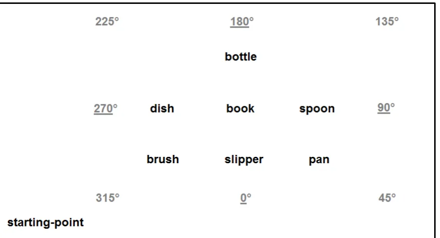
Common objects were arranged inside a roughly rectangular room about 12.5 by 9 m
(see Figure 1). Each object was placed on a 90cm tall stool to facilitate haptic exploration.
Between each stool there was a distance of about 1.5 m. A chair placed about 2 m from the
closest object (brush) was the starting-point of each exploration.
During the JRD task, participant used a LogiTech™ 3DPro joystick connected to a
Dell™ Latitude E5510 laptop running a MatLab™ program that recorded pointing angles and
reaction times. The pointing task took place in a nearby room sized 3 by 2 m. Sighted
participants were blindfolded throughout the experiment by a MindFold™. As no blind
participant had more than light/darkness sensitivity none of them wore the blindfold.
[Figure 1 about here]
Procedure
The experiment consisted of two phases, learning and testing. Sighted participants were
blindfolded. Then participants were guided into the ‘learning’ room where they familiarised
with the use of the joystick. Led by the experimenter, participants began the exploration of
the array. Objects were explored one-by-one, with participants being led along straight routes
back-and-forth from the starting-point to each object (see [12] for similar learning
proceeded by horizontal rows (e.g. starting-point, pan, starting-point, slipper, starting-point,
brush, etc.). To ensure that participants learned the array, after the exploration they were
asked to verbally recall the objects by following the exploration order. This procedure ended
when participants could correctly recall all the objects twice consecutively without help (on
average it took 3-4 attempts, maximum 5).
Led by the experimenter, participants reached the ‘testing’ room where, before using
the joystick, they received a sheet of paper reporting a bird’s eye-view ‘tactile map’ of the
array (see [13]). This was aimed to promote an allocentric spatial representation. Thus, this
simple sheet had seven little holes representing the seven objects arranged as in the learned
array, and one hole representing the starting-point. By using both hands and led by the
experimenter, participants explored the map by following the learning order, and then they
could freely explore the map for about 1 minute.
During the JRD task, the experimenter read the statements that appeared on the
computer screen, for example: “Imagine that you are at the book (brief pause), facing the
bottle (brief pause), point to the pan”. Then a sound indicated that the joystick could be
aimed, as quickly and as accurate as possible, towards the target object. The reaction time
recording began when the sound was emitted. After each pointing a new statement was read
by the experimenter. There were 48 random trials, six for each of the eight headings that had
to be imagined during the task. Four headings were aligned to the routes walked during the
array exploration (315°, 225°, 135° and 45°), whilst four were aligned to the internal
structure of the array (0°, 270°, 180° and 90°), see Figure 1. Horizontal pointing errors in
degrees and reaction times were recorded. The entire experiment took about 50 minutes to
complete.
Average pointing errors and reaction times were analysed in a three-way mixed design
ANOVA with Visual Status (CB, LB, and S) as between-subjects condition, the Reference
Frame underlying the headings (egocentric and allocentric) and the Imagined Headings (0º,
45º, …, and 315º) as within-subjects conditions.
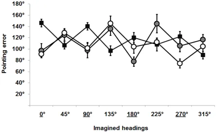
Pointing error analysis revealed no effect of the Visual Status [F(2,27)=1.18, p=.322]
indicating that overall the three groups performed the task equally well. There was a
significant effect of the Reference Frame [F(1,28)=7.84, p=.009] showing that in general
allocentric orientations were better performed (i.e. better performed by both LB and S, see
below). Overall the single Imagined Headings did not produce a significant effect
[F(7,22)=2.68, p=.052]. Interestingly, there was a significant interaction between Visual
Status and Reference Frame [F(4,25)=28.58, p=.000], indicating that CB performed better
egocentric trials, while LB and S were better in allocentric trials (see Figure 2). Additionally,
the Fischer post-hoc analysis showed that: in egocentric trials CB performed better than LB
and S, while in allocentric trials LB and S performed better than CB [all p<.05]. The
interaction between Visual Status and Imagined Heading was not significant [F(10,19)<1]
nor the interaction between the Reference Frame and the Imagined Heading [F(7,22)=1.46,
p=.231] was significant. Finally the interaction across Visual Status, Reference Frame and
Imagined Heading was significant [F(12,17)=3.28, p=.006], showing that for a given group
some particular headings were better performed. Specifically, CB were more accurate with
the 135° and 315° Imagined Headings (which are egocentric), LB were better with the 0° and
90° Imagined Headings (allocentric), while S where more accurate with the 0° and 270°
Imagined Headings (allocentric).
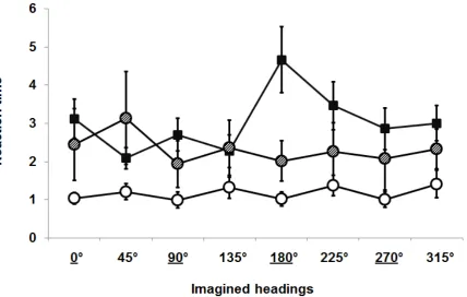
Reaction times were analysed with the same design as pointing errors. There was a
significant effect of the Visual Status [F(2,27)=3.46, p=.046], indicating that S were faster
Overall no single Imagined Heading affected the reaction times [F(7,22)=2.57, p=.060].
Again there was a significant interaction between Visual Status and Reference Frame
[F(4,25)=11.42, p=.000] (see Figure 3). The Fischer post-hoc analysis showed that CB were
faster in egocentric trials than allocentric, while LB were faster in allocentric than egocentric.
Finally S were faster than CB in both allocentric and egocentric trials [all p<.05]. The
interaction between Visual Status and Imagined Heading was significant [F(10,19)=4.04,
p=.001] indicating that some Imagined Headings were faster performed by CB (45° and
135°), others were faster performed by LB (90°, 180° and 270°), and others were faster
performed by S (0°, 90°, 180° and 270°). The interaction between the Reference Frame and
the Imagined Heading was not significant [F(7,22)=1, p=.396] nor the interaction across
Visual Status, Reference Frame and Imagined Heading was significant [F(12,17)=1.20,
p=.310].
Thus, both the results relative to the pointing errors and the reaction times support the
hypothesis that Visual Experience dictates the preference for a given type of spatial
representation. The murkier results associated to the reaction times are likely to be due their
higher variability, which reflect different response ‘styles’ across participants (e.g. more or
less impulsive).
[Figure 2 about here]
[Figure 3 about here]
Discussion
Our results suggest that participants possessing visual experience (LB and S) were able
to extract the structure of the array and to use it during the spatial memory task. Thus they
Conversely, participants without visual experience (CB) were better at using the
self-referenced cues arising from the spatial exploration to perform the memory task (i.e.
idiothetic cues, see [14]). Thus they better performed on trials requiring imagining headings
aligned with the explorative routes they had walked.
Thus we found support for the hypothesis that visual experience is necessary to develop
and use an allocentric spatial representation, which CB find quite difficult to achieve [10]. It
is important to note that here we claim that visual experience determines the preference for a
given type of spatial representation (i.e. it facilitates its adoption). In fact, past studies
reported the CB participants were able to carry out path integration and allocentric spatial
representation [8-9]. Therefore, we may find that by increasing the exposure to the array CB
participants would improve their performance in allocentric trials.
Additionally, our results on sighted participant extend earlier findings on vision [1] to
the somatosensory modality –i.e. that S participants used the intrinsic structure of the array.
Differently from McNamara and colleagues [1] we found that our sighted participants are less
accurate in the pointing task their sighted participants who visually learned the array. This
can be explained by the fact that learning the array by somatosensation represents an effortful
and error prone serial process [15]. In fact, vision has the ability to convey simultaneous
information about different objects, which is particularly advantageous for objects laying
outside the peripersonal space, while by using somatosensation the spatial relations among
objects have to be patiently constructed [15-16].
Finally our results extend a previous study that examined spatial reference frames in
auditory peripersonal space to extra-personal space [17]. In that study, CB, LB, and S
participants judged the spatial occurrence of sounds within peripersonal space (perceptive
of objects in memory. In both experiments, CB participants preferred an egocentric
representation.
Aside its influence on non-visual areas [18], the role of developmental vision on spatial
cognition and on its underlying brain structures can be clarified by the studies investigating
the role of visual experience on brain areas devoted to multisensory integration. For example,
Wallace and colleagues reported that visual experience is necessary to develop multisensory
neurons [19]. Accordingly, Röder and colleagues [20] found that congenitally blind
participants were less affected by an auditory-tactile counting illusion (i.e. tactile taps and
beeps). Crucially, the effect of visual experience on multisensory integration was also found
in spatial tasks, for example CB are not affected by hands crossing in a temporal order
judgement task [21]. Additionally, the role of visual experience was shown in spatial
updating tasks [6, 22]. Recent studies began to report effects of visual experience on the
multisensory brain areas involved in spatial tasks, such as the hippocampus [23-24] and the
posterior parietal cortex [25].
This suggests that during the initial years of the human life visual experience exerts its
effects on the brain areas involved in spatial processing and multisensory integration,
supporting the hypothesis that the use of an allocentric reference frame to remap multisensory
spatial inputs is not innate but its development requires visual experience [10].
Conclusion
In our study we found evidence that visual experience triggers the preference for a given type
of spatial representation. Specifically, although people with visual experience preferentially
represent object locations with an allocentric reference frame, those without visual experience
supported by recent studies reporting an effect of visual experience on brain areas involved in
multisensory integration for spatial cognition. Finally, our results try to provide a broader
interpretation of the contradictory literature on the effect of blindness in spatial cognition by
Acknowledgements
This work was supported by a Marie Curie Intra-European Fellowship (grant number:
PIEF-GA-2010-274163). We thank Dr J. Hodsoll for the technical support, the Royal National
Institute for Blind, and the Royal London Society for Blind for helping us with participant
References
[1] Mou W, McNamara TP. Intrinsic frames of reference in spatial memory. J Exp Psychol
Learn Mem Cogn 2002;28:162-70.
[2] Mou W, Fan Y, McNamara TP, Owen CB. Intrinsic frames of reference and egocentric
viewpoints in scene recognition. Cognition 2008;106:750-69.
[3] Rushmore RJ, Payne BR. Neuroplasticity after unilateral visual cortex damage in the
newborn cat. Behav Brain Res 2004;153:557-65.
[4] Hamilton RH, Pascual-Leone A, Schlaug, G. Absolute pitch in blind musicians.
Neuroreport 2004;15:803-6.
[5] Amedi A, Raz N, Pianka P, Malach R, Zohary E. Early 'visual' cortex activation correlates
with superior verbal memory performance in the blind. Nat Neurosci 2003;6:758-66.
[6] Pasqualotto A, Newell FN. The role of visual experience on the representation and
updating of novel haptic scenes. Brain Cogn 2007;65:184-94.
[7] Rieser JJ, Hill EW, Talor CR, Bradfield A, Rosen S. Visual experience, visual field size,
and the development of nonvisual sensitivity to the spatial structure of outdoor
neighborhoods explored by walking. J Exp Psychol Gen 1992;121:210-21.
[8] Loomis JM, Klatzky RL, Golledge RG, Cicinelli JG, Pellegrino JW, Fry PA. Nonvisual
navigation by blind and sighted: Assessment of path integration ability. J Exp Psychol
Gen 1993;122:73-91.
[9] Passini R, Proulx G. Wayfinding without vision: An experiment with congenitally totally
blind people. Environm Behav 1988;20:227-52.
[10] Pasqualotto A, Proulx MJ. The role of visual experience for the neural basis of spatial
cognition. Neurosci Biobehav Rev 2012;36:1179-1187.
[11] Postma A, Zuidhoek S, Noordzij ML, Kappers AML. Keep an eye on your hands: On
[12] Yamamoto N, Shelton AL. Path information effects in visual and proprioceptive spatial
learning. Acta Psychol 2007;125:346-60.
[13] Heller MA. Tactile picture perception in sighted and blind people. Behav Brain Res
2002;135:65-8.
[14] Mittelstaedt ML, Mittelstaedt H. Idiothetic navigation in humans: Estimation of path
length. Exp Brain Res 2001;139:318-32.
[15] Loomis JM, Klatzky RL, Lederman SJ. Similarity of tactual and visual picture
recognition with limited field of view. Perception 1991;20:167-77.
[16] Ruotolo F, Ruggiero G, Vinciguerra M, Iachini T. Sequential vs simultaneous encoding
of spatial information: A comparison between the blind and the sighted. Acta Psychol
2012;139:382-9.
[17] Röder B, Kusmierek A, Spence C, Schicke T. Developmental vision determines the
reference frame for the multisensory control of action. Proc Natl Acad Sci USA
2007;104:4753-8.
[18] Stevens AA, Weaver KE. Functional characteristics of auditory cortex in the blind.
Behav Brain Res 2009;196:134-8.
[19] Wallace MT, Perrault TJ, Hairston WD, Stein BE. Visual experience is necessary for the
development of multisensory integration. J Neurosci 2004;24:9580-4.
[20] Hötting K, Röder B. Hearing cheats touch, but less in congenitally blind than in sighted
individuals. Psychol Sci 2004;15: 60-4.
[21] Röder B, Rösler F, Spence C. Early vision impairs tactile perception in the blind. Curr
Biol 2004;14:121-24.
[22] Reuschel J, Rösler F, Henriques DYP, Fiehler K. Spatial updating depends on gaze
[23] Kupers R, Chebat DR, Madsen KH, Paulson OB, Ptito M. Neural correlates of virtual
route recognition in congenital blindness. Proc Nat Acad Sci USA 2010;107:12716-21.
[24] Lepore N, Shi Y, Lepore F, Fortin M, Voss P, Chou YY, et al. Pattern of hippocampal
shape and volume differences in blind subjects. Neuroimage 2009;46:949-57.
[25] Fiehler K, Rösler F. Plasticity of multisensory dorsal stream functions: evidence from
Captions
Figure 1: A depiction of the experimental setup with the eight headings. The underlined
headings are the allocentric (0°, 270°, 180° and 90°) while the remaining are the egocentric
(315°, 225°, 135° and 45°).
Figure 2: Mean pointing errors in degrees across the three experimental groups for each of
the eight headings (the underlined headings are the allocentric ones); filled squares indicate
congenitally blind participants; dashed circles are the late blind participants; empty circles are
the blindfolded sighted participants. Error bars represent the standard error.
Figure 3: Mean reaction times in seconds across the three experimental groups for each of the
eight headings (the underlined headings are the allocentric ones); filled squares indicate
congenitally blind participants; dashed circles are the late blind participants; empty circles are
the blindfolded sighted participants. Error bars represent the standard error.
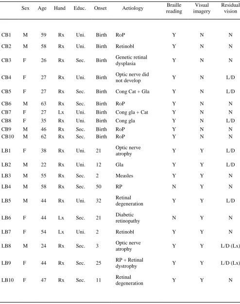
Table 1: Details of the participants; ‘Educ.’ indicates the level of education (University or
Secondary). ‘Y’ means ‘yes’ and ‘N’ means ‘no’, while ‘L/D’ means ‘light/darkness’
sensitivity and, if relevant, in which eye (left of right). Aetiology abbreviations: ‘RoP’,
retinopathy of prematurity; ‘Retinobl’,retinoblastoma; ‘Cong’, congenital; ‘Cat’, cataracts;
Table 1
Sex Age Hand Educ. Onset Aetiology Braille reading
Visual imagery
Residual vision
CB1 M 59 Rx Uni. Birth RoP Y N N
CB2 M 58 Rx Uni. Birth Retinobl Y N N
CB3 F 26 Rx Sec. Birth Genetic retinal
dysplasia Y N N
CB4 F 27 Rx Uni. Birth Optic nerve did
not develop Y N L/D
CB5 F 27 Rx Sec. Birth Cong Cat + Gla Y N L/D
CB6 M 63 Rx Sec. Birth RoP Y N N
CB7 F 27 Lx Uni. Birth Cong gla + Cat Y N N
CB8 F 35 Rx Uni. Birth Cong gla Y N L/D
CB9 M 46 Rx Sec. Birth RoP Y N N
CB10 M 62 Rx Sec. Birth RoP Y N N
LB1 F 38 Rx Uni. 21 Optic nerve
atrophy Y Y L/D
LB2 M 22 Rx Uni. 12 Gla Y Y L/D
LB3 M 55 Rx Sec. 2 Measles Y Y N
LB4 M 58 Rx Sec. 50 RP N Y N
LB5 M 44 Rx Uni. 32 Retinal
degeneration Y Y L/D
LB6 F 44 Lx Sec. 21 Diabetic
retinopathy N Y N
LB7 F 54 Lx Uni. 2 Retinobl Y Y N
LB8 M 24 Rx Sec. 3 Optic nerve
atrophy Y Y L/D (Lx)
LB9 F 44 Rx Sec. 25 RP + Retinal
dystrophy Y Y L/D (Lx)
LB10 F 47 Rx Sec. 11 Retinal