0022-538X/96/$04.0010
Copyrightq1996, American Society for Microbiology
Use of Transdominant Mutants of the Origin-Binding Protein
(UL9) of Herpes Simplex Virus Type 1 To Define
Functional Domains
AJAY K. MALIKANDSANDRA K. WELLER*
Department of Microbiology, University of Connecticut Health Center, Farmington, Connecticut 06030-3205
Received 17 May 1996/Accepted 23 July 1996
UL9, the origin-binding protein of herpes simplex virus type 1, contains six sequence motifs conserved in a large superfamily of RNA and DNA helicases. Single-amino-acid substitution mutations in these motifs inacti-vate UL9 function in vivo (R. Martinez, L. Shao, and S. K. Weller, J. Virol. 66:6735–6746, 1992). Overexpression of wild-type UL9 is inhibitory to plaque formation in a transfection assay which measures viral plaque formation by infectious herpes simplex virus type 1 DNA. Constructs containing mutations in motif I, II, or VI exhibit even stronger inhibitory effects in the same assay and thus can be considered strong transdominant inhibitors of plaque formation by the wild-type virus. The transdominant phenotype can be relieved by introducing a second mutation in the DNA-binding domain or by deleting the N-terminal 35 amino acids of the protein. The inhibitory effects of wild-type UL9 can also be partially relieved by deletion of amino acids 292 to 404. We propose that the N-terminal 35 amino acids of UL9 and residues 292 to 404 may define new functional domains of the UL9 protein.
Most replication systems utilize an initiator protein which specifically recognizes its cognate origin of replication. The DnaA protein of Escherichia coli and large T antigen of the DNA virus simian virus 40 (SV40) provide two paradigms for understanding the functions of initiator proteins. The DnaA protein initiates replication by binding cooperatively to the E.
coli origin of replication, oriC; this results in the localized
unwinding of the AT-rich repeats in the origin. This
DnaA-oriC complex is recognized by the DnaB helicase which further
unwinds DNA, thereby providing a template for priming and replication enzymes (reviewed in reference 20). Large T anti-gen binds the SV40 origin as a double hexamer around the origin DNA. The intrinsic helicase activity of large T antigen leads to origin unwinding in the presence of ATP. In the case of large T antigen, the origin-binding and helicase functions reside in a single polypeptide (reviewed in reference 11).
UL9 is the origin-binding protein of herpes simplex virus type 1 (HSV-1). Like large T antigen, UL9 has both intrinsic heli-case and origin-specific DNA-binding activities (5, 12). The N-ter-minal domain has six canonical motifs shared with superfamily II of DNA and RNA helicases (Fig. 1) (15, 16, 19). In this super-family of helicases, motifs I and II are believed to constitute an ATP-binding fold (48). The functions of the other motifs (III to VI) are not clear for UL9 or any of the other superfamily II helicases. Evidence that the helicase function of UL9 is impor-tant for HSV-1 DNA replication comes from genetic experi-ments. Site-directed mutations in each of these motifs abolish the ability of the UL9 protein to complement a UL9-null mutant virus in an in vivo replication complementation assay (32). Furthermore, the analysis of the binding of purified UL9 to origin DNA by electron microscopy suggests that UL9 may be able to carry out limited unwinding of duplex DNA (28).
The HSV-1 genome contains three origins of replication, oriL (52) and a diploid oriS (35, 43). The minimal HSV-1 origin consists of two UL9-binding sites flanking an AT-rich
region (46). The origin-specific DNA-binding domain has been localized to a 283-amino-acid region within the C-terminal one-third of UL9 (Fig. 1) (1, 8). Full-length UL9 is a dimer in solution (5, 12) and binds cooperativity to origin DNA (9, 17). The N-terminal two-thirds of UL9 is required for cooperative binding to origin DNA (10, 17). The domains of UL9 respon-sible for stable dimer formation and cooperativity have not been identified. Although the biochemical events leading to the duplex unwinding at an HSV-1 origin are not well under-stood, one model proposes that dimers of UL9 bind coopera-tively to the origin, leading to the formation of higher-order structures. It is also possible that other viral and/or host pro-teins participate in origin unwinding in vivo (6, 7).
The study of transdominant mutants provides a genetic ap-proach to map the functional domains of a protein (reviewed in reference 18). Isolation and characterization of transdomi-nant mutants of several HSV-1 proteins, including the trans-activators ICP0, ICP4, and VP16, have facilitated the identifi-cation of regions required for protein-protein interactions (homo- or heterodimerization) (14, 40, 49). Since UL9 is a multifunctional protein, it is particularly suitable for this type of analysis. Thus, it may be possible to isolate mutants of UL9 that are deficient in some but not all wild-type functions. The study of transdominant mutants of UL9 is complicated by the fact that overexpression of wild-type UL9 is inhibitory to HSV-1 infection. We previously reported that in cell lines containing high copy numbers of the UL9 gene (more than 20 copies per haploid genome), virus replication was inhibited, whereas inhibition was not observed in cells with 2 to 10 copies (29). Similar observations have also been made by Stow et al. (45). Inhibitory effects of UL9 have also been observed by Skaliter and Lehman in an in vitro DNA replication system using a partially purified protein fraction from insect cells infected with recombinant baculoviruses expressing HSV-1 essential replication proteins (41). This fraction supported HSV-1 origin-independent replication from a circular plasmid, pUC18, which does not contain an HSV-1 origin of replication (41). UL9 is not required for DNA replication in this assay; in fact, it is inhibitory if the plasmid template also contains an HSV-1 origin (41). The observations that UL9 is essential for * Corresponding author. Mailing address: Department of
Microbiol-ogy, MC3205, University of Connecticut Health Center, 263 Farming-ton Ave., FarmingFarming-ton, CT 06030-3205. Phone: (860) 679-2310. Fax: (860) 679-1239. Electronic mail address: [email protected].
7859
on November 9, 2019 by guest
http://jvi.asm.org/
DNA synthesis and can also be inhibitory if overexpressed may indicate that HSV-1 DNA replication occurs in two or more stages. Recent evidence from a study using temperature-sen-sitive mutations in the UL9 gene indicate that the thermosen-sitive function of the UL9 protein is essential at early but not late times during viral DNA synthesis (2). It is possible that origin-initiated DNA synthesis requiring UL9 gives way to a recombination-based mode of replication which is inhibited by UL9 (reviewed in reference 50).
Despite the fact that wild-type UL9 can inhibit productive infection by wild-type virus, several viral mutants exhibit an even greater inhibitory effect on viral plaque formation. For example, overexpression of the DNA-binding domain of UL9 results in inhibition of HSV-1 DNA replication (39, 44, 45). The mechanism of transdominance presumably involves the titration of one or more of the origins of replication in an inactive complex. Helicase motif mutants provide additional probes with which to study UL9 function, since they map outside the origin-specific DNA-binding domain. Constructs containing mutations in motif I, II, or VI are strong transdomi-nant inhibitors of viral plaque formation. The transdomitransdomi-nant phenotype can be relieved either by introduction of a mutation in the DNA-binding domain or by deletion of the N-terminal 35 amino acids of UL9. Possible models for the mechanism of transdominance will be considered.
(This work constitutes part of the Ph.D. thesis of A.K.M. to be submitted to The University of Connecticut in partial ful-fillment of the requirements for the Ph.D. degree.)
MATERIALS AND METHODS
Cells and viruses.Vero cells (obtained from the American Type Culture Collection) were maintained in culture as previously described (51). The KOS strain of HSV-1 was used as the wild-type virus. hr94, a UL9-null virus containing a lacZ insertion, was previously described (29). hr94 was propagated in 2B-11 cells containing the wild-type version of the UL9 gene (29).
Plasmids.p6UL9-119b contains the UL9 gene expressed from the inducible ICP6 promoter of HSV-1 (29). Plasmids containing point mutations in the helicase motifs (p6UL9-K87A, p6UL9-K87R, p6UL9-E175A, p6UL9-T214S, p6UL9-T214A, p6UL9-F303W, p6UL9-G354A, and p6UL9-R387K) were pre-viously described (Fig. 1) (32). p100-1 contains an HSV-1 origin of replication, oriS, on a 100-bp MspI fragment cloned into the SmaI site of the pUC19 vector (kindly provided by Mark Challberg, National Institutes of Health, Bethesda, Md. [36]). pVP16-9, which expresses the HSV-1 transactivator, VP16, from the murine sarcoma virus long terminal repeat promoter cloned into pTZ19R, was kindly provided by S. Weinheimer (Bristol-Myers Squibb, Wallingford, Conn.). p6UL9-OB, containing an origin-binding insertion mutation (OB mutation) in the UL9 gene (Fig. 1), was created by linearizing p6UL9-119b (propagated in a
dcm1E. coli host) with StuI and inserting a 12-mer BamHI linker (59-CGC-G GA-TCC-GCG). This mutation creates an insertion of four amino acids (Arg-Ile-Arg-Ala [RIRA]) after Ala-591 in the UL9 protein sequence. Double mu-tants containing one motif mutation and the OB mutation (K87A-OB, p6-K87R-OB, p6-E175A-OB, p6-F303W-OB, p6-G354A-OB, and p6-R387K-OB) were constructed by replacing the 805-bp EcoNI-EagI fragment from the UL9 gene in K87A, K87R, E175A, F303W, p6UL9-G354A, and p6UL9-R387K, respectively, with the corresponding fragment from p6UL9-OB, which contains the four-amino-acid RIRA insertion.
p6UL9-D1-35 expresses an N-terminal 35-amino-acid deletion mutation of the UL9 gene. It was constructed from p6UL9-119b by digestion with XhoI, which cleaves at a site between the AUG start codon and the ICP6 promoter (29), and
NheI, which cleaves at a position 96 nucleotides downstream of the AUG start
codon. The resulting overhanging ends were ligated after treatment with the Klenow enzyme. The resultant plasmid, p6UL9-D1-35, is expected to start trans-lation from an in-frame methionine at codon 36 of UL9. TheD1-35 derivative of motif VI mutant, R387K, was constructed from plasmid p6UL9-R387K by a similar deletion strategy. Plasmids expressing internal deletions of UL9,
p6UL9-D98-195, p6UL9-D196-291, p6UL9-D292-404, p6UL9-D405-520, p6UL9-D 521-597, and p6UL9-D598-703, are described elsewhere (30).
p6UL9-D13-536, containing the N-terminal 12 amino acid residues, fused to the DNA-binding domain of UL9 (amino acids 537 to 851), was constructed by deleting the internal BstYI-BamHI fragment in the UL9 gene as follows. p6UL9-119b was digested with HindIII and BamHI to generate 2.2- and 4.3-kbp frag-ments. The 2.2-kbp fragment, containing the ICP6 promoter and the N-terminal two-thirds of UL9, was isolated and was further digested with BstYI, generating a 0.6-kbp fragment. This 0.6-kbp fragment containing the ICP6 promoter and the N-terminal 12 amino acids of UL9 was gel purified and ligated to the 4.3-kbp fragment obtained from the HindIII and BamHI digest (above) to construct plasmid p6UL9-D13-536.
A BamHI site at the 59end of UL9 was introduced into p6UL9-119b by in vitro mutagenesis (22, 32). The mutagenic oligonucleotide corresponding to the non-coding strand was 59-GAA-AGG-CAT-TTC-GGA-TCC-AAC-AGA-CGC-GG C-39. The start codon and the BamHI site are underlined. The resulting plasmid, p6-NBam, also contains a second BamHI site within the UL9 gene at amino acid position 534. p6UL9-D1-534, containing the C-terminal 317 amino acids of UL9; was constructed from p6-NBam by introducing an NcoI linker containing a start codon as follows: p6-NBam was digested with BamHI, and the resulting 4.8-kbp fragment lacking the N-terminal 1 to 534 amino acids was isolated. Following treatment with Klenow enzyme, an NcoI linker (59-AGC-CAT-GGC-T) was insert-ed. The expected protein contains Met and Ala, followed by amino acids 535 to 851. All mutations were verified by the dideoxy-chain termination sequencing method, using a Sequenase kit from United States Biochemical, Cleveland, Ohio.
Viral and plasmid DNA isolation.Infectious HSV-1 viral DNA was made as previously described (53). Plasmids were purified by using an anion-exchange column (Qiagen, Chatsworth, Calif.) followed by phenol-chloroform extraction and ethanol precipitation according to standard protocols. Plasmid DNA was quantified by A260. DNA dilutions were subjected to 1% agarose gel electro-phoresis to verify DNA concentration and to rule out RNA contamination.
Plaque reduction assay.Efficiency of plaque formation was studied by cotrans-fecting 1.53106Vero cells with infectious viral DNA and a 40-fold molar excess of UL9 mutant-expressing plasmids (for example, 2mg of plasmid DNA was used for every 1mg of viral DNA). Generally 0.125 to 0.5mg of viral DNA was used, depending on the infectivity of a particular preparation. Cotransfections were performed as described previously (53), with some modifications. A DNA-CaPO4precipitate of viral and plasmid DNA was incubated with cells in sus-pension at 378C for 30 min, with gentle shaking at 75 rpm. The transfected cells were plated on two 60-mm-diameter culture plates in 5 ml of regular culture media (Dulbecco modified Eagle medium containing 5% fetal bovine serum), and at 2.5 h postplating, monolayers were treated with 15% glycerol in N-2-hydroxyethylpiperazine-N9-2-ethanesulfonic acid (HEPES)-buffered saline for 2 min. Cells were allowed to recover in regular culture media for 3 to 5 h, at which point they were overlaid with approximately 5 ml of 2% (wt/vol) methylcellulose in culture medium. Four days later, plaques were stained by adding 4 ml of 1% neutral red in Tris-buffered saline. Each experiment was repeated three to five times. The mean number of plaques was calculated, and standard errors were determined by using KaleidaGraph software on a Macintosh computer.
Transient transfection-immunoblot assay.For immunoblot analysis of the UL9 mutants, 1.53106Vero cells were transfected with 0.5 to 12mg of plasmid as previously described (53). At 24 h posttransfection, cells were infected with
hr94 at a multiplicity of 10 PFU per cell, and 18 h thereafter, cells were harvested,
washed with phosphate-buffered saline (PBS), resuspended in 200ml of PBS, and lysed by adding 100ml of buffer containing 30 mM Tris-HCl, 15 mM EDTA, and 3% sodium dodecyl sulfate (SDS). The DNA in the samples was sheared by sonication for 30 to 60 s at 48C in a water bath sonicator. Total protein was precipitated by adding 5 volumes of acetone at2208C. The acetone precipitates were pelletted, air dried, and resuspended in approximately 100ml of SDS-polyacrylamide gel electrophoresis (PAGE) loading buffer. The samples were boiled, and 20ml was loaded per lane on an SDS–8% polyacrylamide minigel (Hoefer Scientific, San Francisco, Calif.). Proteins were electroblotted onto an Immobilon-P membrane (Millipore, Bedford, Mass.) and incubated with a 1:5,000 dilution of R250, a rabbit antiserum directed against the C-terminal decapeptide of UL9. R250 was generously provided by Mark Challberg (37). Alkaline phosphatase-conjugated anti-rabbit immunoglobulin G was used for color development (Protoblot system obtained from Promega, Madison, Wis.).
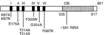
[image:2.612.90.270.69.130.2]Immunofluorescence microscopy.UL9 was expressed from the ICP6 promoter by induction with VP16 (29). A total of 106Vero cells were transfected with 8mg of wild-type or mutant versions of a UL9 expression plasmid and 1mg of VP16 plasmid. Transfections were performed by the CaPO4precipitation method as previously described (53). At 24 h posttransfection, the cells grown on coverslips were washed with PBS and fixed in methanol-acetone (1:1) for 30 min at2208C. Cells were permeabilized with 1% Triton X-100 in PBS for 5 min at room temperature and incubated in 3% normal goat serum (NGS) in PBS for 30 min at room temperature. The primary monoclonal antibody, 17B, recognizes an FIG. 1. Helicase motif and DNA-binding domain mutants of UL9 used in
this study. The black boxes represent six helicase motifs in the N-terminal region. Substitution mutations in UL9 helicase motifs are shown below. The shaded reg-ion represents the 283-amino-acid origin DNA-binding domain of UL9 (1, 8). The four-amino-acid (RIRA) insertion in the origin-binding region (OB) is indicated.
on November 9, 2019 by guest
http://jvi.asm.org/
epitope in the N-terminal 33 amino acids of UL9 (30). 17B was used without dilution. The rabbit antiserum, R250, was used at 1:50 dilution. The secondary antibodies (goat anti-mouse or anti-rabbit rhodamine conjugates; Organon Teknika [Cappel], Durham, N.C.) were used at a 1:200 dilution in 3% NGS. Twenty microliters of each antibody was added to each coverslip for 30 min. Coverslips were washed three to five times with 1% NGS and mounted in glycerol-gelatin mounting solution (Sigma, St. Louis, Mo.) containing 2.5% DABCO (1,4-diazabicyclo [2.2.2] octane) to retard bleaching. Immunofluores-cence images were collected on a Zeiss Axiovert 100TV microscope equipped with a Plan-Neofluar 63X 1.25-numerical aperture oil immersion lens and charge-coupled device camera and controlled by the Innovisions software ISEE. The images were arranged and labeled by using Adobe Photoshop software on a Silicon Graphics workstation.
In vivo transient replication complementation assay.The in vivo transient replication complementation assay was performed as previously described (32). Briefly, 1.53106
Vero cells were transfected with 0.2mg of oriS-containing plasmid (p100-1), 1mg of wild-type or mutant UL9-expressing plasmid, and 11
mg of carrier DNA (sonicated salmon sperm DNA). At 24 h posttransfection, the cultures were infected with the UL9-null virus, hr94, at a multiplicity of 10 PFU per cell. At 18 h postinfection, total cell DNA was prepared, analyzed by diges-tion with EcoRI alone or in combinadiges-tion with DpnI, and then subjected to agarose gel electrophoresis and Southern blotting on a GeneScreenPlus mem-brane (DuPont-NEN, Boston, Mass.). The blots were hybridized with32
P-labeled pBluescript plasmid (Stratagene, La Jolla, Calif.). The replication of oriS-con-taining plasmid was assessed by resistance to DpnI digestion.
RESULTS
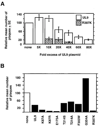
Transdominance of motif mutant versions of UL9. Single-amino-acid substitutions in all six helicase motifs (Fig. 1) abol-ish the ability of UL9 to complement hr94 in the in vivo complementation assay (32). The motif V mutant protein was not detected by transient transfection-immunoblot assay, and thus no conclusion could be drawn about the specific require-ment for motif V (32). To determine whether mutant UL9 protein expressed from a plasmid could inhibit viral plaque formation, it was first necessary to confirm previous observa-tions that wild-type UL9 can be inhibitory (29, 45). Vero cells were cotransfected with viral DNA and various amounts of the wild-type UL9 expression plasmid. In the experiments shown in Fig. 2A, cotransfection of a 20- or 40-fold molar excess of UL9 plasmid resulted in a 30 to 50% reduction in the number of plaques observed; cotransfection with a 60- or 80-fold molar excess of UL9 plasmid resulted in 65 to 80% reduction. These results confirm and extend the observation that overexpression of UL9 is inhibitory to viral plaque formation.
To ascertain whether an inhibitory effect even greater than that obtained with wild-type UL9 could be obtained by the motif mutants and to establish optimal conditions, the motif VI mutation, R387K, was chosen. A plasmid containing the R387K mutation was cotransfected with infectious wild-type HSV-1 DNA at three different molar ratios (10-, 20-, and 40-fold plas-mid excess). As shown in Fig. 2A, a 40-fold molar excess of the motif VI mutant, R387K, resulted in a greater than 90% reduc-tion in plaque formareduc-tion. At this ratio, wild-type UL9 generally results in a 40 to 60% inhibition of plaque formation. In all subsequent experiments, a 40-fold molar excess of plasmid DNA was used.
The entire set of helicase motif mutants were tested at a 40-fold plasmid molar excess in the plaque reduction assay. As shown in Fig. 2B, the two motif III mutants, T214S and T214A, and the motif IV mutant, F303W, showed an inhibition of plaque formation comparable with that of wild-type UL9. The motif V mutant, G354A, apparently potentiates plaque forma-tion (see Discussion). On the other hand, mutaforma-tions in motif I, II, or VI inhibited viral plaque formation by approximately 90% (K87A, K87R, E175A, and R387K in Fig. 2B). Thus, it appears that mutations in motif I, II, or VI are strongly trans-dominant in this assay.
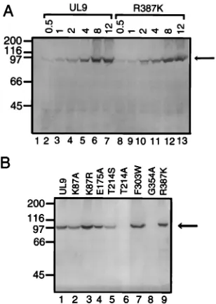
Transdominance of motif mutants is not due to differences in protein expression levels.To determine whether the greater
[image:3.612.349.523.70.290.2]than wild-type inhibition seen with the R387K mutant in Fig. 2A could be explained by differences in protein expression levels, a transient transfection-immunoblot experiment was performed. Vero cells were transfected with 0.5 to 12mg of wild-type UL9 or R387K expression plasmid, and expression was induced by hr94 infection. Total cell protein extracts were resolved by SDS-PAGE, immunoblotted, and analyzed by us-ing the UL9-specific rabbit antiserum, R250. As shown in Fig. 3A, wild-type UL9 and R387K express comparable amounts of protein at each of the plasmid concentrations used. This result suggests that differences in protein expression levels are not a significant factor in the greater than wild-type inhibition seen with the R387K mutant. Similarly, to rule out the possibility that differences in protein expression levels were responsible for the observed transdominant effect of other motif mutants, a transient transfection-immunoblot experiment was per-formed as described above except that cells were transfected with 8mg of expression plasmid. As shown in Fig. 3B, mutants with mutations in motifs III and V (T214S, T214A, and G354A) exhibited decreased levels of protein (Fig. 3B, lanes 5, 6, and 8). Mutations in motif III were not studied further. Given the potentiating effects of transfection with a plasmid bearing a motif V mutation, it was of interest to further exam-ine the expression of this mutant protein. Experiments with an N-terminus-specific monoclonal antibody, 17B, and another polyclonal antibody, RH7, raised against the C-terminal 317 amino acids also failed to detect a protein expressed from the motif V mutant plasmid (data not shown) (see Discussion). On FIG. 2. Effects of plasmids expressing wild-type UL9 or mutants on viral plaque formation by infectious viral DNA. (A) Vero cells were cotransfected with infectious viral DNA and a 5- to 80-fold molar excess of the plasmid expressing wild-type UL9 or R387K as described in Materials and Methods. A histogram depicts the results of four experiments. For comparison, the no-plasmid control was normalized to a value of 100 PFU, and experimental values were adjusted accordingly. The relative mean numbers of plaques6standard errors (SE) are shown. The actual numbers of viral plaques obtained with viral DNA alone (no-plasmid control) were 55 (experiment 1), 65 (experiment 2), 130 (experiment 3), and 59 (experiment 4). These numbers reflect variability of infectivity between different batches of viral DNA and variability in transfections. (B) Plaque reduction assay with plasmids expressing helicase motif mutants of UL9. Vero cells were cotransfected with infectious viral DNA and a 40-fold molar excess of the plasmid expressing the indicated protein. A histogram de-picting normalized relative mean numbers of plaques from two experiments is shown. The actual numbers of viral plaques obtained with viral DNA alone (no-plasmid control) were 295 (experiment 1), and 182 (experiment 2).
on November 9, 2019 by guest
http://jvi.asm.org/
the other hand, cells transfected with plasmids expressing mu-tations in motifs I, II, IV, or VI accumulate levels of protein comparable to the expression from the wild-type UL9 plasmid. Similar results were obtained when cells were transfected with 4 mg of expression plasmid (data not shown). Thus, the strongly transdominant effect of mutations in motif I, II, or VI cannot be explained by higher than expected levels of protein expression compared with wild-type UL9.
Transdominance of motif mutants can be partially relieved by the introduction of a mutation in the DNA-binding domain.
Stow et al. previously showed that inhibition of viral plaque formation by overexpression of wild-type UL9 can be relieved by the introduction of a mutation (i591RIRA) in the origin-binding domain of UL9 (45). This insertion mutation was pre-viously shown to disrupt origin-specific DNA binding (1, 8). In this report, we constructed an identical RIRA insertion mutant (designated the OB mutation) (Fig. 1). To determine whether the introduction of the OB mutation could relieve the inhibi-tory effects of UL9 in our assay system, plasmids expressing wild-type UL9 and the OB mutant form of UL9 were used in a plaque reduction assay. Wild-type UL9 reduced plaque for-mation from a normalized value of 100 PFU in the absence of any cotransfected plasmid to 4066 PFUs (n59). When the OB mutation was used, a normalized value of 108611 PFU (n59) was obtained. Thus, introduction of the OB mutation was able to relieve the inhibitory effect of the wild-type UL9. This finding suggests that the inhibitory effect of overexpressed
wild-type UL9 is likely to be mediated through origin DNA binding (see Discussion).
It is clear that overexpression of wild-type UL9 inhibits viral plaque formation and that mutations in motifs I, II, or VI exert even stronger inhibitory effects. It is possible that the motif mu-tants inhibit plaque formation by the same mechanism as the wild type but more strongly because of the nonfunctional na-ture of the protein containing the motif mutation. Thus, we were interested in whether the addition of an OB mutation to a UL9 gene bearing a mutation in motif I, II, or VI would relieve the strong transdominance effect. Expression plasmids containing double mutations, one motif mutation as well as the OB mu-tation, were constructed. The abilities of the double-mutant constructs to inhibit viral plaque formation were measured and compared with those of the motif mutants. As shown in Fig. 4, the introduction of the OB mutation into the motif mutant background partially or fully relieved inhibition by the trans-dominant motif mutants. Wild-type UL9 and the OB mutant are shown for comparison. The addition of the OB mutation to the motif V mutant even further potentiates plaque formation (see Discussion). In summary, these results suggest that the mo-tif mutants K87A, K87R, E175A, and R387K may sequester or-igin DNA in an inactive complex, and thus introduction of an OB mutation can partially relieve the observed transdominance.
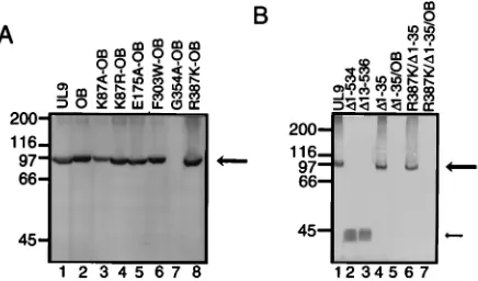
The stability and protein expression levels of double-mutant proteins were assessed in a transient transfection-immunoblot assay as described above. All motif-OB double mutants except the G354A-OB mutant express proteins of the expected sizes and at levels which are comparable to those of wild-type UL9 and the OB mutant (Fig. 5A). The instability of the G354A-OB protein is not surprising, since the G354A mutation itself was previously shown to be unstable (Fig. 3B, lane 8) (32).
[image:4.612.102.256.70.286.2]Transdominance of motif mutants is not due to defective nuclear localization.It is possible that factors other than bind-ing of the origin DNA into an inactive complex also contribute to the transdominance of the motif mutants. For instance, it is possible that the strongly transdominant mutants of UL9 in-hibit the nuclear localization of the wild-type protein. A mu-tation (K128N) in the nuclear localization signal of SV40 large T antigen inhibits the transport of wild-type large T antigen into the nucleus and is a transdominant inhibitor of SV40 replication (24, 25). To test this possibility, cells were cotrans-fected with plasmids expressing either the wild-type or motif FIG. 3. Immunoblot of extracts from cells transfected with wild-type UL9
and motif mutant versions of UL9. Cells were transfected with an expression plasmid, and protein expression was induced by infection with the hr94 virus as described in Materials and Methods. Total cell extracts were resolved by SDS-PAGE and analyzed by immunoblotting using R250, a C-terminal 10-amino-acid-specific rabbit antiserum. Positions of the molecular mass markers (200, 116, 97, 66, and 45 kDa; obtained from Bio-Rad, Hercules, Calif.) are indicated on the left. The arrow on the right indicates the position of wild-type UL9 and mutants. (A) Vero cells were transfected with 0.5, 1, 2, 4, 8, or 12mg of the wild-type or R387K plasmid as indicated at the top (lanes 2 to 13). Lane 1 shows extracts from untransfected cells which were infected with hr94. (B) Vero cells were transfected with 8mg of the wild-type or mutant expression plasmids as indicated. Similar protein expression results were obtained when Vero cells were transfected with 4mg of plasmid and also when immunoblots were probed with the RH7 rabbit antiserum (not shown). The RH7 antiserum was raised against a glutathione S-transferase N-terminal fusion form of the DNA-binding domain (residues 535 to 851) of UL9 (47).
FIG. 4. Plaque reduction assay with plasmids expressing motif mutants con-taining an additional mutation in the DNA-binding domain of UL9 on viral plaque formation. The histogram plots show data obtained from four (A) and five (B) experiments. Vero cells were cotransfected with infectious viral DNA and a 40-fold molar excess of the plasmid expressing the indicated protein as described in Materials and Methods. The number of viral plaques in each experi-ment was normalized with respect to viral DNA alone (no-plasmid control), which was set at 100. (A) The actual numbers of viral plaques obtained with viral DNA alone (no-plasmid control) were 95 (experiment 1), 165 (experiment 2), 234 (experiment 3), and 94 (experiment 4). (B) The actual numbers of viral plaques obtained with viral DNA alone (no-plasmid control) were 76 (experiment 1), 78 (experiment 2), 230 (experiment 3), 174 (experiment 4), and 82 (experiment 5).
on November 9, 2019 by guest
http://jvi.asm.org/
[image:4.612.318.554.71.170.2]mutant proteins from the ICP6 promoter and a VP16 expres-sion clone to activate the ICP6 promoter. Nuclear localization was determined by an indirect immunofluorescence assay using an N-terminus-specific monoclonal antibody, 17B (30). Wild-type UL9 localizes to the nucleus (Fig. 6B). Similar results were obtained when UL9 was expressed from the cytomegalo-virus immediate-early promoter or from the ICP6 promoter induced with ICP0 (30). Thus, as previously reported (29), no
other viral proteins are required for the nuclear localization of UL9. Motif mutants K87R, E175A, F303W, and R387K also localized efficiently to the nucleus (Fig. 6C to F). In summary, it does not appear that inefficient nuclear localization can be responsible for the strong inhibitory effects of mutations in motif I, II, or VI on viral plaque formation.
Use of deletions of UL9 to define important domains. An-other mechanism by which the transdominant motif mutants may exert their inhibitory effects may be through protein-pro-tein interactions, either by the formation of mixed dimers or higher oligomers or by sequestration of other viral and/or cel-lular protein(s). If proteprotein interactions play a role in in-hibition of plaque formation, a deletion or mutation of an inter-action domain may partially relieve the inhibitory effect. The next set of experiments were performed to determine whether an interaction domain could be identified by this strategy.
We constructed six internal deletion mutants of UL9 (D 98-195,D196-291,D292-404,D405-520,D521-597, andD598-703) (30). These deletions were tested in the plaque reduction assay as described above (data not shown). D598-703 relieved the inhibition seen with the wild-type UL9 almost completely (nor-malized value of 97 68 PFU from three experiments). This result confirms that mutations in the DNA-binding domain relieve inhibition. The only other deletion which substantially relieved inhibition wasD292-404, which deletes motifs IV, V, and VI (normalized value of 7566 PFU from three experi-ments); all other deletions inhibited plaque formation compa-rably to the wild type.D292-404 produces mutant UL9 protein in levels comparable to wild-type levels and also localizes to the nucleus (30). Further experiments will be necessary to explain the mechanism responsible for the partial relief of transdomi-nance byD292-404 and to determine if the relief is a result of subtle conformational changes.
Stow and colleagues reported that the C-terminal DNA-binding domain of UL9 when expressed alone inhibits HSV-1 FIG. 5. Immunoblot of extracts from cells transfected with wild-type UL9
[image:5.612.70.288.70.198.2]and mutant versions of UL9. Vero cells were transfected with 8mg of the wild-type or mutant expression plasmids, and protein expression was induced by infec-tion with the hr94 virus as described in Materials and Methods. The expressed proteins were resolved by SDS-PAGE and analyzed by immunoblot analysis using the R250 rabbit antiserum. Positions of the molecular mass markers (200, 116, 97, 66, and 45 kDa) are indicated on the left. The large arrow on the right indicates the position of UL9 and full-length mutants. The small arrow in panel B indicates the position of DNA-binding domain versions of UL9. Similar pro-tein expression results were obtained when Vero cells were transfected with 4mg of plasmid and also when immunoblots were probed with the RH7 rabbit anti-serum (not shown). A Vero cells were transfected with wild-type UL9 (lane 1) and variants of UL9 containing the OB mutation in addition to various motif mu-tations as indicated (lanes 2 to 8). (B) Vero cells were transfected with wild-type UL9 (lane 1) and various deletion mutants of UL9 as indicated (lanes 2 to 7).
FIG. 6. Intracellular localization of wild-type and mutant versions of UL9. Vero cells were transfected with the VP16 plasmid alone (A and G) or cotrans-fected with the plasmids expressing VP16 and UL9 (B and H), K87R (C), E175A (D), F303W (E), R387K (F),D1-35 (I), and R387K/D1-35 (J). The transfected cells were stained with the 17B monoclonal antibody (A to F) or R250 rabbit antiserum (G to J), and immunofluorescence microscopy was performed as described in Materials and Methods.
on November 9, 2019 by guest
http://jvi.asm.org/
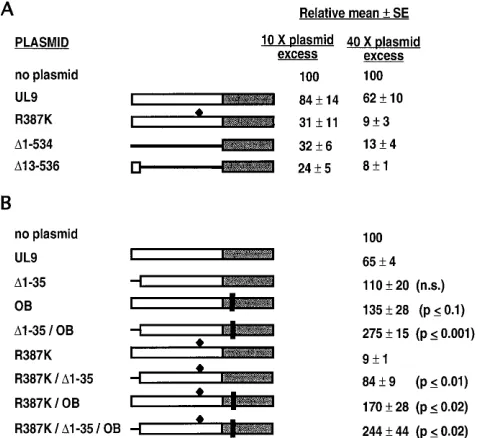
replication (45). The plasmid used by these authors expresses the N-terminal 12 amino acids in addition to the 315-amino-acid C-terminal DNA-binding domain. To address the poten-tial contribution of the N-terminal 12 amino acids to trans-dominance, we constructed plasmids D13-536 and D1-534, expressing the C-terminal DNA-binding domain with and with-out, respectively, the N-terminal 12-amino-acid region. The two mutants express comparable levels of protein in a transient transfection-immunoblot assay (Fig. 5B, lanes 2 and 3). Wild-type UL9, mutant R387K, and the two forms of the C-terminal DNA-binding domain were compared in the plaque reduction assay under the conditions of 10- or 40-fold molar plasmid excess. As shown in Fig. 7A, like R387K, both D1-534 and D13-536 were transdominant at both 10- and 40-fold molar ratios. The apparent differences betweenD1-534 andD13-536 were not statistically significant for either the 10- or the 40-fold molar ratios; nevertheless, there is a trend toward a higher transdominance effect withD13-536.
To ascertain whether the N-terminal region plays a role in the context of the full-length UL9 protein, we constructed versions of the wild type and R387K with an N-terminal 35-amino-acid deletion, D1-35 and R387K/D1-35, respectively (Fig. 7B). Both constructs expressed protein in amounts com-parable to those of wild-type UL9 in a transient transfection-immunoblot assay (Fig. 5B, lanes 1, 4, and 6). An immunoflu-orescence assay was used to demonstrate that these two
mutants also localized to the nucleus (Fig. 6I and J). Figure 6G shows the background staining of cells transfected with VP16 alone, and Fig. 6H shows cells transfected with VP16 and wild-type UL9. In the plaque reduction assay,D1-35 appears to be able to relieve the inhibitory effect of wild-type UL9; how-ever, this relief is not statistically significant (compare UL9 and D1-35 in Fig. 7B). On the other hand, this 35-residue deletion has a more dramatic effect on alleviating the transdominance of the motif VI mutant. R387K/D1-35 showed only a 15% inhibition of viral plaque formation, compared with a 90% inhibition seen with the R387K mutant (R387K and R387K/ D1-35 in Fig. 7B). The difference between R387K and R387K/ D1-35 was statistically significant with P # 0.01 (Fig. 7B). These results indicate that the N-terminal 35 amino acids con-tribute to the transdominance effect of the motif VI mutant.
To determine the combined effect of the OB andD1-35 muta-tions on the inhibitory effect of UL9 or the R387K mutant, we tested plasmids with an additional OB mutation in D1-35 or R387K/D1-35. As shown in Fig. 7B, these constructs further re-lieved the transdominance inhibition (mutants D1-35/OB and R387K/D1-35/OB in Fig. 7B). However, neither theD1-35/OB nor the R387K/D1-35/OB mutant could be detected by a tran-sient transfection-immunoblot assay (Fig. 5B, lanes 5 and 7). Be-cause of the apparent instability of these OB mutation-containing versions ofD1-35 and R387K/D1-35, no conclusions can be drawn about whether the OB andD1-35 mutations are synergistic.
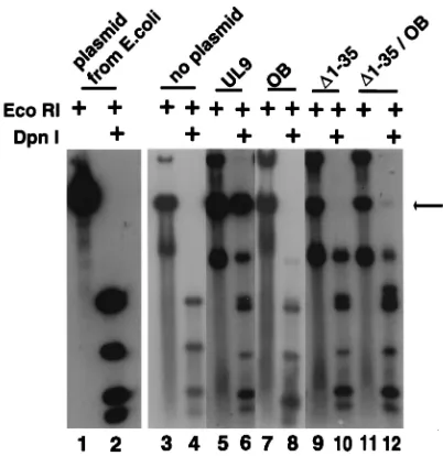
The N-terminal 35-amino-acid deletion (D1-35) was also tested for function in the in vivo complementation assay that measures replication, of plasmid pOriS which is assessed by resistance to DpnI digestion. In this assay, KOS infection results in an amplification of plasmid pOriS (data not shown) where-as no pOriS replication is seen in cells infected with hr94, a UL9-null virus (Fig. 8, lanes 3 and 4). The position of unit-length replicated DpnI-resistant pOriS is marked with an ar-row. Wild-type UL9 expressed from a plasmid complements
hr94 for the replication of pOriS (Fig. 8, lanes 5 and 6),
where-as the OB mutant (lanes 7 and 8), theD1-35 deletion (lanes 9 and 10), and theD1-35/OB double mutant (lanes 11 and 12) were unable to complement hr94. These observations also support the notion that the N-terminal 35 amino acids may define an im-portant domain of UL9 (see Discussion); nevertheless, further experiments will be required to rule out the possibility that this region may be required for proper conformation.
DISCUSSION
The inhibition of plaque formation seen by overexpression of wild-type UL9 in the plaque reduction assay is consistent with previous observations (29, 41, 45). In this report, we con-firm the observations that inhibition by wild-type UL9 can be relieved by the introduction of an OB mutation. This result indicates that binding of DNA is required for the inhibitory effects of UL9. This interpretation is consistent with the ob-servations that UL9 is inhibitory in an in vitro DNA replication system when a plasmid containing an HSV-1 origin of replica-tion is present (41). In this report, we also demonstrate that several of the helicase motif mutants exert even stronger in-hibitory effects than wild-type UL9 and that this transdomi-nance can be relieved either by the introduction of an OB mutation or by deletion of the N-terminal 35 amino acids of UL9. In addition, the inhibitory effects of wild-type UL9 can be partially relieved by deletion of amino acids 292 to 404.
[image:6.612.59.300.71.290.2]Four mutants stimulated viral plaque formation by 50 to 175%: G354A (a motif V mutant), G354A-OB,D1-35/OB, and R387K/D1-35/OB. We were unable to detect these four pro-teins by immunoblot analysis of transiently transfected cells FIG. 7. Plaque reduction assay using the two versions of the DNA-binding
domain and the N-terminal deletion mutations. (A) Inhibitory effects of plasmids expressing two versions of the DNA-binding domain of UL9 (D1-534 andD 13-536). The deleted regions are shown as lines. The R387K mutation is shown by a black diamond. The plaque reduction experiments were performed at 10- and 40-fold molar plasmid excess ratios as described in Materials and Methods. The relative mean numbers of plaques6standard errors (SE) from five experiments are shown. (B) Effect of deletion of the N-terminal 35 amino acids on the inhibitory effect of wild-type UL9 and the transdominance effect of the R387K mutant. The N-terminal deletion is shown as a line, and the vertical black bars represent the OB mutation. The R387K mutation is shown by a black diamond. The plaque reduction experiments were performed at 40-fold molar plasmid excess. The relative mean numbers of plaques6standard errors from four exper-iments are shown. The statistical difference between the mutants was determined by the unpaired Student t test, and the results are indicated on the right.D1-35, OB, and
D1-35/OB mutants were compared with wild-type UL9. R387K/D1-35, R387K/OB, and R387K/D1-35/OB mutants were compared with R387K. The difference be-tween R387K/D1-35 and R387K/OB was P#0.05. n.s., not significant.
on November 9, 2019 by guest
http://jvi.asm.org/
with three different antisera: R250 against the C-terminal 10 amino acids, another polyclonal antibody, RH7, raised against the C-terminal 317 amino acids, and the N-terminus-specific monoclonal antibody, 17B. Despite the inability to detect these mutant proteins with three different antisera, it remains pos-sible that a specific degradation intermediate of these mutants interacts with wild-type UL9 molecules and target them for degradation. A decreased level of UL9 in the later phase of replication may allow for increased replication and plaque formation in our assay.
Mutants containing mutations in motif I, II, or VI display a transdominant phenotype. Another mutation in motif I, K87E, was previously shown to inhibit viral plaque formation (45). The fact that the transdominance of mutants in motif I, II, or VI could be relieved by introducing a DNA-binding mutation lends support for the model that transdominance occurs by binding origin DNA into an inactive complex. The observation that only mutants in motif I, II, or VI are strongly transdomi-nant is of interest. Motifs I and II are believed to be involved in ATP binding and hydrolysis, since they contain the estab-lished ATP-binding consensus sequences (32, 48). The role of motif VI in UL9 function is not known. A mutation in the corresponding motif in eIF-4A, a helicase superfamily II mem-ber, is deficient in ATPase binding and/or hydrolysis (38). There-fore, by analogy, it is possible that motif VI of UL9 also contrib-utes to ATP binding and/or hydrolysis. If motifs I, II, and VI are involved in ATP binding, it is possible that mutations in these motifs alter the DNA-binding affinities of these mutants, and this may partially explain the strongly transdominant phenotype. UL9 is capable of several protein-protein interactions, one or more of which may contribute to the transdominance of UL9 by mediating interactions through the N-terminal do-main. The known UL9-UL9 interactions are the dimerization (5, 12) and ability to bind cooperatively to origin DNA (9, 17). Although the domains of UL9 responsible for dimer formation and cooperativity have not been identified, both interactions require the presence of the N-terminal two-thirds of UL9 (534
amino acids) (10, 13, 17, 42). UL9 interactions with other proteins have also been reported. The C-terminal 27 amino acids have been shown to interact with the HSV-1 single-stranded DNA-binding protein, ICP8 (3, 4). The N-terminal 534 amino acids of UL9 interact with UL8, a member of the heterotrimeric helicase-primase complex (34), and UL9 has recently been reported to interact with the 180-kDa catalytic subunit of cellular polymerasea-primase (26). With the excep-tion of ICP8, the domains for other protein-protein interac-tions have not been finely mapped.
The N-terminal domain of UL9 contains four leucine resi-dues spaced seven resiresi-dues apart (33) and is similar to leucine zipper motif often associated with protein-protein interactions (21, 23). This putative leucine zipper (residues 147 to 171) has been postulated to play a role in dimerization or cooperative binding to the origin (8, 17, 32); however, a closer look at the overall composition of the zipper casts some doubt on this hypothesis. Most leucine zippers exist in an amphipathic a helix in which hydrophobic residues occupy positions on one face of the helix (positions a and d). In contrast, this proposed motif in UL9 contains a proline in the b position of the helix, which is not entirely consistent with a-helix formation (al-though in rare instances, prolines may be tolerated [27]), and a glycine in the a position, which should significantly decrease the hydrophobic nature of this face of the putative zipper. Hazuda et al. constructed a four-amino-acid insertion muta-tion in this motif and tested for cooperative binding to a ra-diolabeled origin DNA in an electrophoretic mobility shift assay (17). These authors concluded that the putative leucine zipper is responsible for cooperative binding of UL9. However, they have not ruled out the possibility that the insertion can cause conformational alterations in the protein.
We constructed two mutants (L12 and L34) in which the leucine zipper region was disrupted by the substitution of leucine residues in positions 1 and 2 and positions 3 and 4, respectively) with valine residues (31). If the putative leucine zipper plays a role in protein-protein interactions contributing to plaque reduction, mutations which disrupt the leucine zip-per may relieve inhibition of wild-type plaque formation. How-ever, disruption of the putative leucine zipper leads to a highly transdominant phenotype instead of relieving the inhibition (31). It is possible that the leucine zipper mutants exert their transdominant effects by binding origin DNA in an inactive complex. The L12 and L34 mutants were expressed in E. coli or translated in vitro and were tested for dimerization in a veloc-ity sedimentation assay; both putative leucine zipper mutant proteins sedimented as dimers (31). This finding implies that the putative leucine zipper is not likely to specify a dimeriza-tion domain of UL9. Further experiments will be required to determine whether this region contributes to cooperativity.
The deletion of the N-terminal 35 amino acid residues was able to relieve the transdominance of the motif VI mutant, and deletion of residues 292 to 404 was able to partially relieve inhibition by wild-type UL9. It is possible that these regions play roles in proteprotein interactions, either UL9-UL9 in-teractions or inin-teractions of UL9 and another viral or cellular protein(s). The abilities of various forms of UL9 to form dimers, bind origin DNA cooperatively, and interact with other proteins are currently being examined in order to distinguish between these possibilities.
ACKNOWLEDGMENTS
We gratefully acknowledge Mark Challberg for the R250 antiserum, p100-1, and discussions regarding the manuscript. We also thank Dan Tenney and Robert Hamatake for RH7 antiserum and Steve Wein-FIG. 8. In vivo transient replication complementation assay. The in vivo
complementation assay was performed as described in Materials and Methods. Lanes 1 and 2 contain plasmid pOriS propagated in E. coli. The arrow indicates the position of the DpnI-resistant DNA corresponding to a monomer of pOriS. Lanes 3 and 4 contain cellular DNA isolated from cells transfected with pOriS alone and infected with hr94. Lanes 5 to 12 were transfected with the plasmids indicated at the top along with pOriS and infected with hr94.
on November 9, 2019 by guest
http://jvi.asm.org/
[image:7.612.76.277.68.275.2]heimer for plasmid pVP16-9. We also are also grateful to members of our laboratory for critically reviewing the manuscript.
This investigation was supported by Public Health Service grant A121747 from the National Institutes of Health. S.K.W. was the re-cipient of an American Heart Association-Genentech Established In-vestigator Award during the initial stages of this work.
REFERENCES
1. Arbuckle, M. I., and N. D. Stow. 1993. A mutational analysis of the DNA-binding domain of the herpes simplex virus type 1 UL9 protein. J. Gen. Virol. 74:1349–1355.
2. Blumel, J., and B. Matz. 1995. Thermosensitive UL9 gene function is re-quired for early stages of herpes simplex virus type 1 DNA synthesis. J. Gen. Virol. 76:3119–3124.
3. Boehmer, P. E., M. C. Craigie, N. D. Stow, and I. R. Lehman. 1994. Asso-ciation of origin binding protein and single strand DNA-binding protein, ICP8, during herpes simplex virus type 1 DNA replication in vivo. J. Biol. Chem. 269:29329–29334.
4. Boehmer, P. E., and I. R. Lehman. 1993. Physical interaction between the herpes simplex virus 1 origin-binding protein and single-stranded DNA-binding protein ICP8. Proc. Natl. Acad. Sci. USA 90:8444–8448. 5. Bruckner, R. C., J. J. Crute, M. S. Dodson, and I. R. Lehman. 1991. The
herpes simplex virus I origin binding protein: a DNA helicase. J. Biol. Chem.
266:2669–2674.
6. Dabrowski, C. E., P. J. Carmillo, and P. A. Schaffer. 1994. Cellular protein interactions with herpes simplex virus type I oriS. Mol. Cell. Biol. 14:2545–2555. 7. Dabrowski, C. E., and P. A. Schaffer. 1991. Herpes simplex virus type 1 origin-specific binding protein: oriS-binding properties and effects of cellular proteins. J. Virol. 65:3140–3150.
8. Deb, S., and S. P. Deb. 1991. A 269 amino acid segment with a pseudo leucine zipper and a helix turn helix motif codes for the sequence specific DNA binding domain of herpes simplex virus type I origin binding protein. J. Virol. 65:2829–2838.
9. Elias, P., C. M. Gustafsson, and O. Hammarsten. 1990. The origin binding protein of herpes simplex virus 1 binds cooperatively to the viral origin of replication oriS. J. Biol. Chem. 265:17167–17163.
10. Elias, P., C. M. Gustafsson, O. Hammarsten, and N. D. Stow. 1992. Struc-tural elements required for the cooperative binding of the herpes simplex virus origin binding protein to oriS reside in the N-terminal part of the protein. J. Biol. Chem. 267:17424–17429.
11. Fanning, E., and R. Knippers. 1992. Structure and function of simian virus 40 large tumor antigen. Annu. Rev. Biochem. 61:55–85.
12. Fierer, D. A., and M. D. Challberg. 1992. Purification and characterization of UL9, the herpes simplex virus 1 origin binding protein. J. Virol. 66:3986–3995. 13. Fierer, D. S., and M. D. Challberg. 1995. The stoichiometry of binding of the herpes simplex virus type 1 origin binding protein, UL9, to oriS. J. Biol. Chem. 270:7330–7334.
14. Friedman, A. D., S. J. Triezenberg, and S. L. McKnight. 1988. Expression of a truncated viral trans-activator selectively impedes lytic infection by its cognate virus. Nature (London) 335:452–454.
15. Gorbalenya, A. E., E. V. Koonin, A. P. Donchenko, and V. M. Blinov. 1988. A novel superfamily of nucleoside triphosphate-binding motif containing proteins which are probably involved in duplex unwinding in DNA and RNA replication and recombination. FEBS Lett. 235:16–24.
16. Gorbalenya, A. E., E. V. Koonin, A. P. Donchenko, and V. M. Blinov. 1989. Two related superfamilies of putative helicases involved in replication, re-combination, repair and expression of DNA and RNA genomes. Nucleic Acids Res. 17:4713–4730.
17. Hazuda, D. J., H. C. Perry, and W. L. McClements. 1992. Cooperative interactions between replication origin-bound molecules of herpes simplex virus origin-binding protein are mediated via the amino terminus of the protein. J. Biol. Chem. 267:14309–14315.
18. Herskowitz, I. 1987. Functional inactivation of genes by dominant negative mutations. Nature (London) 329:219–222.
19. Hodgman, T. C. 1988. A new superfamily of replicative proteins. Nature (London) 333:22–23.
20. Kornberg, A., and T. A. Baker. 1992. DNA replication, 2nd ed. W. H. Freeman & Co., San Francisco.
21. Kouzarides, T., and E. Ziff. 1988. The role of the leucine zipper in the fos-jun interaction. Nature (London) 336:646–651.
22. Kunkel, T. A. 1985. Rapid and efficient site-specific mutagenesis without phenotypic selection. Proc. Natl. Acad. Sci. USA 82:488–492.
23. Landschultz, W. H., P. F. Johnson, and S. L. McKnight. 1988. The leucine zipper: a hypothetical structure common to a new class of DNA binding proteins. Science 240:1759–1764.
24. Lanford, R. E., and J. S. Butel. 1980. Inhibition of nuclear migration of wild-type SV40 tumor antigen by a transport-defective mutant of SV40-adenovirus 7 hybrid virus. Virology 105:303–313.
25. Lanford, R. E., and J. S. Butel. 1984. Construction and characterization of an SV40 mutant defective in nuclear transport of T antigen. Cell 37:801–813. 26. Lee, S. S.-K., Q. Dong, T. S.-F. Wang, and I. R. Lehman. 1995. Interaction
of herpes simplex virus 1 origin-binding protein with DNA polymerase alpha. Proc. Natl. Acad. Sci. USA 92:7882–7886.
27. MacArthur, M. W., and J. M. Thornton. 1991. Influence of proline residues on protein conformation. J. Mol. Biol. 218:397–412.
28. Makhov, A. M., P. E. Boehmer, I. R. Lehman, and J. K. Griffith. 1996. The her-pes simplex virus type 1 origin-binding protein carries out origin specific DNA unwinding and forms unwound stem-loop structures. EMBO J. 15:1742–1750. 29. Malik, A. K., R. Martinez, L. Muncy, E. P. Carmichael, and S. K. Weller. 1992. Genetic analysis of the herpes simplex virus type 1 UL9 gene: isolation of a LacZ insertion mutant and expression in eukaryotic cells. Virology
190:702–715.
30. Malik, A. K., L. Shao, J. D. Shanley, and S. K. Weller. Intracellular local-ization of the herpes simplex virus type-1 origin binding protein, UL9. Vi-rology, in press.
31. Malik, A. K., and S. K. Weller. Unpublished data.
32. Martinez, R., L. Shao, and S. K. Weller. 1992. The conserved helicase motifs of the herpes simplex virus type I origin-binding protein UL9 are important for function. J. Virol. 66:6735–6746.
33. McGeoch, D. J., M. A. Dalrymple, A. Dolan, D. McNab, L. J. Perry, P.
Taylor, and M. D. Challberg.1988. Structures of herpes simplex virus type 1 genes required for replication of virus DNA. J. Virol. 62:444–453. 34. McLean, G. W., A. P. Abbotts, M. E. Parry, H. S. Marsden, and N. D. Stow.
1994. The herpes simplex virus type I origin-binding protein interacts spe-cifically with the viral UL8 protein. J. Gen. Virol. 75:2699–2706. 35. Mocarski, E. S., and B. Roizman. 1982. Herpesvirus-dependent
amplifica-tion and inversion of cell-associated viral thymidine kinase gene flanked by viral a sequences and linked to an origin of viral DNA replication. Proc. Natl. Acad. Sci. USA 79:5626–5630.
36. Olivo, P. D., N. J. Nelson, and M. D. Challberg. 1988. Herpes simplex virus DNA replication: the UL9 gene encodes an origin-binding protein. Proc. Natl. Acad. Sci. USA 85:5414–5418.
37. Olivo, P. D., N. J. Nelson, and M. D. Challberg. 1989. Herpes simplex virus type 1 gene products required for DNA replication: identification and over-expression. J. Virol. 63:196–204.
38. Pause, A., and N. Sonenberg. 1992. Mutational analysis of a DEAD box RNA helicase: the mammalian translational initiation factor eIF-4A. EMBO J. 11:2643–2654.
39. Perry, H. C., D. J. Hazuda, and W. L. McClements. 1993. The DNA binding domain of herpes simplex virus type 1 origin binding protein is a transdomi-nant inhibitor of virus replication. Virology 193:73–79.
40. Shepard, A. A., P. Tolentino, and N. A. DeLuca. 1990. trans-dominant inhi-bition of herpes simplex virus transcriptional regulatory protein ICP4 by heterodimer formation. J. Virol. 64:3916–3926.
41. Skaliter, R., and I. R. Lehman. 1994. Rolling circle DNA replication in vitro by a complex of herpes simplex virus type 1-encoded enzymes. Proc. Natl. Acad. Sci. USA 91:10665–10669.
42. Stabell, E. C., and P. D. Olivo. 1993. A truncated herpes simplex virus origin binding protein which contains the carboxy terminal origin binding domain binds to the origin of replication but does not alter its conformation. Nucleic Acids Res. 21:5203–5211.
43. Stow, N. D. 1982. Localization of an origin of DNA replication within the TRS/IRS repeated region of the herpes simplex virus type 1 genome. EMBO J. 1:863–867.
44. Stow, N. D. 1992. Herpes simplex virus type 1 origin-dependent DNA rep-lication in insect cells using recombinant baculoviruses. J. Gen. Virol. 73: 313–321.
45. Stow, N. D., O. Hammarsten, M. I. Arbuckle, and P. Elias. 1993. Inhibition of herpes simplex virus type 1 DNA replication by mutant forms of the origin-binding protein. Virology 196:413–418.
46. Stow, N. D., and E. C. McMonagle. 1983. Characterization of the TRS/IRS origin of DNA replication of herpes simplex virus type 1. Virology 130:427–438. 47. Tenney, D., and R. Hamatake. Unpublished data.
48. Walker, J. E., M. Saraste, M. J. Runswick, and N. J. Gay. 1982. Distantly related sequences in theaandb-subunits of ATP synthase, myosin, kinases and other ATP-requiring enzymes and a common nucleotide binding fold. EMBO. J. 1:945–951.
49. Weber, P. C., and B. Wigdahl. 1992. Identification of dominant-negative mutants of the herpes simplex virus type 1 immediate-early protein ICP0. J. Virol. 66:2261–2267.
50. Weller, S. K. 1995. Herpes simplex virus DNA replication and genome maturation, p. 189–213. In G. M. Cooper, R. G. Temin, and B. Sugden (ed.), The DNA provirus: Howard Temin’s scientific legacy. American Society for Microbiology, Washington, D.C.
51. Weller, S. K., K. J. Lee, D. J. Sabourin, and P. A. Schaffer. 1983. Genetic analysis of temperature-sensitive mutants which define the gene for the major herpes simplex virus type 1 DNA-binding protein. J. Virol. 45:354–366. 52. Weller, S. K., A. Spadaro, J. E. Schaffer, A. W. Murray, A. M. Maxam, and
P. A. Schaffer.1985. Cloning, sequencing, and functional analysis of oriL, a herpes simplex virus type 1 origin of DNA synthesis. Mol. Cell. Biol. 5:930–942. 53. Zhu, L., and S. K. Weller. 1988. UL5, a protein required for HSV DNA synthesis: genetic analysis, overexpression in Escherichia coli, and generation of polyclonal antibodies. Virology 166:366–378.