and Research, Food and Drug Administration,
Bethesda, Maryland 20892
Received 11 November 1994/Accepted 9 February 1995
In latently infected neurons, herpes simplex virus type 2 (HSV-2) expresses one abundant family of
tran-scripts, the latency-associated transcripts (LATs). We demonstrate here that the sequence lying about 700 bp
upstream of the 5
*
end of the HSV-2 major LAT acts as a very strong promoter in transient expression assays
in both neuronal and nonneuronal cells. Transcription starts about 27 to 32 bp downstream of a functional
TATA box. The proximal fragment from
2
102 to
1
34 includes the basal promoter and accounts for constitutive
transcriptional activity in various cell lines. The distal region from
2
392 to
2
103 contributes to particularly
strong promoter activity in neuronal cell lines and involves multiple
cis
-acting elements. A functional activating
transcription factor/cyclic AMP (cAMP) response element binding protein motif lies just upstream of the
TATA. By DNase I footprint and methylation protection assays, we identified several additional
protein-binding sites upstream of the activating transcription factor/cAMP response element protein-binding protein motif. A
GC-rich element, termed LAT-3, was located between bases
2
128 to
2
102. A 2-bp substitution in LAT-3
markedly reduced promoter activity and abolished protein-binding ability in vitro. Gel retardation assay
showed no competition for protein binding to LAT-3 by other GC-rich elements. LAT-3 appears to be a novel
cis
-acting element that may contribute to the neuronal responsiveness of the HSV-2 LAT promoter.
Herpes simplex virus type 2 (HSV-2) causes genital
infec-tions (12, 28) that recur by virtue of reactivation of virus that
persists in sensory neurons from the time of first infection (1,
6). The mechanisms by which HSV-2 or other
alphaherpesvi-ruses establish and maintain latent infection and reactivate
periodically are not fully understood.
In latently infected human or animal sensory neurons,
HSV-1 and HSV-2 express a single, abundant family of
tran-scripts (7, 8, 9, 35, 36). These latency-associated trantran-scripts
(LATs) map to the long genomic repeats and arise from the
strand complementary to and overlapping that which encodes
a major viral immediate-early transcriptional regulatory
pro-tein, ICP0 (7, 13, 14, 19, 26, 36, 37).
The most abundant, or major, HSV-2 LATs are about 2.2 kb
in length (7, 19, 26). A minor, less abundant, population of
HSV-2 LATs of about 8 to 9 kb in length initiates about 700 bp
upstream of the major LATs (Fig. 1), extends beyond the ICP0
gene through the gene encoding a neurovirulence determinant,
ICP34.5, and terminates near the 3
9
end of the ICP4 gene (19,
26).
Since the discovery of these LATs, their properties,
biolog-ical function, and regulation have been the subject of
consid-erable work and speculation. LATs are not proven to encode
protein products. Nevertheless, much of the accumulated
evi-dence suggests that they facilitate reactivation of latent virus
(4, 20, 22, 31, 34, 38). As the only abundant viral transcripts
detected in latency, the LAT’s regulation must differ from that
of all other HSV genes.
Most knowledge concerning the LAT promoter derives from
studies of HSV-1. An active polymerase II promoter was
iden-tified upstream of the HSV-1 minor LAT (10, 39). The
tran-scription start site and TATA box (10, 41), cis-acting elements
such as the activating transcription factor/cyclic AMP (cAMP)
response element binding protein motif (ATF/CREB) (18, 23),
LAT promoter-binding factor/upstream stimulating factor
(LPBF/USF) (40), and neuron-responsive regions (2, 3, 41)
have been localized within the region as well. Recently, an
atypical TATA-less promoter was identified just upstream of
the major LAT start site (15, 29).
Unlike the major LATs themselves, the upstream
untran-scribed regions of the HSV-1 and HSV-2 LAT genes are highly
conserved (19, 25). Sequence comparisons showed that there
are two cis elements within the putative HSV-2 LAT promoter
(2LATP) region identical to those found in the HSV-1 LAT
promoter (1LATP) region, namely, the TATA box and the
ATF/CRE, but no LPBF/USF sequence. By transient
expres-sion assay and primer extenexpres-sion, Krause et al. identified an
active polymerase II promoter domain upstream of the 5
9
end
of the major LAT (19) and later more precisely localized it to
a NotI-NotI fragment in that region (19a). Deletion of both
copies of this fragment from the virus did not appear to alter
its in vitro and in vivo growth or virulence but markedly
im-paired spontaneous virus reactivation in the guinea pig model
of genital herpes (20).
To further understand the nature and transcriptional
regu-lation of the HSV-2 LAT gene, we generated and evaluated a
series of truncation, deletion, and site-specific mutations of the
2LATP domain, with which we were able to characterize its
basal promoter elements, constitutively active region, and
neu-ronal responsiveness. By footprint, methylation protection,
site-specific mutation, and gel mobility shift assays, we
identi-fied eight protein-binding sites within the 2LATP, including a
novel GC-rich cis-acting element, which we termed LAT-3.
* Corresponding author. Mailing address: LCI, NIAID, NIH, Bldg. 10, Room 11N228, Bethesda, MD 20892. Phone: (301) 496-5221. Fax: (301) 496-7383.
2873
on November 9, 2019 by guest
http://jvi.asm.org/
MATERIALS AND METHODS
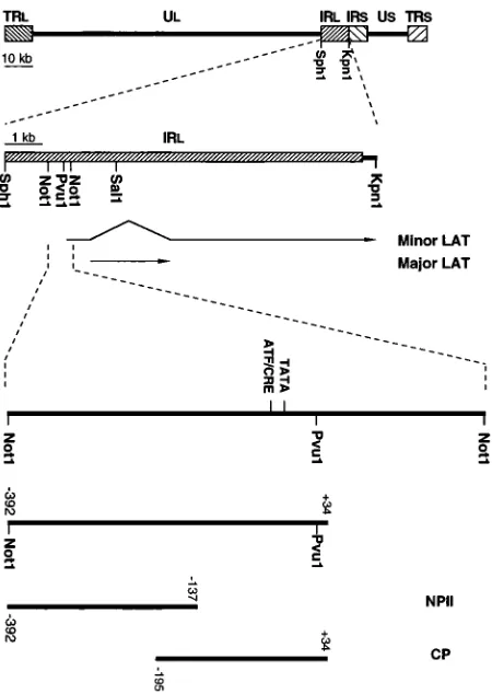
Plasmid constructs.A 632-bp DNA fragment encompassing the putative
2LATP region from2392 to1240 relative to the transcription start site (see below) was released by NotI cleavage (Fig. 1) from a larger SphI-SalI plasmid clone of the inverted long repeat region of HSV-2 strain 333 genomic DNA. This
NotI fragment was subcloned into the plasmid vector pCATzBasic (Promega, Madison, Wis.) between SalI and XbaI sites upstream of the chloramphenicol acetyltransferase (CAT)-coding sequence and designated pCATzNN. A deriv-ative of this fragment from2392 to134 (Fig. 1) relative to the start site of transcription was also cloned into pCATzBasic and designated p-392. A series of
59-end deletions of p-392 from base 2392 were generated by PCR with
pCATzNN as the template and cloned into the pCATzBasic vector at its PstI and XbaI sites. The names of the plasmids are based on the positions of their 59 ends relative to the transcription start site (see Fig. 4).
Site-specific point mutations were introduced into the wild-type plasmid p-392 with the Mut-a-gene kit (Bio-Rad, Richmond, Calif.) following the manufactur-er’s instruction.
pCATzControl (Promega) is a plasmid in which the CAT gene is driven by the simian virus 40 (SV40) early promoter and enhancer; for convenience, we refer to it here as pSV40CAT. To make pICP0CAT, a DNA fragment spanning the
HSV-1 ICP0 gene promoter region from 2807 to 1120 was released from
p110pKS (a gift from Liyanage P. Perera) and inserted into pCATzBasic at the
HindIII and XbaI sites. pCMVCAT was created with a DNA fragment
encom-passing the human cytomagalovirus (CMV) major immediate-early promoter from2582 to17 released from pBKzCMV (Stratagene, La Jolla, Calif.) with
NsiI and NheI and cloned into pCATzBasic at the PstI and XbaI sites (Fig. 2) after the ends had been blunted with T4 DNA polymerase.
All sequences of the fragments generated from PCR and mutagenesis were confirmed by chain termination sequencing with a Sequenase version 2.0 DNA sequencing kit (United States Biochemical, Cleveland, Ohio). All of the plasmids
used for transient expression assays were purified as described previously (30) with two cycles of ultracentrifugation in cesium chloride.
Cells.The rat pheochromocytoma cell line PC12 (American Type Culture
Collection, Rockville, Md.) was cultured in RPMI 1640 medium supplemented with 10% horse serum (all sera were heat inactivated), 5% fetal calf serum, and 1% GASP (a combination of glutamine, aureomycin, streptomycin, and penicil-lin). The human neuroblastoma cell line IMR32 (American Type Culture Col-lection) was cultured in minimal essential medium (MEM) supplemented with 10% fetal calf serum and 1% GASP. HeLa cells were cultured in MEM with 10% fetal calf serum and 1% GASP. The mouse neuroblastoma cell line, C1300 (American Type Culture Collection), was cultured in MEM-199 medium (1:1) with 10% fetal calf serum and 1% GASP.
Transfection and CAT assay.Two days before transfection, cells were plated
onto 60-mm-diameter tissue culture dishes and maintained at 378C in 5% CO2.
Either 5mg of DNA (for PC12, IMR32, and HeLa cells) or 1mg of DNA (for
C1300 cells) was used for transfection by the calcium phosphate coprecipitation method of Graham and Van Der Eb (16). Two days after transfection, cells were harvested and extracted in 0.25 M Tris-Cl (pH 7.4) with three cycles of freezing on dry ice and thawing at 378C.
CAT assays were performed as described by Shaw (33). Briefly, cell extracts were mixed with 2ml (50 nCi) of [14C]chloramphenicol (Amersham, Arlington Heights, Ill.)–20ml of 4 mM acetyl coenzyme A and 0.25 M Tris-Cl (pH 7.4) to make a volume of 150ml. The mixtures were incubated at 378C for 1 h. Reactions were stopped by extraction with 1 ml of cold ethyl acetate, and the organic phase was recovered and lyophilized. The pellets were resuspended in 30ml of ethyl acetate and subjected to thin-layer chromatography. The radioactivity on the thin-layer chromatography sheet was quantitated by a System 200 imaging scan-ner (Bioscan, Inc., Washington, D.C.). To insure that the CAT assays were conducted within the linear range of the test, in each set of CAT assays the amounts of cell extracts were adjusted so that the acetylation of the p-392 construct was about 20 to 50%. The percent acetylation in each experiment was then converted into percent CAT activity relative to that of the construct p-392 in the same experiment. The data presented in Fig. 2, 4, and 7 represent the averages of the results of three or four independent transfections.
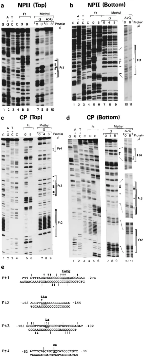
DNase I footprint assay.Nuclear extracts used for the footprint assay were
prepared as described by Montminy and Bilezikjian (27). The DNA fragments
NPII and CP shown in Fig. 1 were 39 end labeled with Klenow enzyme and
[a-32P]deoxynucleoside triphosphates or 59end labeled with T4 polynucleotide kinase and [g-32P]ATP. The footprint assay was conducted as described by Jones et al. (17). Briefly, 16 to 32mg of nuclear extracts was mixed with 23104
cpm
of DNA probes and 1mg of nonspecific competitor poly(dI-dC)-poly(dI-dC)
(Bethesda Research Laboratories, Inc., Gaithersburg, Md.) in a 45-ml volume with a final concentration of 25 mM Tris-Cl (pH 7.9)–6.25 mM MgCl2–50 mM KCl–0.5 mM EDTA–0.5 mM dithiothreitol–10% glycerol and incubated on ice for 15 min. Samples were then equilibrated in a 228C water bath for 2 min, and 5ml of DNase in 5 mM CaCl2was added to each tube. After a 90-s incubation, the reaction was stopped with 100ml of stop buffer (200 mM NaCl, 1% sodium
dodecyl sulfate, 20 mM EDTA, 100mg of tRNA per ml), and the reaction
mixture was subjected to phenol-chloroform extraction and ethanol precipita-tion. The pellet was resuspended in 2ml of TE (10 mM Tris-Cl [pH 8.0], 1 mM EDTA) and 3ml of sequencing stop buffer (United States Biochemical), dena-tured at 908C for 3 min, and resolved on an 8% acrylamide sequencing gel. FIG. 1. Schematic representation of HSV-2 genome and LAT promoter
(2LATP) region studied. A 632-bp NotI-NotI fragment, bases 5476 to 6106 according to McGeoch et al. (25), was subcloned, as was its left-most two-thirds derivative, a 426-bp fragment spanning bases2392 to134 relative to the tran-scription start site. The later (construct p-392) was used for extensive transient
expression studies. Further cleavages of the2392 to134 segment provided
fragments NPII (2392 to2137) and CP (2195 to134) for footprinting and
[image:2.612.65.290.68.385.2]methylation protection studies.
FIG. 2. Comparison of 2LATP activity with that of other promoters in tran-sient expression assays. The promoter activities of p-392 in PC12 and HeLa cells were compared with those of the SV40 early promoter plus enhancer, the CMV immediate-early promoter, and the HSV-1 ICP0 promoter. The percent CAT activity was normalized relative to that for the p-392 construct in PC12 cells (solid bar; 21.7% average acetylation in four experiments) and in HeLa cells (hatched bar; 19.1% average acetylation in four experiments). To the left, the solid bars represent the promoter sequences placed upstream of CAT, the thinner bars indicate the plasmid sequences, and the hatched box shows the SV40 enhancer placed downstream of CAT.
on November 9, 2019 by guest
http://jvi.asm.org/
Methylation protection assay.DNA probes (23104
cpm per reaction) were mixed and incubated with nuclear extracts and other reagents as in the footprint
assay but in a volume of 45ml. The mixtures were then subjected to DNA
methylation and either cleavage at G residues or strong cleavage at A and weak cleavage at G (A.G) residues as described by Maxam and Gilbert (24). The final products were dissolved in 2ml of TE and 3ml of sequencing stop buffer, denatured at 908C for 3 min, and resolved on an 8% acrylamide sequencing gel. DNA sequencing ladders were generated by the Maxam and Gilbert method (24) with the same probe used in the methylation protection assay.
Gel mobility-shift assay.Synthesized oligonucleotides spanning the regions of
footprint 3 (Ft3) were used as probes. To serve as a probe, two complementary oligonucleotides were annealed and end labeled with T4 polynucleotide kinase and [g-32P]ATP to a specific activity about 0.0025 to 0.005 pmol/23104cpm. Labeled probes were purified on 6% acrylamide nondenaturing gels. For each reaction, 23104cpm of the probe was mixed, with or without specific unlabeled oligonucleotide competitors as required, in 17ml of reaction buffer [0.5mg poly(dI-dC)-poly(dI-dC), 25 mM Tris-Cl (pH 7.9), 6.25 mM MgCl2, 50 mM KCl, 0.5 mM EDTA, 0.5 mM dithiothreitol, 10% glycerol] with 1ml (about 4mg of protein) of nuclear extract, and the mixture was incubated on ice bath for 15 min. The reactions were resolved on 1.5-mm-thick, 6% acrylamide–0.253TBE gels, run at 12.5 V/cm at 48C about 1.5 h while the buffer was recirculated. The gels were dried and autoradiographed. The specific oligonucleotide competitors were synthesized on a Gene Assembler Plus (Pharmacia LKB, Piscataway, N.J.), purified on 15% denaturing acrylamide–urea gels, and quantitated by measuring their absorbance at 260 nm with a DU-40 spectrophotometer (Beckman Instru-ments, Inc., Fullerton, Calif.). The names and sequences of the oligonucleotide competitors are shown in Fig. 8b.
Primer extension.To prepare RNA for primer extension, C1300 cells were
transfected, as described above, but 100-mm-diameter dishes and 12mg of DNA per dish were used. Two days after transfection, cells were harvested, and total cellular RNAs were purified with RNAzol (Tel-Test, Inc., Friendswood, Tex.) following the manufacturer’s instruction.
The structure of the plasmid for transfection and the primer are shown in Fig. 3. Primer CAT2281R was used for studies of RNA expressed from cells trans-fected with plasmid p-392 in that it anneals to the sequence from vector pCATzBasic at bp 2281 to 2310, which is 74 bp downstream of the putative 2LATP TATA box. Primer NN438R was used for RNA from cells transfected with plasmid pCATzNN as it anneals to the HSV-2 sequence 73 bp downstream
the presence of a potential polymerase II promoter several
hundred bp upstream of the 5
9
end of the major HSV-2 LAT.
By transient expression, CAT assay, mutagenesis, and in vivo
study in the guinea pig, Krause et al. confirmed the presence of
an active LAT promoter between two NotI enzyme recognition
sites (19, 20). To characterize this promoter and its cis-acting
elements, the 632-bp NotI fragment and a 426-bp truncated
derivative of it spanning bases
2
392 to
1
34 relative to the
transcription start site (Fig. 1), as determined below, were
cloned into plasmid pCAT
z
Basic and designated pCAT
z
NN
and p-392, respectively.
When transfected into rat neuronal PC12 cells, p-392
dis-played two- to fourfold more CAT activity than pCAT
z
NN.
The quantities of CAT-specific mRNA, however, were the
same for each transfection (data not shown), indicating that
the pCAT
z
NN mRNA may be translated less efficiently.
Therefore, we chose p-392 as the parental construct for our
subsequent analyses.
On transient expression, the p-392 promoter proved to be
very strong in neuronal cells. To more precisely define strength
of 2LATP, we compared its activity with that of other potent
promoters: the human CMV immediate-early promoter, the
SV40 early promoter plus enhancer, and the HSV-1 ICP0 gene
promoter. The CMV immediate-early promoter is a very active
one in many different tissues (5, 32); the SV40 early promoter
has high levels of activity in monkey cell lines, less activity in
human cell lines, and particularly low levels of activity in
neu-ronal cells (11).
The promoter activities of the CMV, SV40, and ICP0
con-structs and the 2LATP construct p-392 were compared in both
PC12 and HeLa cells by transient expression and CAT assay.
The results of four independent experiments are summarized
in Fig. 2. In PC12 cells, the p-392 construct yielded an average
of 21.7% acetylation, almost as active as pCMVCAT. In HeLa
cells p-392 exhibited an average of 19.1% acetylation, which
was about one-half to one-third the activity of pCMVCAT.
The activity of the SV40 promoter construct, pSV40CAT, was
only 2.8 and 14.7% of that of p-392 in PC12 and HeLa cells,
respectively. pICP0CAT exhibited only 4.5 and 14.1% of the
activity seen with p-392 in PC12 and HeLa cells, respectively.
These studies showed that the 2LATP DNA fragment
span-ning bases
2
392 to
1
34 has very strong transcriptional activity,
particularly in neuronal cells.
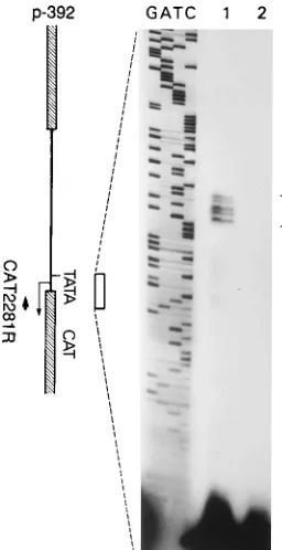
Identification of transcription start site.
The transcription
start site of 2LATP was mapped with primer extension. Since
transfected C1300 cells afforded even higher levels of CAT
activity of p-392 than the other cell lines (see below), we
used C1300 cell RNA for these studies. RNA from
untrans-fected as well as p-392-transuntrans-fected C1300 cells was tested with
primer CAT2281R that anneals within the CAT sequence 74
bp downstream of the predicted TATA box in p-392. The
labeled primers were hybridized with the RNAs, extended with
avian myeloblastosis virus reverse transcriptase, and resolved
on a denaturing acrylamide gel. A sequencing ladder was
gen-FIG. 3. HSV-2 LAT start site as determined by primer extension assay. (Leftpanel) Structure of the p-392 construct with its putative TATA box and tran-scription start site. The primer CAT2281R used in the assay is indicated as a bold arrow facing upstream. Total cellular RNA was purified from C1300 cells trans-fected with p-392 as well as from untranstrans-fected C1300 cells. Extension products from the reaction were loaded on lanes 1 and 2, respectively. Lanes labeled as GATC reveal the sequencing ladders generated with p-392 as the template and CAT2281R as the primer. The open bracket on the right indicates the primer-extended products.
on November 9, 2019 by guest
http://jvi.asm.org/
[image:3.612.111.239.70.319.2]erated with the p-392 as the template and CAT2281R as the
primer and loaded side-by-side with the primer extension
re-action (Fig. 3). No extended bands were detected from
un-transfected control cell RNA (lane 2). A set of nested bands
were detected from the reaction with transfected cell RNA
(lane 1), with the largest comigrating with a T, 27 bp
down-stream of the 5
9
end of the putative TATA box.
The result was confirmed by using RNA purified from C1300
cells transfected with the larger 2LATP construct, pCAT
z
NN,
and a primer that anneals to the viral sequence 73 bp
down-stream of the putative TATA box (data not shown).
The powerful promoter activity and transcription from a
specific start site suggested that the putative 2LATP TATA
box is functional. It was necessary to prove with site-specific
mutation that the putative TATA element is active, but before
doing so, we developed a fuller appreciation of the regulatory
elements situated further upstream of it.
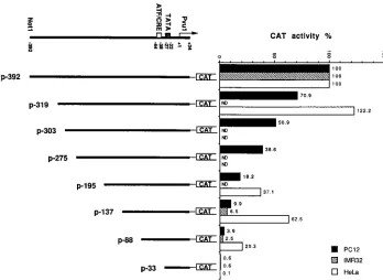
Characterization of neuron-responsive and constitutive
reg-ulatory regions.
To detect functional elements upstream of the
TATA box, we generated a series of 5
9
-end deletional
deriva-tives of p-392 and analyzed their promoter activities in both
PC12 and HeLa cells. Deletion of the sequence from
2
392 to
2
137 reduced the promoter activity by 90% in PC12 but by
only 38% in HeLa cells (Fig. 4), implying that some portions of
the distal (5
9
-most) region of 2LATP are particularly neuron
responsive. Successive, smaller deletions to positions
2
319,
2
303,
2
275, and
2
195 reduced CAT activity in PC12 cells by
29, 49, 61, and 82%, respectively (Fig. 4). These data indicate
that the whole distal region, extending to position
2
392,
con-tributes to the promoter activity in PC12 cells, probably
be-cause of the presence of multiple cis-acting elements.
The further 5
9
-end deletion of bases
2
137 to
2
88 reduced
2LATP activity an additional two- to threefold in both PC12
and HeLa cell lines (Fig. 4), indicating the presence of crucial
cis-acting elements in this region. Deletion of the sequence
from bases
2
88 to
2
33, just upstream of the TATA, further
reduced the promoter activity in PC12 and HeLa cells to
un-detectable levels. By analogy to the 1LATP sequence, this
region should contain an ATF/CREB element, which may
con-tribute activity to 2LATP (23).
To confirm that differences in promoter activity in rat PC12
and human HeLa cells are tissue specific and not merely
spe-cies specific, key 2LATP constructs were also tested in the
human neuroblastoma IMR32 cell line. The promoter activity
pattern was more similar to that of PC12 cells than to that of
HeLa cells (Fig. 4).
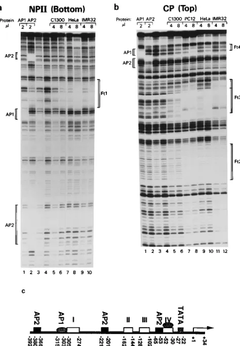
Localization and characterization of individual 2LATP
cis
-acting elements.
We conducted DNase I footprint, methylation
protection, and gel mobility shift assays to identify individual
protein-binding elements in 2LATP. For the DNase I footprint
and methylation protection assays, two overlapping DNA
frag-ments, NPII and CP (Fig. 1), were end labeled and used as
probes. Nuclear extracts from multiple cell lines were used, as
were purified trans-activator proteins AP1 and AP2, because
sequences highly homologous to their recognition sites are
found in 2LATP. When the labeled NPII probes were
incu-bated with crude nuclear extracts from HeLa, PC12, C1300, or
IMR32 cells, bases
2
305 to
2
274 were protected from DNase
I digestion (Fig. 5a, lanes 5 to 10,) and designated Ft1. When
NPII was incubated with purified AP1, a footprint was seen
spanning bases
2
315 to
2
303 (Fig. 5a, lane 1). AP2 protected
two regions of NPII, from bases
2
380 to
2
366 and
2
221 to
[image:4.612.130.478.71.325.2]2
201 (Fig. 5a, lane 2). Crude nuclear extracts protected three
regions of probes from CP, designated Ft2 (
2
162 to
2
144),
Ft3 (
2
128 to
2
102) and Ft4 (
2
52 to
2
30), which spans the
putative ATF/CREB site (Fig. 5b, lanes 5 to 12). An
AP1-binding region overlapped Ft4. An AP2-AP1-binding site was also
found within the CP region at
2
65 to
2
53 (Fig. 5b, lane 2).
FIG. 4. Schematic representation of structure and CAT activity of p-392 and a series of its 59-end deletion products in different cell lines. The percent CAT activity is normalized relative to that for the p-392 construct in PC12 cells (21.3% average acetylation in four experiments), in IMR32 cells (11.7% average acetylation in two experiments), or in HeLa cells (15.7% average acetylation in four experiments). The structure of the viral insert in plasmid p-392 is shown on the top. Each plasmid is named according to the base position of its 59end relative to the transcription start site (11). ND, not done.on November 9, 2019 by guest
http://jvi.asm.org/
The above experiments located at least eight cis-acting
ele-ments which can interact with nuclear proteins or purified AP1
or AP2 in vitro (Fig. 5c). When these data are considered
together with the results of the CAT assays using sequential 5
9
deletions, it would appear that some of the footprints, Ft3 and
Ft4 for example, may be particularly important to 2LATP
activity. Since the nuclear extracts from neuronal cells seem to
protect a longer sequence at the Ft3 region than extracts from
HeLa cells, Ft3 may also contribute to the neuronal
respon-siveness of 2LATP.
Mutational analyses of footprints.
To delineate the
func-tional relevance of each of the above footprints more precisely,
it was necessary to create site-specific mutations within them.
Because the footprints were large, methylation protection
as-says were first used to identify the bases in closest proximity to
the bound nuclear proteins and, hence, the best targets for
mutagenesis.
[image:5.612.61.298.69.411.2]The results of methylation protection assays are shown in
Fig. 6. According to the bases which appeared to be enhanced
or protected from methylation, we chose a series of 2- to 3-base
pared with that of the parental p-392 in both PC12 and HeLa
cells (Fig. 7). In PC12 cells the activity of p-392Ft1M2 was
57.6% of that of the parental construct; p-392Ft2M1 was
37.2%, p-392Ft3M1 was 19.8%, p-392CREM1 was 20.4%, and
p-392TATAM1 was 8% of that of p-392. In HeLa cells their
activities proved to be 57.1, 98.5, 54.3, 26.1, and 15%,
respec-tively, of that of p-392. This demonstrated that the TATA box
and ATF/CREB element or Ft4 are important in both cell
lines. The elements in Ft2 and Ft3 are more important for the
promoter activity in neuronal cells than in nonneuronal cells.
These data also verified the earlier suggestion that multiple
elements contribute to the neuronal responsiveness of the
2LATP.
Since mutational analyses proved the ATF/CREB sequence
in 2LATP to be active, its response to cAMP was tested in
PC12 cells by using plasmid p-195 and its derivative,
p-195CREM1. The mutation in p-195CREM1 was the same as
that in p-392CREM1. The basal activity of the mutant was
about 10% of that of the parental construct, but the response
to 1 mM dibutyryl cAMP in the culture medium was the same
as that of the parental DNA. That is, the activities of both
plasmids increased two- to fourfold in the presence of 1 mM
dibutyryl cAMP (data not shown). This may indicate that the
protein binding to this region is an ATF family member which
is not responsive to cAMP. That the purified AP1 protein
protected this region supports this possibility. Alternatively,
the 2LATP sequence between bases
2
195 to
1
34 may include
a different CRE, as recently suggested for HSV-1 by Kenny et
al. (18).
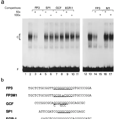
Novel
cis
element in 2LATP.
Because Ft3 seemed so
impor-tant to the overall activity of 2LATP, we sought to characterize
it more fully. The sequence of Ft3 is GC rich. A computer
search of the database sites from the National Center for
Biotechnology Information revealed Ft3 to have a high degree
of homology with the binding sites for nuclear proteins SP1,
GCF, and EGR-1, as shown in Fig. 8b. We, therefore, used gel
mobility shift competition assays to investigate the binding
ability of those known elements to the protein binding to Ft3.
Two complementary 30-mer oligonucleotides encompassing
Ft3 were annealed and termed FP3 (Fig. 8b). When
end-labeled FP3 was incubated with nuclear extracts from PC12
cells, a major retarded complex was seen (Fig. 8a, lane 2). This
complex could be competed with effectively by 10- to 100-fold
excesses of unlabeled FP3. FP3M1, a mutant oligonucleotide
containing the same 2-bp alteration as that in p-392Ft3M1, lost
the protein-binding activity and no longer competed for the
formation of the major complex (Fig. 8a, lanes 16 and 17). This
suggests that the major retarded complex is the functional
complex formed on Ft3. None of the oligonucleotides specific
for binding of SP1, GCF, or EGR-1 efficiently competed for
formation of the complex (Fig. 8a, lanes 5 to 10). Thus, Ft3
spans a novel cis element, designated here LAT-3.
FIG. 5. Results of DNase I footprint assays. (a and b) DNase I protection by binding of 2ml of purified nuclear proteins AP1 and AP2 or of 4 or 8ml of nuclear extracts from C1300, PC12, HeLa, or IMR32 cells, as indicated above the lanes. Lanes 3 and 4, probe only (no added binding proteins) digested with various amounts of DNase I. In panel a, the probe, NPII (Bottom), was the NPII fragment (bases2392 to2137) labeled on the 39end of the bottom strand. In panel b, the probe, CP (Top), was the CP fragment (bases2195 to134) labeled on 59end of the top strand. Footprints created by binding of AP1 or AP2 are shown as brackets to the left of each panel, while footprints generated by nuclear extracts are bracketed as Ft1 to Ft4 to the right of each panel. (c) Summary of the footprints and their locations in 2LATP.
on November 9, 2019 by guest
http://jvi.asm.org/
DISCUSSION
We conducted a detailed study of cis elements that may
regulate the expression of the HSV-2 LAT gene. By specific
mutational and transient expression assays, the TATA
se-quence of 2LATP was localized and proven to be functional.
Transcription starts at about 27 to 32 bp downstream of this
TATA box, similar to that seen with the 1LATP. Alteration of
the TATA sequence abrogated 85 to 92% of the activity of this
promoter. These findings suggest that the basal transcription
activity of the LAT gene depends largely on a classical
poly-merase II promoter. The residual promoter activity of the
TATA mutant construct may be due to the remaining activity
of an impaired TATA element or, alternatively, initiation
with-out a TATA box.
Upstream of this basal promoter is a very potent enhancer
region composed of multiple elements. By direct comparisons
with other promoter constructs, the sequence from bases
2
392
to
1
34 of 2LATP, which spans both the basal promoter and
enhancer regions, displays very strong activity in both neuronal
and nonneuronal cell lines. The strength of this promoter
seems surprisingly great since the concentration of the minor
LAT, the primary product of the gene in latently infected
neurons, is very low (19, 41). This suggests either that LAT
expression in neurons is actually very low because of the
in-hibitory influence of some regulatory elements contained in
sequences beyond that which we tested here or that the level of
LAT expression in vivo is high but the minor LAT is very
labile.
The sequence spanning bases
2
392 to
2
137 seems
particu-larly important for promoter activity in neuronal cells, similar
to the observations of Batchelor and O’Hare (3) and
Zwaag-stra et al. with 1LATP (40). The progressive reduction in
promoter activity with sequential deletions towards the TATA
box predicted that there must be multiple cis-acting elements
in this region. With DNase I footprint and methylation
pro-tection assays, we documented multiple footprints generated
by binding of proteins in nuclear extracts or purified AP1 and
AP2 proteins.
Just upstream of the TATA box, we demonstrated an ATF/
CREB binding motif that contributes constitutively to 2LATP
activity. Its mutation reduced promoter activity by 76 to 80%,
depending on the type of cells transfected. Site-specific
alter-ation of this element, however, did not impair its
responsive-ness to cAMP, in contrast to the observations of Leib et al.
(23), with this element in 1LATP. Nevertheless, we
demon-strated that crude nuclear protein extracts or purified AP1, a
member of the ATF family, bound to this element and
pro-tected it from DNase digestion in vitro. It has been reported
that the ATF/CREB binding motif can bind proteins of both
the ATF and CREB families, resulting in differential
regula-tion of transcripregula-tion (21).
[image:6.612.62.297.69.640.2]In addition to localizing the TATA box and ATF/CREB
element, we identified seven other upstream elements in
2LATP, including some that have not been reported for
1LATP. Three AP2, one AP1, and three still incompletely
characterized binding sites, termed Ft1, Ft2, and Ft3, were
documented. Ft1 and Ft2 were located in the region that was
FIG. 6. Methylation protection assays of 2LATP segments. (a to d) Lanes: 1to 4 DNA sequencing ladders; 5 and 6, DNase I footprints Ft1 to Ft4 generated by binding of 8 ml of nuclear extracts; 7 and above, results of methylation protection assays. Brackets are used to delineate the footprint regions, arrows show bases protected from methylation, arrowheads show areas of enhanced methylation, and dotted lines in panels b and d connect corresponding regions in lanes from different gels. In panel a, the probe, NPII (Top), was the NPII fragment labeled at the 39end of the top strand. In panel b, the probe, NPII (Bottom), was NPII fragment labeled at the 39end of its bottom strand. In panel c, the probe, CP (Top), was the fragment CP labeled at the 59end of its top
strand. In panel d, the probe, CP (Bottom), was the CP fragment labeled at the 39end of the bottom strand. (e) Summary of all methylation protection data for footprints Ft1 to Ft4. The flanking numbers indicate the positions of the 59and 39ends of footprints on the top strand. Vertical bars indicate methylation pro-tected bases; arrowheads show bases subject to enhanced methylation. Specific bases mutated in later studies are underlined, and the substituting bases are indicated above each of the sequences.
on November 9, 2019 by guest
http://jvi.asm.org/
particularly neuron responsive. Site-specific mutation studies
found Ft1 to have a modulatory effect on 2LATP activity.
Alteration of the Ft2 sequence, however, reduced promoter
activity by 63% in PC12 cells but by only 2% in HeLa cells.
Transient expression revealed that Ft3 contributed
substan-tially to the neuronal responsiveness of the
2
392 to
1
34
frag-ment. Its mutation reduced promoter activity in neuronal cells
by 80% but only within the context of an intact upstream
region. Ft3 proved less able to influence the activity of the
promoter constructs in which sequences upstream of it were
deleted (Fig. 4). Thus, Ft3 does not directly enhance the
neu-ronal responsiveness of 2LATP. Rather, it may do so in
coop-eration with elements upstream of it.
More refined analyses showed that a protein recognition site
within Ft3 that we termed LAT-3 spans an incomplete
palin-drome from bases
2
121 to
2
107 (CGGGCGCCGTGCCCG).
Mutation of two G’s in this region at positions
2
119 and
2
118
reduced 2LATP activity by 80% in transient assays.
Competi-tion gel shift studies showed LAT-3 to be a novel GC-rich
element that differs from other GC-rich elements which bind
SP1, GCF, and EGR-1.
The present studies uncovered evidence of a complex
inter-play of cis-acting elements, yielding the in vitro phenotype of
2LATP. The roles that these elements play in LAT expression
in vivo and their influence on viral latency and reactivation are
now being studied by creation of targeted viral mutants and
analyses of them in experimentally infected animals.
ACKNOWLEDGMENTS
We gratefully acknowledge Michael Lenardo, Jeffrey Cohen, and Liyanage Perera for helpful discussion and review of the manuscript.
REFERENCES
1. Baringer, R. 1974. Recovery of herpes simplex virus from human sacral ganglions. N. Engl. J. Med. 291:828–830.
2. Batchelor, A. H., and P. O’Hare. 1990. Regulation and cell-type-specific activity of a promoter located upstream of the latency-associated transcript of herpes simplex virus type 1. J. Virol. 64:3269–3279.
3. Batchelor, A. H., and P. O’Hare. 1992. Localization of cis-acting sequence requirements in the promoter of the latency-associated transcript of herpes simplex virus type 1 required for cell-type-specific activity. J. Virol. 66:3573– 3582.
4. Bloom, D. C., G. B. Devi-Rao, J. M. Hill, J. G. Stevens, and E. K. Wagner. 1994. Molecular analysis of herpes simplex virus type 1 during epinephrine-induced reactivation of latently infected rabbits in vivo. J. Virol. 68:1283– 1292.
5. Boshart, M., F. Weber, G. Jahn, K. Dorsch-Hasler, B. Fleckenstein, and W.
[image:7.612.133.481.73.263.2]Schaffner.1985. A very strong enhancer is located upstream of an immediate
FIG. 7. Schematic representation of structure and CAT activity of the p-392 segment and its derivatives mutated at the specific bases shown in Fig. 6e. The positions of footprints Ft1 to Ft3, Ft4 (ATF/CRE), and the TATA box are shown on the top. Arrowheads indicate the sites of specific base substitutions. The percent CAT activity is normalized relative to that for p-392 construct in PC12 cells (46.6% average acetylation in four experiments) or HeLa cells (25.2% average acetylation in four experiments).
FIG. 8. Electrophoretic gel mobility shift competition analysis of Ft3 region of 2LATP. (a) Radiolabeled probe FP3 encompassing Ft3 was reacted with nuclear extracts from PC12 cells with (1) or without the addition of a 10-fold (103) or 100-fold (1003) excess of unlabeled oligonucleotides FP3, FP3M1 (a 2-bp substitution mutant of FP3), and binding sites for SP1, GCF, and EGR-1, as indicated above each lane. Lanes: 1 and 12, probe only; 2, 11, and 13, probe plus nuclear extract but no competitor. (b) Sequences of top strands of probe FP3, mutant competitor FP3M1, and the competitors of known GC-rich ele-ments, with GC-rich regions underlined.
on November 9, 2019 by guest
http://jvi.asm.org/
[image:7.612.57.296.398.647.2]early gene of human cytomegalovirus. Cell 41:521–530.
6. Corey, L., and P. G. Spear. 1986. Infections with herpes simplex viruses. N. Engl. J. Med. 314:686–691.
7. Croen, K. D., J. M. Ostrove, L. Dragovic, and S. E. Straus. 1991. Charac-terization of herpes simplex virus type 2 latency-associated transcription in human sacral ganglia and in cell culture. J. Infect. Dis. 163:23–28. 8. Croen, K. D., J. M. Ostrove, L. J. Dragovic, J. E. Smialek, and S. E. Straus.
1987. Latent herpes simplex virus in human trigeminal ganglia: detection of an immediate early gene ‘‘anti-sense’’ transcript by in situ hybridization. N. Engl. J. Med. 317:1427–1432.
9. Deatly, A. M., J. G. Spivack, E. Lavi, and N. W. Fraser. 1987. RNA from an immediate early region of the HSV-1 genome is present in the trigeminal ganglia of latently infected mice. Proc. Natl. Acad. Sci. USA 84:3204–3208. 10. Dobson, A. T., F. Sederati, G. Devi-Rao, W. M. Flanagan, M. J. Farrell, J. G.
Stevens, E. K. Wagner, and L. T. Feldman. 1989. Identification of the
latency-associated transcript promoter by expression of rabbit beta-globin mRNA in mouse sensory nerve ganglia latently infected with a recombinant herpes simplex virus. J. Virol. 63:3844–3851.
11. Donis, J. A., M. Ventosa-Michelman, and R. L. Neve. 1993. Comparison of expression of a series of mammalian vector promoters in the neuronal cell lines PC12 and HT4. BioTechniques 15:786–787.
12. Dowdle, W. R., A. J. Nahmias, R. W. Harwell, and F. P. Pauls. 1967. Asso-ciation of antigenic type of herpesvirus hominis with site of viral recovery. J. Immunol. 99:974–980.
13. Galloway, D. A., C. Fenoglio, and J. K. McDougall. 1982. Limited transcrip-tion of the herpes simplex virus genome when latent in human sensory ganglia. J. Virol. 41:686–691.
14. Galloway, D. A., C. Fenoglio, M. Shevchuk, and J. K. McDougall. 1979. Detection of herpes simplex RNA in human sensory ganglia. Virology 95: 265–268.
15. Goins, W. F., L. R. Sternberg, K. D. Croen, P. R. Krause, R. L. Hendricks,
D. J. Fink, S. E. Straus, M. Levine, and J. C. Glorioso.1994. A novel
latency-active promoter is contained within the herpes simplex virus type 1 ULflanking repeats. J. Virol. 68:2239–2252.
16. Graham, F. L., and A. J. Van Der Eb. 1973. A new technique for the assay of infectivity of human adenovirus 5 DNA. Virology 52:456–467. 17. Jones, K. A., K. R. Yamamoto, and R. Tjian. 1985. Two distinct transcription
factors bind to the HSV thymidine kinase promoter in vitro. Cell 42:559–572. 18. Kenny, J. J., F. C. Krebs, H. T. Hartle, A. E. Gartner, B. Chatton, J. M.
Leiden, J. P. Hoffler, P. C. Weber, and B. Wigdahl.1994. Identification of a
second ATF/CREB-like element in the herpes simplex virus type 1 (HSV-1) latency-associated transcript (LAT) promoter. Virology 200:220–235. 19. Krause, P. R., J. M. Ostrove, and S. E. Straus. 1991. The nucleotide
se-quence, 59end, promoter domain, and kinetics of expression of the gene encoding the herpes simplex virus type 2 latency-associated transcript. J. Virol. 65:5619–5623.
19a.Krause, P. R., and S. E. Straus. Unpublished data.
20. Krause, P. R., L. R. Stanberry, N. Bourne, B. Connelly, J. F. Kurawadwala,
A. Patel, and S. E. Straus.1995. Expression of the herpes simplex virus type
2 latency-associated transcript enhances spontaneous reactivation of genital herpes in latently infected guinea pigs. J. Exp. Med. 181:297–306. 21. Lee, K. A. W., J. S. Fink, R. H. Goodman, and M. R. Green. 1989.
Distin-guishable promoter elements are involved in transcriptional activation by E1a and cyclic AMP. Mol. Cell. Biol. 9:4390–4397.
22. Leib, D. A., C. L. Bogard, M. Kosz-Vnenchak, K. A. Hicks, D. M. Coen, D. M.
Knipe, and P. A. Schaffer.1989. A deletion mutant of the latency-associated
transcript of herpes simplex virus type 1 reactivates from the latent state with reduced frequency. J. Virol. 63:2893–2900.
23. Leib, D. A., K. C. Nadeau, S. A. Rundle, and P. A. Schaffer. 1991. The promoter of the latency-associated transcripts of herpes simplex virus type 1 contains a functional cAMP-response element: role of the latency-associated
transcripts and cAMP in reactivation. Proc. Natl. Acad. Sci. USA 88:48–52. 24. Maxam, A. M., and W. Gilbert. 1977. A new method for sequencing DNA.
Proc. Natl. Acad. Sci. USA 74:560–564.
25. McGeoch, D. J., C. Cunningham, G. McIntyre, and A. Dolan. 1991. Com-parative sequence analysis of the long repeat regions and adjoining parts of the long unique regions in the genomes of herpes simplex viruses types 1 and 2. J. Gen. Virol. 72:3057–3075.
26. Mitchell, W. J., S. L. Deshmane, A. Dolan, D. J. McGeoch, and N. W. Fraser. 1990. Characterization of herpes simplex virus type 2 transcription during latent infection of mouse trigeminal ganglia. J. Virol. 64:5342–5348. 27. Montminy, M. R., and L. M. Bilezikjian. 1987. Binding of a nuclear protein
to the cyclic-AMP response element of the somatostatin gene. Nature (Lon-don) 328:175–178.
28. Nahmias, A. J., Z. M. Naib, and W. E. Josey. 1971. Herpesvirus hominis type 2 infection—association with cervical cancer and perinatal disease. Perspect. Virol. 7:73–89.
29. Nicosia, M., S. L. Deshmane, J. M. Zabolotny, T. Valyi-Nagy, and N. W.
Fraser. 1993. Herpes simplex virus type 1 latency-associated transcript
(LAT) promoter deletion mutants can express a 2-kilobase transcript map-ping to the LAT region. J. Virol. 67:7276–7283.
30. Sambrook, J., E. F. Fritsch, and T. Maniatis. 1989. Molecular cloning: a laboratory manual, 2nd ed. Cold Spring Harbor Laboratory, Cold Spring Harbor, N.Y.
31. Sawtell, N. M., and R. T. Thompson. 1992. Herpes simplex virus type 1 latency-associated transcription unit promotes anatomical site-dependent establishment and reactivation from latency. J. Virol. 66:2157–2169. 32. Schmidt, E. V., G. Christoph, R. Zeller, and P. Leder. 1990. The
cytomeg-alovirus enhancer: a pan-active control element in transgenic mice. Mol. Cell. Biol. 10:4406–4411.
33. Shaw, W. 1975. Chloramphenicol acetyltransferase from resistant bacteria. Methods Enzymol. 53:737–754.
34. Steiner, I., J. G. Spivack, R. P. Lirette, S. M. Brown, A. R. MacLean, J.
Subak-Sharpe, and N. W. Fraser.1989. Herpes simplex virus
latency-asso-ciated transcripts are evidently not essential for latent infection. EMBO J.
8:505–511.
35. Steiner, I., J. G. Spivack, D. R. O’Boyle II, E. Lavi, and N. W. Fraser. 1988. Latent herpes simplex virus type 1 transcription in human trigeminal ganglia. J. Virol. 62:3493–3496.
36. Stevens, J. G., E. K. Wagner, G. B. Devi-Rao, M. L. Cook, and L. T. Feldman.
1987. RNA complementary to a herpesvirusagene mRNA is prominent in
latently infected neurons. Science 235:1056–1059.
37. Suzuki, S., and J. R. Martin. 1989. Herpes simplex virus type 2 transcripts in trigeminal ganglia during acute and latent infection in mice. J. Neurol. Sci.
93:239–251.
38. Trousdale, M. D., I. Steiner, J. G. Spivack, S. L. Deshmane, S. M. Brown,
A. R. MacLean, J. H. Subak-Sharpe, and N. W. Fraser.1991. In vivo and in
vitro reactivation impairment of a herpes simplex virus type 1 latency-asso-ciated transcript variant in a rabbit eye model. J. Virol. 65:6989–6993. 39. Zwaagstra, J., H. Ghiasi, A. B. Nesburn, and S. L. Wechsler. 1989. In vitro
promoter activity associated with the latency-associated transcript gene of herpes simplex virus. J. Gen. Virol. 70:2163–2169.
40. Zwaagstra, J. C., H. Ghiasi, A. B. Nesburn, and S. L. Wechsler. 1991. Identification of a major regulatory sequence in the latency associated tran-script (LAT) promoter of herpes simplex virus type 1 (HSV-1). Virology
182:287–297.
41. Zwaagstra, J. C., H. Ghiasi, S. M. Slanina, A. B. Nesburn, S. C. Wheatley, K.
Lilycrop, J. Wood, D. S. Latchman, K. Patel, and S. L. Wechsler.1990.
Activity of herpes simplex virus type 1 latency-associated transcript (LAT) promoter in neuron-derived cells: evidence for neuron specificity and for a large LAT transcript. J. Virol. 64:5019–5028.