0022-538X/94/$04.00 0
Copyright ©) 1994,AmericanSociety for Microbiology
Helicase-Primase Complex of
Herpes
Simplex Virus Type
1:
a
Mutation in the UL52 Subunit Abolishes Primase
Activity
DONNA K. KLINEDINST AND M. D. CHALLBERG*
Laboratoryof ViralDiseases, NationalInstituteof Allergy andInfectious Diseases, Bethesda, Maryland20892
Received29December 1993/Accepted 14March 1994
The UL52 gene product of herpes simplex virus type 1 (HSV-1) comprises one subunit of a 3-protein
helicase-primasecomplex that is essential for replication of viral DNA. Thefunctions of the individual subunits ofthe complex are not known with certainty, although it is clear that the UL8 subunit is notrequiredforeither helicase or primase activity. Examination ofthe predicted amino acid sequence of the UL5 gene reveals the existence of conserved helicasemotifs;it seemslikely, therefore,that UL5 isresponsiblefor the helicaseactivity
ofthe complex. We have undertaken mutational analysis of UL52 in an attempt to understand thefunctional
contribution of this protein to the helicase-primase complex. Amino acid substitution mutations were introduced into five regions of the UL52 gene that are highly conserved among HSV-1 and the related herpesviruses equine herpesvirus 1, human cytomegalovirus, Epstein-Barr virus, andvaricella-zostervirus. Of seven mutantsanalyzed byaninvivoreplicationassay, three mutants, in three differentconservedregions of the protein, failed to support DNAreplication. Within one of the conserved regions is a 6-amino-acid motif
(IL)(VIM)(LF)DhD (where h is a hydrophobic residue), which is also conserved in mouse, yeast, and T7 primases.Mutagenesis of the first aspartate residue of the motif, located at position 628 of the UL52 protein, abolished the ability of the complex to support replication of an origin-containing plasmid in vivo and to
synthesize oligoribonucleotideprimers in vitro. The ATPase and helicase activities wereunaffected,as wasthe ability of the mutant enzyme to support displacement synthesis on a preformed fork substrate. These results provide experimental support for the idea that UL52 is responsible for the primase activity of the HSV helicase-primasecomplex.
Herpessimplex virustype 1 (HSV-1)encodes sevenproteins
that arerequired for viral DNA replication (7-11, 24, 34, 36, 44, 53-56). The functions of several of these polypeptidesare nowwellestablished. UL9bindsspecificsequenceswithin the
origins of replication of HSV (19, 20, 39) and also possesses
helicase activity (5, 21). UL29 (ICP8) is a single-stranded DNA-binding protein (42, 54). UL30 and UL42 compose a
heterodimericDNApolymeraseinwhich UL30is thecatalytic subunit and UL42 acts to increase the processivity of the
enzyme(10-12, 22, 23, 26, 27, 41, 43). Finally, UL5, UL8, and
UL52 form a heterotrimeric complex with both 5' to 3'
helicase and primase activities(13, 15).
The roles of the individual subunits of the UL5-UL8-UL52 complex havenotbeen definedclearly. The complex has been
purified
from insect cellstriply
infected with baculovirusesexpressing
each of thesepolypeptides
and has DNA-depen-dentATPase,helicase,andprimase activities identicaltothose obtainedfrom herpesvirus-infected mammalian cells (17, 46). Likewise, a subcomplex containing only UL5 and UL52 has been purified from insect cells and retains DNA-dependentATPase and helicase activity (6, 18, 46) and the ability to
synthesize primers (18, 46).
These results indicate that thecoreprimase and helicase activities of the complexare not
depen-denton UL8.
Examination of the primary structure of the UL5 gene suggeststhat it is the helicase ofthe complex.The amino acid
*Correspondingauthor. Mailingaddress: Laboratoryof Viral Dis-eases, National Institute of Allergy and Infectious Diseases, 9000 Rockville Pike, Bethesda, MD 20892. Phone: (301) 496-8274. Fax: (301)402-2622.
sequenceofthe UL5subunit containssix conserved motifs that arefound in many DNA and RNAhelicases (25, 29). Twoof these motifs define a nucleotide-binding site (52), consistent with the known ability of UL5-UL52tohydrolyzeATP.All six conserved motifs have been showntobecriticaltothe function
ofthe UL5 protein, asmutations within these motifs abolish the ability of an origin-containing plasmid to replicate in a
transient replication assay (56). Because the UL5 protein contains the amino acid sequence motifs conserved among other helicases, it seems likely that it contains the helicase
active site. Yet UL5 expressed from recombinant
baculovi-rusesin insect cells in theabsence of UL52 had lessthan 1% of the ATPase activity of either the UL5-UL8-UL52 or the UL5-UL52 complex(6, 46).
Because UL5-UL52 cansynthesize primersintheabsenceof
UL8 and UL5 contains all of the motifs found in known
helicases, UL52is thelikely candidate fortheprimase subunit. Experimental evidence for this functional assignment has, however,been difficult toobtainbecause of the lowsolubility of this subunit. Unlike with UL5, examination ofthe
primary
amino acidsequenceofUL52 failedto uncoveranysignificant
degree ofhomology
tootherproteins
in the data basesexceptfor homologs of UL52 found in other herpesviruses. As an alternateapproachtothequestionof the function ofUL52,we have undertaken a mutational analysis ofthis
polypeptide
toobtain mutants
lacking
one, but not both, of the activities of the UL5-UL52 complex. We analyzed in detail one mutant,UL52(D6280).
Biochemicalanalysis
of thepurified
UL5-UL8-UL52(D628Q) complex
demonstrated that this mutationcom-pletely abolished the ability of the complex to
synthesize
primers, while it had no effect on the helicaseactivity
ofthecomplex.These results
provide
the firstexperimental
evidence3693
on November 9, 2019 by guest
http://jvi.asm.org/
3694 KLINEDINST AND CHALLBERG
for the assignment of the primase function to the UL52
subunit.
MATERIALSAND METHODS
Cells and viruses. Spodoptera frugiperda (Sf9) cells were
grown in TMNFH medium (GIBCO) containing 10% fetal
bovine serum, 50
[Lg
of gentamicin per ml, and 2.5p.g
of amphotericin B (Fungizone) per ml. Recombinantbaculovi-ruses werepropagated aspreviously described (50).
Buffers. BufferB contained20 mM HEPES (N-2-hydroxy-ethylpiperazine-N'-2-ethanesulfonic acid; pH 7.6), 0.5 mM
dithiothreitol (DTT), 0.5 mM phenylmethylsulfonic acid, 0.5 mM MgCl2, 10 mMNaHSO3, and 2
pug
each ofleupeptin and pepstatin Aper ml. BufferC contained 20mM HEPES (pH 7.6), 0.5 mMEDTA,0.5 mMDTT, 0.5mM phenylmethylsul-fonic acid,2p.g
each ofleupeptin andpepstatinAperml, and 10% glycerol.Enzymeassays. (i) dependent ATPase assays.
DNA-dependent ATPaseassays wereperformed asdescribed previ-ously (46).
(ii) Assays for lagging-strand synthesis. Assaymixtures for lagging-strand synthesis(25-,u reactionmixtures) contained 25
mM TrisHCl (pH 7.5); 5 mM MgCl2; 25 mMNaCl; 0.1 mM
each dGTP, dATP, and dTTP; 0.025 mM
dCTP;
33 nM[OL-32P]dCTP
(6,000 Ci/mmol); 0.05 mMeachGTP, CTP, and UTP; 2 mM ATP; 1 mM DTT; 25 fmol of single-stranded pBS(+)DNA(molecules); 1pmol ofHSVDNA polymerase-UL42(pol-42)
complex; various amounts ofUL5-UL8-UL52or UL5-UL8-UL52(D628Q); and 15
pmol
ICP8. Reaction mixtureswerepreincubated withpol-42 for 15 min at30°Ctoelongateanyself-primed linear molecules intheDNA
prepa-ration, thenchilledonice for thesequential addition of various
amountsof thewild-type or mutanthelicase-primase complex andICP8, and incubatedafurther2h at30°C. Reactionswere
terminated by addition of an equal volume of
1%
sodium dodecylsulfate(SDS),40 mMEDTA,tRNA(200
,ug/ml),andproteinase K(1 mg/ml) and incubation at 37°C for 1 h. The reaction productswereprecipitated in ethanol andanalyzed by electrophoresis ina0.8% neutralagarose
gel
and autoradiog-raphy. For control reactions, a primer(PBO)
homologous
tobase pairs 826 to 855 of pBS was annealed to the
single-stranded DNA, andtreatment wasthesameasfor theexper-imental reactions.
(iii) Direct primase assays. Direct primase assay mixtures (25-,ul reactionmixtures) contained 25mMTrisHCl
(pH 7.5),
5 mM
MgCl2,
25 mMNaCl, 5pLg
ofacetylated bovineserumalbumin
(BSA),
2 mMATP, 1 mMGTP, 0.1mMCTP,2 FM UTP, 130 nM[o-32P]UTP
(3,000
Ci/mmol),
1mM DTT, 600fmol ofsingle-strandedpBS(+)DNA(molecules), and various
amountsof UL5-UL8-UL52or
UL5-UL8-UL52(D628Q).
The reaction mixtures were incubated at 30°C for 1.5h,
and the resulting primers were purified on a NENsorb 20 column (DupontNENResearchProducts, Boston,Mass.)
accordingtothe manufacturer's recommendations, eluted in
50%
MeOH, dried, resuspended in50% formamide, and analyzed byauto-radiography following electrophoresis through an 18%
Hy-drolink Long Rangergel(AT Biochem, Malvern, Pa.) contain-ing 7 M urea.
(iv) Helicase assays. Helicase assay mixtures (20 ,lI)
con-tained 20mMTris
HCl
(pH 7.5), 5mM
MgCl2,
2mMATP,2pug
ofacetylated BSA, 5 mM DTT, 10% glycerol, 25 fmol of single-strandedM13mpl8
DNA (molecules) that was singly primed with a 5'-end-labeled oligodeoxyribonucleotide(45-mer, 22 bases annealed, 23-base 3' tail) (14), and various
amountsof UL5-UL8-UL52or
UL5-UL8-UL52(D628Q).
Thereaction was allowed to proceed for 1 h at 37°C and then stopped by the addition of SDS to0.1% and EDTAto 20 mM.
The reaction products were separated on a 15% nondenatur-ingpolyacrylamide gel and visualized by autoradiography.
(v) Leading-strand replication assays. Leading-strand rep-lication assay mixtures (25
pd)
contained 25mM Tris HCl (pH 7.5); 2 mM MgCl2; 2 mM ATP; 25 mM NaCl; 0.1 mM each dGTP, dATP, and dTTP; 0.025mMdCTP; 33nM[(x-32P]dCTP (6,000 Ci/mmol); 0.05 mM each GTP, CTP, andUTP; 1 mM DTT; 25 fmol ofpBS(+) single-stranded DNA (molecules) singly primed with the deoxyribonucleotide primer PB32 (62-mer, homologous to bases 826 through 855 of pBS, with a32-base nonhomologous 5' tail); 0.25 pmol of HSV pol-42; various amounts of UL5-UL8-UL52 or UL5-UL8-UL52 (D628Q); and 15 pmol of ICP8. The reaction mixtures were preincubated with pol-42 for 15 min at 30°C to produce the preformed fork substrate(open duplex circular plasmid witha
free 5' tail) and were placed on ice prior to the sequential addition of thewild-type or mutant helicase-primase complex andICP8. The reactionmixtures were incubated forafurther 1.5 h at 30°C,andreactions were terminated by the addition of an equal volume of 1% SDS, 40 mM EDTA, tRNA
(200
,ug/ml), and proteinase K (1 mg/ml). Following incubation at370
for1h, the reaction mixtureswereethanolprecipitated
and the products were separated on a 0.8% alkaline agarosegel.
The gel was neutralized, dried and exposed for autoradiogra-phy.Invivo replication assays.The in vivoreplicationprocedure is a modification of the method of Stow
(47).
Sf9 cells in Grace'smedium (GIBCO) containing 10% serum weretrans-fected with CaPO4
(50)
with 750 ng ofplasmid pMC110(9),
which contains the minimal sequences oforis
required for replication, and incubated for 4 h at27°C.
The cells werewashed and then infected at a multiplicity of infection of10
each with recombinant baculoviruses
expressing
thewild-type
or mutant versions of UL52 and eachof the six othergenes required for HSV DNAreplication (UL5, UL8, UL9, UL29, UL30, andUL42) for1h. The mediumwaschanged, and cells were incubated at 27°C in TMNFH medium for 48 h. Total cellular DNA was prepared. Five micrograms of DNA was
digested with
EcoRI
andDpnI,
and the presenceorabsence of unitlength(2.7-kb)
DpnI-resistantDNA wasassayedby
South-ern blot with radiolabeled pUC18DNA as aprobe.
Construction of UL52 mutants. The SphI site in the polylinker of
pGEM5Zf(-) (Promega, Madison, Wis.)
waschanged to a
BglII
sitebyoligonucleotide-directed mutagene-sis (32)toyield pGEM5Bgl. The 5' end of thewild-type UL52 genewasmodified by oligonucleotide-directedmutagenesissothat an NcoI site was created at the site of the initiator methionine codon. The UL52 genewasthen inserted between the NcoI and EcoRV sites ofpGEM5Bgl. The UL52
coding
sequence of the resultant plasmid, pGEM/UL52, wasse-quencedinitsentiretyandcomparedtothecoding sequence of the UL52 gene of pNN5 (55), which contained the
original
gene derived from the KOS strain of HSV-1. The twose-quences were
identical,
although there were 16 single-nucle-otide substitutions compared with thepublished
sequence ofHSV-1 strain 17 (37). Single-amino-acid substitutions were
introduced intothe UL52 coding sequence by oligonucleotide-directedmutagenesis
(32).
Theoligonucleotides used (comple-mentary to the sense strand of the UL52gene), along
with the resultant amino acid substitutions in UL52, are listed in Table 1.Each pGEM/UL52 mutant was sequenced to verify the presence of the correct basechanges. Eachmutant allelewas
ligated into thepolylinker of the baculovirus vector, pVL1392,
J. VIROL.
on November 9, 2019 by guest
http://jvi.asm.org/
TABLE 1. Oligonucleotidesused
Sequence' Amino acid
mutation"
CCAGGCTTTTTT11GTCCGGCCC... E239Q
GAATATCTCGTTGAGATTCACGTAC... R612L
GCCGTTGAATATCTGGTTGCGATTCAC... E614Q
GGCGATGTCGAGCTGCAGGATGATG... D628Q
CATGCGCTTTCGAAAAAATAACAGG... K677E
CCCGCAGTCCAATATTGTCGGTGCAGG... K759N
GGGCATGCACACCTGCAGTCCGATC... R763Q
CAAAATACGGCAAGAGCAGGCTGTG... R823L
a The underlined base indicates theposition at which the sequence was altered toyieldtheindicated amino acid change.
" The first letter designates the amino acid subject tomutation, the number indicates its position, and the second letter designates theamino acid to which it ischanged.
and was recombined into theAutographa califomica nuclear polyhedrosis virus genome with BaculoGold linearized bacu-lovirus DNA (PharMingen, San Diego, Calif.) as the target DNA. At least two rounds of plaque purification were per-formed on each recombinant virus.
Protein purification. The purification of UL5-UL8-UL52 was essentially as previously described(46), except that the size exclusion chromatography stepwas omitted. The proteinwas
estimated to be about 95% homogeneous by SDS-polyacryl-amide gel electrophoretic analysis. The mutant UL5-UL8-UL52(D628Q) proteinwaspurified by two different methods. The complex was purified as previously described for the wild-type complex and was also purified by a modification of themethod of J. Crute (1 la). Briefly, 40 225-cm2 flasks of Sf9 cellswereinfectedat amultiplicity of infection of 10 each with recombinantbaculovirusescontaining the genes for UL5, UL8 and UL52(D628Q). Cytoplasmic extract (350 mg of total protein) was prepared by the lysis of the cells inbufferB.The cytoplasmic extract was spun at 70,000 x g for 15 min and combined withanequal volume of buffer C with 200mMNaCl and2 M(NH4)2SO4onice for4h. The resultantprotein pellet (18 mg) was resuspendedinbuffer Ccontaining200 mMNaCl, dialyzed to equilibrium against buffer C containing 100 mM
NaCl, andapplied to a MonoQ HR5/5 column (Pharmacia). Proteins were eluted witha20-mllinear gradient of0.1 to 1 M
NaCl in buffer C. The fractions containing UL5-UL8-UL52(D628Q) protein (eluting at approximately 400 mM
NaCl)
were identified by Coomassie blue staining of anSDS-polyacrylamide gel.
These fractions(containing
4.5 mgofprotein)
werepooled, concentratedto1/4 volume(0.5 ml),
and appliedto a size exclusion column(Biosil
SEC250; Bio-Rad Laboratories) in buffer C (pH 7.0) containing 250mM NaCl and0.01% NonidetP-40.The fractionscontaining the peak ofUL5-UL8-UL52(D628Q) protein (3 mg)
were pooled andfrozen at -80°C.
Western immunoblot
analysis.
Western immunoblot analy-siswasperformed with antipeptideseraspecific for UL5, UL8, and UL52proteinsaspreviously described(40).
Computerized generation of figures.
Autoradiograms
and photographs were scannedwith the AgfaArcus Plus scanner and an Apple Macintosh computer. Images were generated with AdobePhotoshop, version 2.5.RESULTS
Mutagenesis of UL52. To
identify
potentially important
regions within UL52totargetformutagenesis,
the sequenceof the HSV-1 UL52 genewascompared with itshomologs
in fourA
HSV-1 UL52
VZVgene 6 HCMVUL70 EBV BSLF-1 EHV-1 ORF7
II m IV V
1058 1083 1063 874 1081
B
HSVUL52 VZVgene 6 EHV-1 ORF7EBVBSLF-1
HCMV UL70
HSVUL52
VZVgene 6 EHV-1 ORF7
EBV BSLF-1
HCMV UL70
HSV UL52
VZVgene 6 EHV-1 ORF7
EBV BSLF-1 HCMV UL70
HSV UL52 VZVgene 6 EHV-1 ORF7
EBVBSLF-1
HCMV UL70
HSV UL52
VZVgene 6 EHV-1 ORF7
EBVBSLF-1
HCMV UL70
Yeast Primase 1 MousePrimase (p4 Phage T7 Primase
22 212 22 15 25 67 71 69 54 67
27 SPFWFLSKFGPDEKSLVLTTRYY 249 22 SPFWYIVRFGPSEKTLVLATRYY 244 27 TPFWFASKFGPGZREIVLATRYY 249
54 SARWFISTFGSHEAQFVLVTAAY 176
58 SPSWFISVFGHTZGQVLLTMAYY 280
72 PCYFFKSACRP 682 13 PCYFYKTACPE 723
96 PCYFYITSCPP 706
44 PVYFFXSACPP 554 72 PVYFFKSACPP 682
* **1*1I1* E239Q K677E K759N R763Q R823L R612L E614Q D628Q 752 PVCSCTDKIGLRVCMPVPA 770
791 LPCNCKEKIGFRVCVPIPN 809 789 DACECTEXMGFRVTVPVPP 807 565 PFCICTGKLGFRVITPLPG 583 745 PYCRCKGKIGLRIITPFPA 763
* * 1*1*1*1 * *
810 LIDTGVYAHGHSLRLPYF 827 849 FIDTGVYSHGHSLRLPFF 866 847 FIDTGVYSHGRSLRLPFF 864 624 LFDSGVYHAGRCIRLPHT 641 803 SLDTGIYHHGRSVRLPYM 820
I* *I* *l ***
607 QMYVNRNEIFNGALAITNIILDLD 630
648 QLYINRNULFNINLIITNLILDVD 671
631 QMYINRNEIFNSSLAVSNIILDVD 654
482 QLFVSRHZYFNPRLPVCNLVLDLD 505 612 QFYYTRHEVFNERLPVFNFVADFD 635 107 LVFDID 113
49) 105 LVFDID 111
140 LMFDMD 146
FIG. 1. Regions of homology between HSV-1 UL52 and its ho-mologsin otherherpesviruses. (A)Thepositionsofregionsspanning at least 10 amino acids and possessing 50% or greater homology between HSV-1 and the related herpesviruses varicella-zoster virus (VZV), equine herpesvirus1(EHV-1), Epstein-Barrvirus(EBV),and human cytomegalovirus (HCMV) are indicated by the variously hatched boxes. TheseregionsofUL52 include amino acids 227to249 (I),607to630(II),672to682 (III),752to770 (IV),and 810to827 (V). Four regionsof UL52 of greater than 20 amino acids with no counterpartin EBVareindicated inparentheses. (B) Alignmentof the five conserved regions shown in panel A. At the bottom of each
alignment,invariant residuesareindicated byanasterisk and conser-vativechangesareindicatedbyavertical line.Targetedamino acids in each region are indicated in boldface, and the actual amino acid changes introduced in each region are shown tothe rightwith the
originalamino acid (single-letterdesignation) precedingtheposition
number and thereplacementfollowingthepositionnumber.The motif thatis conserved between UL52 and otherprimasesis also shown. The numbersprecedingandfollowingeach sequence show thepositionsof theregionsintheirrespective proteins.
herpesviruses:
varicella-zostervirus, Epstein-Barr virus,
humancytomegalovirus,
andequine
herpesvirus
1.HSV-1,
varicella-zostervirus,
andequine herpesvirus
1 arealphaherpesviruses
andappearto beclosely
relatedevolutionarily,
while humancytomegalovirus
isabetaherpesvirus
andEpstein-Barr
virus is agammaherpesvirus.
We identified fiveregions
ofat least 10amino acids
possessing
30%identity
and .50%homology
(i.e.,
identical orstrongly
conservedresidues)
between HSV UL52 and each of the other four viralhomologs. Figure
lA showsdiagrammatically
thelocation of these conservedregions
in thefive
proteins.
Inanattempttodefinemoreprecisely potentially
I
on November 9, 2019 by guest
http://jvi.asm.org/
3696 KLINEDINST AND CHALLBERG
important amino acid residues within these regions, the se-quences of known viral, prokaryotic, and eukaryoticprimases
were searched for homology to theconservedregions of UL52. One potential motif that is imperfectly conserved between phage T7 primase, yeast primase 1, the p49subunit of mouse primase, and the five herpesvirushomologs was identified. This 6-amino-acid motif,(IL)(VIM)(LF)DhD (where h is a hydro-phobic residue), is located at amino acids 625 to 630 of UL52, within conserved region II (Fig. IB). Single-amino-acid substi-tutions wereintroduced into each of thefive conserved regions of UL52, as illustrated in Fig. lB.Recombinant baculoviruses carrying each mutated UL52 gene were identified by their expression of full-length, immunoreactive UL52 protein. The protein expressed by each mutant UL52 recombinant was stable,showing steady-state levels of UL52 protein comparable to thatexpressed by the wild-type recombinant whenanalyzed
by Westernblot (data not shown).
The biological activity of UL52 mutants. The baculovirus
recombinants expressing the mutated versions of UL52 were used toassess the ability of the mutantpolypeptides tosupport DNA synthesis in vivo. A plasmid containing a functional origin of replication from HSV-1
(orij)
can be amplifiedfollowing transfection of Sf9 cells when the cells are subse-quently infected with a mixture of baculovirus recombinants
each encoding one of the seven essential HSV-1 replication
proteins (47). DNA replication in this system isdependent on each of the seven genes that are necessaryfor viralreplication
in mammalian cells (9, 55), and the system is useful for assessing the biological activities of mutants of these genes (47). To analyze the abilities of the UL52variants to support DNA synthesis, Sf9 cells weretransfectedwith pMCl10(9),an
oris-containing
plasmid, and subsequently infected with a mixture ofrecombinant baculoviruses containing the mutated UL52 gene and each of the other six herpesvirus genes required for replication. After 48 h of infection, the plasmid DNA was isolated and digested with the restriction enzyme DpnI. Replication of the transfected plasmid DNA results in the loss of adenine methylation and consequent resistance toDpnl digestion. The infected cells were also examined by
Western blot analysis for the steady-state levels of UL52 polypeptides; as expected, the mutantUL52polypeptideswere expressed in the coinfected cells at levelsequal to that of the
wild-type polypeptide (data not shown).
The mutant proteins fell into three classes with respect to their ability to support plasmid replication in this system (Fig.
2). Mutants E239Q and R612L supportedreplicationaswellas the wild-type UL52. Conversely, mutants D628Q, R763Q,and R823L completely failed to support replication. Mutants E614Q, K677E, and K759N had an intermediate phenotype,
supporting replication about 25 to40%aswell asthewild type. Because mutant D628Q was totally defective in supporting
replication in the in vivo assay and because it contained the
mutation within the motif we identified in known primases, this protein variant was chosen for further biochemical
character-ization.
TheD628Q mutation has no effect on helicase function. Sf9 cells were triply infected with recombinant baculoviruses ex-pressing wild-typeUL5andUL8 and the
D628Q
UL52 variant. Aheterotrimeric complex containing these three polypeptides was purified from the infected cells according to two differentpurification protocols asdiscussed in Materials and Methods. The results of experiments using proteins purified with each protocol were identical (data not shown). Figure 3A shows a silver-stained gel of fractions from the final step in the
purificationofUL5-UL8-UL52(D628Q). We estimate that the
protein is80% pure.Theprotein concentration of the mutant
N 't co
cn
z
"a,)
X
' N.- LnatCD0 N
ND_
(N 0 (0 (0
'(._N 0
[image:4.612.335.516.74.230.2]a b c d e*fg h
FIG. 2. Ability of UL52 mutants to support DNA replication in vivo.Sf9 cells weretransfected withpMC110,which containsonecopy oforisof HSV-1. The cellsweresubsequentlyinfected withamixture of baculovirusesexpressingUL5, UL8,UL9,UL29,UL30,UL42,and either the wild-type (WT)orthe indicated mutantvariant of UL52. After 48h, totalDNA wasisolated and5
p.g
ofDNAfrom eachsample was digested with EcoRI and DpnI. Following electrophoresis and transferto aGeneScreen Plusmembrane, theimmobilized DNAwas probed with 32P-labeled pUC18 DNA.The presence ofafull-length 2.7-kbDpnI-resistantband(indicated by thearrowhead)indicates that theplasmidwasamplified in the insect cells. The data shown in lanes a toh and in lanes i andjarederived fromtwodifferentexperiments.complex was determined by dye binding and confirmed in
comparison with thewild-type protein by
quantitative
Westernblot analysis (data not shown).
As all of the helicase motifs lie in the UL5
subunit,
weanticipated that the D628Q mutant complex
might
retain helicase and ATPase activities. Therefore, individual columnfractions from the final purificationstep were alsoassayed for theseenzymatic activities.Asillustratedin
Fig. 3,
bothATPaseactivity and helicase activity were
readily
detected and both precisely coeluted with the peak ofUL5-UL8-UL52(D628Q)
protein, stronglysuggesting that theobserved helicaseactivity
is due to an intrinsic activity of the mutant UL5-UL8-UL52 complex. To determine whether the mutation has a quantita-tive orqualitative effectonthehelicaseactivity
ofthecomplex,
we performed a detailed comparison of the activities of the wild-typeand D628Qcomplexesinthreeassayswith
increasing
degrees of complexity. Thus, we measured specific ATPaseactivity, the ability of the complex to
displace
short oligonu-cleotide primers from single-stranded DNA(helicase
assay),
and the ability ofthe complexto support strand
displacement
synthesis ofDNA at a preformedreplication
fork in conjunc-tion with pol-42 and ICP8(leading-strand
assay).
Wecomparedthe ATPase
specific
activities of thewild-type
and mutant enzyme complexes by a colorimetric assay thatmeasures theproduction of freephosphate. Five
picomoles
of wild-type or mutant D628Q complex was incubated in thepresence of ATP and denatured calfthymus DNAfor 1 h at
37°C. Table 2 shows the average ATPase activity of each
preparation as determined from duplicate experiments. The specific ATPase activity ofthe mutant complex is
98%
of thewild-type activity.
To compare the helicase activities of the mutant and
wild-typecomplexes, 0.25 and Ipmol of eachcomplexwereassayed
in a standard oligonucleotide displacement assay
(14).
Quan-titation of the radioactivity present in thefaster-migrating
bandscorresponding to released oligonucleotide(Fig.
4)
indi-J. VIROL.
on November 9, 2019 by guest
http://jvi.asm.org/
A
a
;PIo >-m
0
*4
0 X) e, 0o- c"X et in W r O 0
2Nr - N C 4N N CM CM X' C- C4 Cn
UL52* OM
UL5 *
-UL8 *
-inapm we-
-0
15 20 25 30
Fraction
FIG. 3. Gelfiltration chromatography ofUL5-UL8-UL52(D628Q) protein. (A) The indicated fractions were analyzed by SDS-polyacryl-amidegel electrophoresis. Proteins in the gelwerevisualized by silver stain.The void volume of the column (V) and the positions of elution of thyroglobulin (Thy) with a molecular mass of 670 kDa and immunoglobulin G (IgG) with a molecular mass of 158 kDa were determined by elution of protein standards using the same conditions. (B) ATPaseactivities and helicase activities of the indicated fractions were determined as described in Materials and Methods. ATPase activity is expressed in relative optical density (OD) units. After electrophoretic separation of the products, helicase activitywas quan-titated onthe Betagen Betascope 603 and isexpressedascountsper minute of the displaced oligonucleotide.
cated that the UL5-UL8-UL52(D628Q) complex exhibited 90% of the helicase activity of the wild-type complex (Table 2).
Each of these assays measures a property of the helicase-primase that is independent of the requirement for other
HSV-1 replication proteins. The results indicate that
UL5-UL8-UL52(D628Q)iscapable of catalyzingthesereactions to
TABLE 2. ATPase and helicase activities of UL5-UL8-UL52 andUL5-UL8-UL52(D628Q)
Activity of: Ratio of
Enzyme mutant to
UL5-UL8-UL52 UL5-UL8-UL52(D628Q) wild type Helicase" 1.16fmol/pmol/h 1.04fmol/pmol/h 0.90
ATPaseh 2.9nmol/pmol/h 2.86 nmol/pmol/h 0.98 "Femtomolesofoligomerreleased perpicomoleof enzyme perhour. "Nanomoles ofPireleased perpicomoleof enzyme per hour.
a b c d e f
0.4 FIG. 4. Comparison of the helicase activities of wild-type
UL5-UL8-UL52 (WT) and UL5-UL8-UL52(D628Q). UL5-UL8-UL52 (0.25
orI pmol)orUL5-UL8-UL52(D628Q) (0.25or1pmol)wasincubated 0.3 at
37°C
for 1 h in thepresenceof single-strandedM13mpl8DNAthat had been annealed to a 32P-labeled oligonucleotide as described in ° Materials and Methods. Following electrophoresis through a 15% 0.2Om
nondenaturing polyacrylamide gel, the products were visualized bycL autoradiography.The reaction mixture in lanea wasincubated without
added protein, and that in lane fwas heated to 100°C just priorto
0.1 electrophoresis.
the same extent as the wild-type enzyme. It has been shown previously that the UL5-UL52 complex, lacking UL8, also carries out these two core reactions as well as the wild-type
heterotrimer. Amorecomplex reaction that dependsonother HSV proteins in addition to UL5 and UL52 is an assay for
leading-strand synthesis utilizing a preformed fork substrate
(25a). Thisassayrequires, in additiontothehelicase functions measured by the simpler reactions, the ability of the helicase-primase complextounwindlong stretches of duplex DNA and,
verylikely, the abilitytoparticipate in protein-protein interac-tions that havenotyetbeen characterized indetail.Unlike the simpler ATPase and helicase assays, the leading-strand assay
requires UL8. To determine the extent to which
UL5-UL8-UL52(D628Q)couldparticipate inthiscomplex reaction, 0.25
or1 pmol of UL5-UL8-UL52orUL5-UL8-UL52(D628Q)was
incubated with thepreformed fork substrate in thepresenceof
pol-42andICP8asdetailed in Materials andMethods, and the
products of the reaction were separated on a 0.8% alkaline
agarose gel. The wild-type helicase-primase was capable of
supporting rolling-circle replication, as evidenced by the
for-mation oflong DNAsingle strands exceeding 20 kb in length (Fig. 5, lane d). A reaction containing only ICP8 and the polymerase holoenzyme showed only unit-length molecules (Fig. 5, lane a). The products of reactions containing the UL5-UL8-UL52(D6280) complexwereessentiallyidenticalto those with thewild-type complex, showingthe same range of sizesand thesamedistribution ofproducts. Thus,these results indicatethat the UL5-UL8-UL52(D628Q) complexcan carry
outall of the functionsrequiredforleading-strand replication
asefficiently as UL5-UL8-UL52.
TheD628Q mutation eliminates primase activity. The
pri-mase activity of the UL5-UL8-UL52(D628Q) complex was
compared with that of the wild-type complex by two different
assays. Inthe first assay, primers synthesized by the
helicase-WT D628Q
Loo L_o CV
CNl
+ 200
0
0
v-* 97.4
4- 69
4 46
B
.
C)
0ni
CBs
Fraction
on November 9, 2019 by guest
http://jvi.asm.org/
[image:5.612.69.303.74.409.2] [image:5.612.402.485.76.267.2] [image:5.612.65.306.651.706.2]3698 KLINEDINST AND CHALLBERG
WT D628Q
0 0.5 1 2
0.5
1
2
23.1 * 9.4 * 6.6 -*
4.4 -+
2.3 2.0*
a b c d e f g
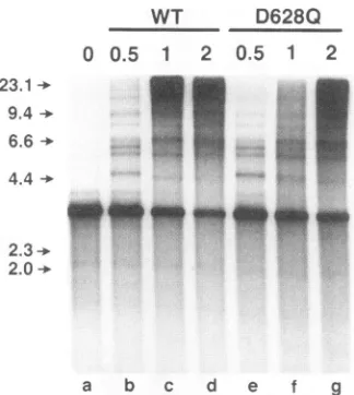
FIG. 5. Comparison of the abilities of UL8-UL52 and UL5-UL8-UL52(D628Q) tosupportleading strand replication. UL5-UL8-UL52 (WT) or UL5-UL8-UL52(D628Q) (0.5 to 2 pmol) was
incu-bated with apreformed fork substrate[single-stranded pBS(+) singly primed witholigonucleotide PB32 and extended with pol-42toyield double-stranded pBS with a free 5' tail] in the presence of pol-42, ICP8, [z-32P]dCTP, the other three dNTPs, and the four ribonucleo-side triphosphates for 1.5 h at 30°C as detailed in Materials and
Methods. Following alkaline agarose gel electrophoresis, the single-strandedproducts werevisualized by autoradiography. The positions
of molecular size markers(in kilobases)are indicatedtothe left.
primase complex on ICP8-coated single-stranded DNA are
elongated by the HSV-encoded pol-42 complex to result in fully duplex circular molecules (46). Inthe secondassay,RNA primer synthesiswas analyzed directly.
To assess the ability of the D628Q complex to participate withpol-42 and ICP8inRNA-primed DNA synthesis (lagging-strand assay), single-stranded pBS(+) DNA was incubated
with pol-42, ICP8, and UL5-UL8-UL52 or
UL5-UL8-UL52(D628Q) and the resultant products were analyzed by
electrophoresis through a 1% neutral agarose geland autora-diography (Fig. 6). With 0.25 and I pmol of the wild-type
complex (Fig. 6,lanes c and d),adose-dependent production
of double-stranded circular molecules was evident, while 2
pmol of the D628Q complex (lane e) resulted in only a
background level of double-stranded circles. These results indicated that while thewild-type enzyme was ableto synthe-size primers that could be elongated by the HSV-1 DNA polymerase, the mutant enzymewas completely incapable of performing this function. This result would be obtainedifthe UL5-UL8-UL52(D628Q) complex did not synthesize primers
orifitsynthesized primers that couldnotbeelongated by the DNApolymerase.
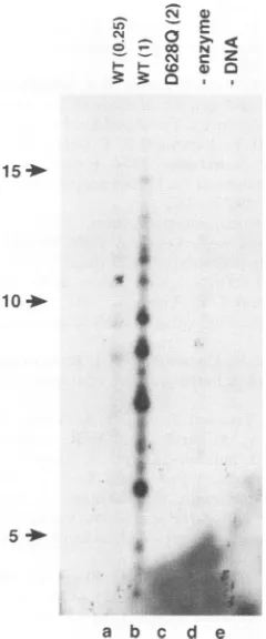
To assay directly for the capacity of the mutant enzyme to
carry out primer synthesis, RNA primers weresynthesized in
thepresenceof[oL-32P]UTP and visualized byautoradiography
after separation on a sequencing-type denaturing
polyacryl-amide gel. The results are shown in Fig. 7. The wild-type
enzymeproduced primersapproximately 5to14nucleotides in
length(predominantly 8to9nucleotides) inadose-dependent
manner.Incontrast,therewas nodetectable synthesis of RNA
primers with the UL5-UL8-UL52(D628Q) enzyme.
Inboththe direct andthe coupledassay,themutantenzyme
complex exhibited less than 12% of the activity ofwild-type UL5-UL8-UL52, while in the threeassaysforhelicase activity,
the twocomplexes were equivalent. The most likely
explana-WT D628Q
00.25 1 2
*0
a b c d e
FIG. 6. Comparison of the abilities of UL8-UL52 and UL5-UL8-UL52(D6280)to carry out RNA-primed DNAsynthesis. UL5-UL8-UL52 (WT) (0.25 or I pmol) or UL5-UL8-UL52(D628Q) (2 pmol)wasincubated withsingle-stranded pBS(+) DNA,ICP8,pol-42, four ribonucleoside triphosphates, [0x32P]dCTP, and the other three dNTPsasdescribed in Materials and Methods. Thereaction products wereseparatedby neutralagarosegel electrophoresis andvisualized by autoradiography. Inlane a,thepBStemplate DNA wasprimed with the deoxyoligonucleotide PBO (see Materials and Methods) and the reaction was carried out with all of the components added except helicase-primase. The arrowhead shows the position of the product formed byprimingof thesingle strand and extension by polymerase (double-stranded form II circular DNA).
tion forthese results is that UL52 is the primase subunitofthe helicase-primase complex.
DISCUSSION
UL5, UL8, and UL52 are three of the sevengenes that are essential for HSV-1 DNA replication. The products of these
three genesform a heterotrimericcomplex with helicase and
primase activity (13, 15). Given its known roles, it is likely that
the complex functions in both leading- and lagging-strand replication. The helicase presumably acts to unwind duplex
DNA ahead of the progressing replication fork, thereby
pro-ducing the open configuration needed for both continuous-and discontinuous-strand synthesis. By analogy to other
pri-mases, the primase of HSV-1 presumably initiates both
con-tinuous DNA synthesis near the origin of replication and
discontinuous DNAsynthesisonthelagging strandby provid-ingthe oligoribonucleotide primersthat are elongated bythe
polymerase.
The helicase activity of the UL5-UL52 subcomplex is as
efficient as that of the UL5-UL8-UL52 complex when mea-sured by an assayin which relatively short 3'-tailed oligonu-cleotides aredisplaced from single-stranded DNA(6, 18, 46).
In addition, the UL5-UL52 subcomplex can also synthesize oligoribonucleotide primers (18, 46). Therefore, UL8 is not
required for the core helicase and primase activities of the
complex.Arequirementfor UL8 in bothleading-and
lagging-strand replication has, however, been demonstrated. UL8 appears toberequiredfor theefficient utilizationofprimers by polymerase, thereby increasingtheefficiencyoflagging-strand synthesis (46). Additionally, it has been shown that UL8
stimulates leading-strand synthesis in vitro by a mechanism that is distinct fromanypossible role inprimase activity. UL8
is required for the synthesis of long (>20-kb) DNA single strands in a reaction utilizinga circular preformed fork
sub-strate and requiring six of the seven essential replication proteinsofHSV(no UL9) (25a).Theroleof UL8 inthislatter assay is unclear; one possibility is that UL8 is required to J.VIROL.
on November 9, 2019 by guest
http://jvi.asm.org/
[image:6.612.371.481.72.205.2] [image:6.612.91.253.73.254.2]cs 0
(
- Go N
;: CN c z w 0
tD@
15
10
5
i
a b c d e
FIG. 7. Comparison of the abilities of UL5-UL8-UL52 and
UL5-UL8-UL52(D628Q) to synthesize primers. UL5-UL8-UL52 (WT) (0.25or1 pmol)orUL5-UL8-UL52(D628Q) (2 pmol)was incubated
withsingle-stranded pBS DNA in thepresenceof[ox-32P]UTPand the
otherthreeribonucleoside triphosphatesasdetailedin Materials and Methods.Theproductswereisolatedby NENsorb20chromatography, separatedon an 18%HydrolinkLong Ranger gel with7 Murea,and visualized byautoradiography. The positions of5' end-labeled
oligo-nucleotides U5,
UIO,
and U15 used as molecular size markers areindicated to the left andwere determined in a separate experiment
with theproducts of wild-typeprimase run inaparallellane. Lanes d
andeshowtheeffectsof omittingenzyme ortemplate DNA,
respec-tively.
increase the processivity of the helicase on long regions of
duplexDNA.
Examination of the amino acid sequence of UL5 strongly
suggests that it is the helicase of the complex, as it shares
conservedsequence motifswith other known helicases(25,29) andmutationswithin these motifs abolish the abilityofUL5to
supportreplication ofan
oris-containing
plasmid in anin vivoassay(56). Formally, however, the question of whether UL5is
thehelicaseremainsopen,asUL5 alone hasnotdemonstrated convincing levels of helicaseactivity in the absenceof UL52 (6, 46). IfUL5 is,asseemslikely, the helicase, thenanimportant
question that remains to be understood is how UL52 stimu-lates its helicase activity in the formation ofthe UL5-UL52 complex. One possibilityis that UL5 undergoes an activating
conformational change upon UL52 binding that results inan
increasedaffinityfor DNAornucleoside triphosphate (NTP).
Another possibility is that UL52 contributes essential amino acidsto thestructureof the catalyticsite. In favor ofthefirst
possibility is the fact that UL5 itself contains all six motifs associatedwith otherhelicases.
The association of helicase with primase is not unique to HSV, although thispairingofactivities ismore reminiscentof
prokaryotic than eukaryotic systems. In phage T4, gene 61 primase and gene 41 helicase each stimulate the activity of the other in vitro assays (51). Therefore, these proteins have been shown to be functionally, if not physically, associated. In phage T7, the helicase and primase activities are encoded by a single gene (28, 45,49). The products of phage T7 gene 4 exist in a longer (63-kDa) form with both activities (38) and a shorter (56-kDa) form with only helicase activity (4, 38). These two forms can be isolated as acomplex in a
1:1
ratio (38), or theymay also be expressed individually, in which case the 56-kDa
form stimulates theprimase activity of the 63-kDa form up to 100-fold (38). In
Escherichia
coli, the helicase activity of DnaB and the primase activity of DnaG also appearcoordinated (1, 33). The mechanisms responsible for the mutual stimulationof the activities of the helicase and primase have not been determined for any of these systems.The data in this paper provide the first experimental evi-dence that UL52 functions as a primase. A single-amino-acid mutation (changing the aspartate at position 628 to glutamine) completely abolished the ability of the UL5-UL8-UL52 com-plex to support replication of an origin-containing plasmid in vivo, to support lagging-strand DNA synthesis in vitro, and to synthesize oligoribonucleotide primers in vitro. In contrast, the mutation had no effect on any measurable aspect of leading-strand replication: ATPase, helicase, or displacement DNA synthesis from a preformed fork in conjunction with ICP8, DNA polymerase, and UL42.
Although the most straightforward explanation for our results is that UL52 contains the catalytic site for primase activity, there are other possible explanations. One possibility is that UL5 contains both helicase and primase activities and that UL52 is required to stimulate both of these activities but itself does not play a role in primer synthesis. Another alternative possibility is that the UL52 subunit contains some but not all of the sequences required for primase activity and that bothUL5 and UL52 contain domains that must physically interact in such a way as to create the primase catalytic sites. If this were the case, the mutation at amino acid 628 could either interfere with this interaction or form part of a defective catalytic site. No direct evidence exists to distinguish among these possibilities as yet. Biochemical characterization of other UL52 mutants and of UL5 mutants may help to clarify this issue. Recently, partially purified preparations of UL52 alone have been shown to retain primase activity (1 Ia).These results suggest that UL52 contains all of the elements comprising the primase active site.
In the attempt to identify short sequence similarities be-tween UL52 and known primases, only one potential con-served motif, (IL)(VIM)(LF)DhD, was found. This motif is located within a conserved region of the small subunit of mouse primase and yeast primase 1, as well as in phage T7 primase. The significance of this motif is underscored by the fact that similar sequences are also found in most classes of nucleic acid polymerases, including DNA-dependent DNA polymerases, reverse transcriptases, and RNA-dependent RNA polymerases (2), and in other prokaryotic primases, including E. coli and Salmonella
typhimurium
DnaG, Bacillus subtilis DnaE, T4gp6l,T3 gp4 (30), and the primases of some conjugative plasmids (48). Among these various classes of polymerases, the only invariant residues are DD or DXD. However, the DNA and RNA polymerases seem to have in common a secondary structure in which the aspartates are flanked by hydrophobic residues that form aP
hairpin struc-ture, leaving the aspartates on an exposed loop (2). The protein secondary structure near the aspartates in the primases is less clear. There is a tendency toward hydrophobicityon November 9, 2019 by guest
http://jvi.asm.org/
[image:7.612.125.245.75.363.2]3700 KLINEDINST AND CHALLBERG
proximal to the motif, but the C-terminal flanking region is somewhat less hydrophobic than in the other classes of poly-merases.
The DD or DXD motif in the polymerases has been proposed to play a role inMg2+orNTP binding or possibly to play adirect role at the polymerase catalytic site(2).Mutations have been made in the proposed hairpin loop of several polymerases, with a variety of results. Poliovirus RNA poly-merase mutants with single-amino-acid substitutions for the glycine in the TGDD motif had reduced or undetectable
polymerase activity (31).Asimilar mutationthat changed the glycine to alanine at the YGDTD sequence of HSV-1 DNA polymerase was lethal in the context ofthe virus (35).
Muta-tions within the sequence YCDTD of B. subtilis
429
DNA polymerase had effects both on protein-primed initiation of DNA synthesis and on elongation, depending on the specificmutation and its position (3). Finally, conservative aspartate-to-glutamate mutations at either position of the sequence GDMD of rat DNA polymerase ( caused diminishedbinding
of the polymerase totheprimer/template, leadingtodrastically
reduced DNApolymerase activity (16).
Our results indicate that mutation of the first aspartate in the sequence IILDLD of HSV-1 UL52 eliminatesany
detect-able primer synthesis by the
UL5-UL8-UL52(D628Q)
com-plex. Further biochemical characterization of this and otherUL52 mutants may yield information concerning the
mecha-nism of the defect in primersynthesis,whichcould bein metal
binding, NTP binding, template binding, or adirect defect in catalysis. In this regard, conservative mutations of the aspar-tates atpositions 628 and630,suchasD->Emutations, might exhibit diminished but measurable primase activity, thereby
allowing kinetic analyses of various steps of the primase
reaction. Likewise, partiallydefectivemutants inotherregions of the protein,suchasK677E,mightpermit the localization of the domains of the UL52 protein that are responsible for
various steps in the reaction.
ACKNOWLEDGMENTS
We aregrateful to James Crute, Daniel Tenney, RobertHamatake, John Gottlieb, and Daniel Fierer for helpful discussions and sugges-tions during the course of the work and to Thomas Kristie for useful comments on themanuscript.
REFERENCES
1. Arai, K., and A. Kornberg. 1979. A general priming system employing only dnaB protein and primase for DNA replication. Proc. Natl. Acad. Sci. USA 76:4308-4312.
2. Argos, P. 1988. A sequence motif in many polymerases. Nucleic Acids Res. 16:9909-9916.
3. Bernad, A., L. Blanco, and M. Salas. 1990.Site-directed mutagen-esis of the YCDTDS amino acid motif of the phage 29 DNA polymerase. Gene 94:45-51.
4. Bernstein, J. A., and C. C. Richardson. 1988. A 7-kDa region of the bacteriophage T7gene 4 protein isrequired for primase but not for helicaseactivity. Proc. Natl. Acad. Sci. USA 85:396-400. 5. Bruckner, R. C., J. J. Crute, M. S. Dodson, and I. R. Lehman.
1991.The herpes simplex virus 1 origin binding protein: a DNA helicase. J. Biol. Chem. 266:2669-2674.
6. Calder, J. M., and N. D. Stow. 1990. Herpes simplex virus helic-se-primase: the UL8 protein is not required for DNA-dependent ATPase and DNA helicase activities. Nucleic Acids Res. 25:3573-3578.
7. Carmichael, E. P., M.Kosovosky,and S. K. Weller. 1988.Isolation and characterization of herpes simplex virus type 1 host range mutants defective in viral DNA synthesis. J. Virol. 62:91-99. 8. Carmichael, E. P., and S. K. Weller. 1989. Herpes simplex virus
type I DNA synthesis requires the product of the UL8 gene: isolation and characterization ofanICP6::lacZ insertion mutation.
J.Virol. 63:591-599.
9. Challberg, M. D. 1986.Amethod for identifyingthe viral genes required for herpesvirus DNA replication. Proc. Nati.Acad. Sci. USA 83:9094-9098.
10. Chartrand, P.,C. S. Crumpacker,P.A. Schaffer, and N. M. Wilkie. 1980. Physical and genetic analysis of the herpes simplex virus DNA polymerase locus. Virology 103:311-326.
11. Coen, D. M., D. P. Aschman, P. T. Gelep, M. J. Retondo, S. K. Weller, and P. A. Schaffer. 1984. Fine mapping and molecular cloning of mutations intheherpessimplex virusDNApolymerase locus. J.Virol.49:236-247.
11a.Crute, J. J.Personal communication.
12. Crute, J. J., and I. R. Lehman. 1989. Herpes simplex-I DNA polymerase.Identificationof an intrinsic 5' to 3'exonuclease with ribonuclease H activity. J. Biol. Chem. 264:19266-19270. 13. Crute, J. J., and I. R. Lehman. 1991. Herpes simplex virus-I
helicase-primase. Physical and catalytic properties. J.Biol. Chem. 266:4484-4488.
14. Crute, J. J., E. S. Mocarski, and I. R. Lehman. 1988. A DNA helicaseinduced by herpessimplex virus type 1. Nucleic Acids Res. 16:6585-6596.
15. Crute, J. J., T. Tsurumi, L. Zhu, S. K. Weller, P. D.Olivo, M. D. Challberg, E. S. Mocarski, and I. R. Lehman. 1989. Herpes simplex virus 1 helicase-primase: a complex of three herpes-encoded gene products. Proc. Nati. Acad. Sci. USA86:2186-2189. 16. Date, T., S. Yamamoto, K. Tanihara, Y. Nishimoto, and A. Matsukage. 1991. Asparticacid residuesatresidues190and 192 of ratDNApolymerase
P
areinvolved inprimerbinding. Biochem-istry30:5286-5292.17. Dodson, M. S., J.J. Crute, R. C. Bruckner, and I. R. Lehman. 1989. Overexpression and assembly ofthe herpes simplex virus type-I helicase-primase ininsect cells. J. Biol. Chem. 264:20835-20838.
18. Dodson, M. S., and I. R. Lehman. 1991. Association of DNA helicase and primase activities witha subassembly of the herpes simplexvirus 1 helicase-primase composedof the UL5 and UL52 geneproducts. Proc. Natl. Acad. Sci. USA 88:1105-1109. 19. Elias, P., andI. R. Lehman. 1988. Interaction oforigin binding
protein with an origin of replication of herpes simplex virus 1. Proc.Natl. Acad. Sci. USA85:2959-2963.
20. Elias, P., M. E. O'Donnell, E. S. Mocarski, and I. R. Lehman. 1986. ADNAbinding proteinspecificforan origin ofreplication of herpes simplex virus type 1. Proc. Natl. Acad. Sci. USA 83:6322-6326.
21. Fierer, D. S., and M. D. Challberg. 1992.Purification and charac-terizationof UL9, the herpessimplex virustype 1 origin-binding protein.J. Virol. 66:3986-3995.
22. Gallo, M. L., D. I. Dorsky, C. S. Crumpacker, and D. S. Parris. 1989.The essential65-kilodalton DNA-binding protein ofherpes simplex virus stimulates the virus-encoded DNA polymerase. J. Virol. 63:5023-5029.
23. Gallo, M. L., D. H. Jackwood, M. Murphy, H. S. Marsden, and D. S.Parris. 1988.Purification of the herpes simplex virustype 1 65-kilodalton DNA-binding protein: properties of theprotein and evidence of its association with the virus-encoded DNA poly-merase.J. Virol. 62:2874-2883.
24. Goldstein,D. J.,and S. K. Weller.1988. AnICP6::lacZinsertional mutagen is used to demonstrate that the UL52 gene of herpes simplex virus type I is required for virus growth and DNA synthesis. J.Virol. 62:2970-2977.
25. Gorbalenya, A. E., E. V. Koonin, A. P. Donchenko, and V. M. Blinov. 1989. Two related superfamilies of putative helicases involved in replication, repair andexpression ofDNAand RNA genomes. Nucleic Acids Res. 17:4713-4730.
25a.Gottlieb,J., and M. D.Challberg. Unpublished data.
26. Gottlieb, J., A.I.Marcy, D. M. Coen, and M. D. Challberg. 1990. The herpes simplexvirus type 1 UL42 geneproduct:asubunit of DNApolymerase thatfunctions to increase processivity. J.Virol. 64:5976-5987.
27. Hernandez, T. R., andI. R.Lehman. 1990. Functional interaction between the herpes simplex-I DNA polymerase and UL42 pro-tein. J. Biol. Chem. 265:11227-11232.
28. Hinkle, D. C., and C. C. Richardson. 1975. Bacteriophage T7 J. VIROL.
on November 9, 2019 by guest
http://jvi.asm.org/
deoxyribonucleic acid replication in vitro. Purification and prop-erties of the gene 4 protein of bacteriophage T7. J. Biol. Chem. 250:5523-5529.
29. Hodgman, T. C. 1988. A newsuperfamily of replicative proteins. Nature(London) 333:22-23.
30. Ilyina, T. V., A. E.Gorbalenya, and E. V. Koonin. 1992. Organi-zation and evolution of bacterial and bacteriophage primase-helicase systems. J. Mol. Evol. 34:351-357.
31. Jablonski, S. A., M. Luo, and C. D. Morrow. 1991. Enzymatic activity ofpoliovirus RNA polymerase mutants with single amino acid changes in the conserved YGDD amino acid motif. J. Virol. 65:4565-4572.
32. Kunkel, T. A., J. D. Roberts, and R. A. Zakour. 1987.Rapid and efficient site-specific mutagenesis without phenotypic selection. MethodsEnzymol. 155:166-180.
33. LeBowitz, J. H., and R. McMacken. 1986. TheEscherichia coli dnaB replication protein is a DNA helicase. J. Biol. Chem. 261:4738-4748.
34. Marchetti, M. E., C. A. Smith, and P. A. Schaffer. 1988. A temperature-sensitive mutation in a herpes simplex virus type 1 generequiredfor viral DNAsynthesis maps to coordinates 0.609 through0.614 in UL. J. Virol. 62:715-721.
35. Marcy, A. I., C. B. C. Hwang, K. L. Ruffner, and D. M. Coen. 1990. Engineeredherpes simplex virus DNA polymerasepointmutants: the most highly conserved region shared among a-like DNA polymerases is involved in substrate recognition. J. Virol. 64:5883-5890.
36. Martinez, R., L. Shao, and S. K. Weller. 1992. The conserved helicase motifs of the herpes simplex virus type I origin-binding protein UL9 are important for function. J. Virol. 66:6735-6746. 37. McGeoch, D. J., M. A. Dalrymple, A. Dolan, D. McNab, L. J.
Perry, P.Taylor, and M. D.Challberg.1988. Structuresof herpes simplex virus type 1 genes required for replication of virus DNA. J. Virol.62:444-453.
38. Mendelman, L. V., and C. C. Richardson. 1991.Requirements for primersynthesisby bacteriophageT763-kDa gene 4 protein. J. Biol. Chem.266:23240-23250.
39. Olivo, P. D., N. J. Nelson, and M. D. Challberg. 1988. Herpes simplex virusDNAreplication: the UL9 gene encodes an origin binding protein.Proc. Natl.Acad. Sci. USA 85:5414-5418. 40. Olivo, P. D., N. J. Nelson, and M. D. Challberg. 1989. Herpes
simplexvirus type 1 geneproducts requiredfor DNAreplication: identificationandoverexpression. J.Virol.63:196-204.
41. Parris, D. S., A. Cross, L. Harr, A. Orr, M. C. Frame, D. J. Murphy,D.J. McGeoch, and H. S. Marsden. 1988. Identification of the gene encodingthe 65-kilodalton DNA-binding proteinof herpessimplexvirus type 1. J. Virol.62:818-825.
42. Powell, K. L., E. Littler, andD.J. M.Purifoy. 1981. Nonstructural proteins of herpes simplex virus. II. Majorvirus-specific DNA-binding protein.J.Virol. 39:894-902.
43. Purifoy, D. J. M., R. B. Lewis, and K. L. Powell. 1977. Identifica-tion of the herpes simplex virus DNA polymerase gene. Nature (London) 269:621-623.
44. Purifoy, D. J. M., and K. L.Powell. 1981. Temperature-sensitive mutantsin twodistinct complementation groups of herpes simplex virus type 1 specify thermolabileDNA polymerase. J. Gen. Virol. 54:219-222.
45. Scherzinger, E., E. Lanka, G. Morelli, D. Seiffert, andA. Yuki. 1977. Bacteriophage-T7-induced DNA-priming protein. A novel enzyme involved in DNA replication. Eur. J. Biochem. 72:543-558.
46. Sherman, G., J. Gottlieb, and M. D. Challberg. 1992. The UL8 subunit of the herpes simplex virus helicase-primase complex is required for efficient primer utilization. J. Virol. 66:4884-4892. 47. Stow, N. D. 1992. Herpes simplex virus type 1 origin-dependent
DNAreplicationin insect cells using recombinant baculoviruses. J. Gen. Virol. 73:313-321.
48. Strack, B., M.Lessl,R.Calendar, and E. Lanka. 1992.Acommon sequence motif, -E-G-Y-A-T-A-, identified within the primase domains ofplasmid-encoded I- and P-type DNA primases and the
aproteinof the Escherichia colisatellitephage P4. J. Biol. Chem. 267:13062-13072.
49. Studier,F.W. 1972.BacteriophageT7.Science 176:367-376. 50. Summers, M. D., and G.E.Smith.1987. A manualof methods for
baculovirusvectorsandinsect cellprocedures.TexasAgricultural Experimental Station, Bulletin 1555. Texas A&M University, College Station, Tex.
51. Venkatesan, M.,L. L. Silver, and N. G. Nossal. 1982. Bacterio-phage T4 gene 41 protein, required for the synthesis ofRNA primers, is also a DNA helicase. J. Biol. Chem. 257:12426-12434. 52. Walker, J. E., M.Saraste,M.J.Runswick, and N. J.Gay. 1982. Distantly related sequences in the a and b subunits of ATP synthase,myosin, kinases and otherATP-requiringenzymes anda commonnucleotidebindingfold. EMBO J. 1:945-951.
53. Weller, S. K., E.P.Carmichael, D.P.Aschman,D.J.Goldstein, and P.A.Schaffer.1987.Geneticandphenotypic characterization of mutants in four essential genes that map tothe left half of HSV-1 UL DNA.Virology 161:198-210.
54. Weller, S. K.,K.J.Lee, D. J.Sabourin, and P. A. Schaffer. 1983. Genetic analysis of temperature-sensitive mutants which define the genefor themajor herpes simplexvirus type 1 DNA-binding protein.J.Virol. 45:354-366.
55. Wu, C. A., N. J. Nelson, D. J. McGeoch,and M. D. Challberg. 1988. Identification ofherpes simplexvirus type 1 genesrequired fororigin-dependentDNAsynthesis.J. Virol. 62:435-443. 56. Zhu, L., and S. K.Weller.1992.The six conservedhelicase motifs
of the UL5 geneproduct,acomponentof theherpessimplexvirus type 1 helicase-primase, are essential for its function. J. Virol. 66:469-479.