Sensitive and Specific Method for Rapid Identification of
Streptococcus pneumoniae
Using Real-Time Fluorescence PCR
JAMES C. M
CAVIN,
1PATRICIA A. REILLY,
2ROBERT M. ROUDABUSH,
1WILLIAM J. BARNES,
1ANN SALMEN,
3GLEN W. JACKSON,
3KATHLEEN K. BENINGA,
3ALICIA ASTORGA,
1FERNE K. M
CCLESKEY,
3WILLIAM B. HUFF,
4DEBRA NIEMEYER,
5ANDKENTON L. LOHMAN
1*
Molecular Epidemiology Branch,
1Clinical Microbiology Branch,
3and Epidemiology Surveillance Division,
4Air Force Institute for
Environment and Occupational Health Risk Analysis/Epidemiology Surveillance Division,
Brooks Air Force Base, San Antonio, and Clinical Microbiology, Wilford Hall Medical Center, Lackland
Air Force Base, Lackland,
2Texas, and Medical NBC Sciences and Technology Directorate,
Office of the Air Force Surgeon General, Bolling Air Force Base, Washington, D.C.
5Received 22 March 2001/Returned for modification 16 May 2001/Accepted 18 July 2001
Molecular surveillance of pathogens has shown the need for rapid and dependable methods for the
identi-fication of organisms of clinical and epidemiological importance. As the leading cause of community-acquired
pneumonia,
Streptococcus pneumoniae
was used as a model organism to develop and refine a real-time
fluo-rescence PCR assay and enhanced DNA purification method. Seventy clinical isolates of
S. pneumoniae, verified
by latex agglutination,
were screened against 26 negative control clinical isolates employing a TaqMan assay
on a thermocycler (LightCycler). The probe, constructed from the
lytA
gene, correctly detected all
S. pneumoniae
genomes without cross-reaction to negative controls. The speed and ease of this approach will make it
adaptable to identification of many bacterial pathogens and provide potential for adaptation to direct detection
from patient specimens.
Streptococcus pneumoniae
is the leading cause of
communi-ty-acquired pneumonia, meningitis, and otitis media in the
United States (2). While traditional antimicrobial therapy has
proven an effective treatment in the past, the emergence of
penicillin- and multidrug-resistant strains has resulted in an
increasing number of cases of illnesses and fatalities (4, 18).
Pneumococcal isolation and identification are complicated by
antimicrobial suppression of growth in culture and
contamina-tion by normal flora alpha-streptococci. Deteccontamina-tion by classical
techniques, culture, and serological methods can be
time-con-suming and indeterminate. Sensitive and specific assays that
can be completed quickly in the clinical laboratory are essential
for early diagnosis and effective therapy. Molecular assays are
inherently valuable because detection can be achieved with
enhanced sensitivity and specificity, and detection is not
dimin-ished with nonviable organisms.
Various molecular methods have been employed to assist
investigations (8, 11, 23). These methods include restriction
fragment length polymorphism (RFLP)-based protocols and
fingerprinting. PCR-based assays for the detection of
S.
pneu-moniae
with primers specific to repetitive regions and genes
encoding rRNA (12, 14, 21), pneumococcal surface adhesion A
molecule (22), pneumolysin (20, 27, 30, 37), penicillin-binding
protein (5, 6, 7, 24, 31), and autolysin (10, 25, 26) have been
employed with various degrees of success. Autolysin, encoded
by the
lytA
gene, is required for
S. pneumoniae
pathogenesis
and is a well-characterized virulence marker (1). The
lytA
gene
has been shown to have restricted allelic variation and
there-fore makes an ideal target for specific identification in clinical
and epidemiological studies (9, 35). Sequencing of the
lytA
locus and high-resolution DNA typing of
S. pneumoniae
dem-onstrated that the gene is highly conserved within the species
(32, 33), and it has been shown that
lytA
separates
S.
pneu-moniae
from the genotypically similiar species
Streptococcus
mitis
and
S. oralis
(19, 34).
Real-time PCR with sequence-specific primers and a
fluo-rescent TaqMan probe allows continuous monitoring of in
vitro DNA amplification, eliminating the need for gel
electro-phoresis. The TaqMan probe is an oligonucleotide designed to
a sequence within the target DNA (38). Hybridization of the
probe allows continuous monitoring of the PCR through a dual
fluorophore-labeled system and prevents false-positives due to
the absence of nonspecific amplification product fluorescence.
The reporter dye is quenched by fluorescent resonance energy
transfer by a quencher dye until the reporter is released during
primer elongation through 5
⬘
-to-3
⬘
exonuclease activity of
Taq
polymerase. Acquisition of the resulting fluorescence measures
the number of copies of target DNA product based on linear
regression analysis of a standard curve generated with known
genomic equivalents of the organism detected. Real-time
flu-orescence PCR was conducted on a LightCycler (39).
Ampli-fication and detection occur in closed glass capillaries,
prevent-ing amplicon cross-contamination. The LightCycler has a
32-sample capacity and achieves high-speed thermal cycling
through fan-driven air rather than heat block conduction. In
this work, results were obtained rapidly (
⬍
1 h) and were highly
reproducible, and the range of detection was from fewer than
10 organisms to more than 4 million. We found that
commer-cial, quality-controlled reagents to be proved than those that
were home-brewed, more consistent and in some cases
sensi-tivity was improved by several logs.
In this work, we describe the application of an enhanced
sample preparation method and a sequence-specific
fluores-* Corresponding author. Mailing address: Molecular Epidemiology,
AFIERA, 2601 Westgate Rd., Building 930, Brooks AFB, San
Anto-nio, TX 78235. Phone: (210) 536-2639. Fax: (210) 536-2638. E-mail:
[email protected].
3446
on May 15, 2020 by guest
http://jcm.asm.org/
cent probe system that achieves rapid identification of cultured
S. pneumoniae
. Ultimately, it is our hope to adapt this method
for direct detection from clinical specimens.
MATERIALS AND METHODS
Strain identification, cultivation, and DNA stocks.AnS. pneumoniaetype strain (ATCC 33400; American Type Culture Collection, Rockville, Md.) and 70 confirmed isolates ofS. pneumoniaefrom patient samples were used to deter-mine the PCR in vitro assay sensitivity and test sensitivity and to validate the enhanced DNA purification protocol. Specificity was determined with 27 nega-tive control organisms representing oral and respiratory flora selected from laboratory bacterial stocks. Each isolate was randomly assigned a sample number (U1 to U97). Identical experiments were conducted in two separate laboratories under identical conditions. All isolates were originally identified asS. pneu-moniaeby latex agglutination (LA) using the Pneumoslide method (BBL Pneu-moslide test forStreptococcus pneumoniae; BBL Microbiology Systems, Cock-eysville, Md.). Clinical isolates were recovered from frozen cultures and restreaked to single colonies on blood agar plates. Cultures for positive-control genomic DNA isolation were grown in TSAII (Remel Inc., Lenexa, Kans.). Both plates and broth cultures were grown at 37°C under CO2using MGC
Anaer-oPack System (Mitsubishi Gas Chemical Company, Inc., New York, N.Y.). Isolates showing no growth on MacConkey agar and mucoid, alpha-hemolytic colonies were found to be gram-positive cocci on Gram stain. Subsequent to the double blind study, samples with discordant PCR and LA results were identified to the species level by biochemical analyses using Phadebact, RapID Strep, and the GPI card on the Vitek system. Phadebact (Remel Inc.) is a coagglutination test using specific antipneumococcal antibodies coupled to protein A. RapID STR system (Remel Inc.) is a combination of conventional biochemical tests and single-substrate chromogenic test. The GPI card is used in the Vitek Automi-crobic system (Biomerieux-Vitek Inc., Hazelwood, Mo.). Negative-control or-ganisms were cultured under conditions appropriate for each species.
S. pneumoniaetype strain (ATCC 33400) was used for the PCR in vitro assay sensitivity optimization. A cross-reactivity panel of 44 species representing di-verse bacterial genera was established for specificity testing from DNA stocks previously purified in our laboratory and human genomic DNA (Roche Molec-ular Biochemicals, Mannheim, Germany). Quantification and determination of quality of DNA stock were conducted spectrophotometrically and by agarose gel electrophoresis. All clinical isolates and negative-control organisms were col-lected over several years prior to this study at either the Clinical Microbiology Laboratory, Air Force Institute for Environment and Occupational Health Risk Analysis/Epidemiology Surveillance Division, Brooks Air Force Base (AFB) or Wilford Hall Medical Center, Clinical Microbiology Laboratory, Lackland AFB. For each specimen, the source, date of collection, and location are on file.
Chromosomal DNA isolation: positive-control organism.Genomic DNA from the positive control,S. pneumoniaeATCC 33400, was isolated from overnight cultures with a modified version of the Puregene DNA isolation kit (Gentra Systems, Minneapolis, Minn.). Cell lysis was completed by adding six 1.0-ml aliquots of cell suspension (from 8.0 ml of overnight liquid culture) to six sterile 1.5-ml microcentrifuge tubes that were placed in an ice block. The tubes were placed in a tabletop microcentrifuge and spun at 12,000⫻gfor 60 s to pellet the cells. The supernatant was removed using a pipette, and 600l of sterile H2O
was added to each cell pellet and gently pipetted up and down until the cells were resuspended. To each tube, 10.0l of lysostaphin (2 U/5l) was added, and the tubes were inverted 25 times to mix and incubated at 37°C for 60 min to digest the cell walls. The preceding step was added to the nominal kit protocol to augment cell lysis but has subsequently been corrected to use lysozyme (1 mg/ml) as the enzyme of choice for streptococcal cell wall digestion. The tubes were inverted occasionally during the incubation period. The samples were centri-fuged at 12,000⫻gfor 60 s to pellet the cells, and the supernatant was removed. Cell lysis was continued by adding 600l of cell lysis solution to each cell pellet and gently pipetting up and down. To each cellular lysate, 6.0l of RNase A solution (4 mg/ml) was added, and the tubes were inverted 25 times and incu-bated at 37°C for 1.5 h. Following RNase A treatment, 10l of proteinase K (14.4 mg/ml) was added and incubated for 1 h at 37°C.
Protein precipitation was completed by cooling the samples to room temper-ature and adding 200l pf protein precipitation solution to the cell lysate. The tubes were vortexed vigorously at high speed for 20 s to mix the protein precip-itation solution uniformly with the cell lysate. The samples were centrifuged at 12,000⫻gfor 3 min. The precipitated proteins formed a tight white pellet. Precipitation of the DNA was completed by pipetting the supernatant into six sterile 1.5-ml microcentrifuge tubes containing 600l of 100% isopropanol (2-propanol) and mixing the samples by inverting gently 50 times. The tubes were
centrifuged at 12,000⫻gfor 1 min, and the supernatant was pipetted off and allowed to drain for 15 min on clean absorbent paper. To each tube, 600l of 70% ethanol was added, and the tubes were inverted several times to wash the DNA pellet and centrifuged at 12,000⫻gfor 1 min. The ethanol was carefully pipetted off the pellet, and the tubes were inverted on clean absorbent paper to air dry for 15 min. The DNA pellets were combined in 100l of DNA hydration solution, and the solution was placed in a 1.5-ml sterile microcentrifuge tube, hydrated overnight at 4°C, and stored at⫺20°C. Nucleic acid concentration was determined spectrophotometrically, and genome copy number concentration was calculated. (Refer to the Puregene DNA isolation kit for the manufacture’s upgraded protocol for isolation of gram-positive DNA.)
Chromosomal DNA isolation: solid-phase DNA purification protocol. Chro-mosomal DNA from theS. pneumoniae(ATCC 33400) positive-control organ-ism,S. pneumoniaeclinical isolates, and a panel of negative-control organisms were isolated by a modified version of the Generation Capture Disk protocol for DNA purification from 3l of gram-negative bacteria (Gentra Systems Inc.). The capture disk is a solid-phase DNA purification system comprised of a 3-mm-diameter paper disk shipped in a sterile 2.0-ml microcentrifuge tube (spin tube) within a removable, inner basket assembly. The nominal capture disk protocol calls for a 3-l suspension containing at least 600,000 cells from an overnight liquid culture. In the modified protocol, colonies were selected after 18 h of growth (eliminating overnight, liquid growth) and directly transferred using a sterile loop to 50l of sterile H2O in a 1.5-ml microcentrifuge tube. The
suspension was vortexed vigorously, and the entire volume was pipetted onto a disk in a spin tube. The disk and bacterial suspension were allowed to incubate at room temperature for approximately 10 min and centrifuged at 12,000⫻gfor 20 s to remove excess fluid. To each sample, 200l of generation DNA purifi-cation solution was carefully added down the side of the spin tube so that the disk was immersed in the entire volume. The disk was incubated for 60 s in the DNA purification solution, and the spin tube was centrifuged at 12,000⫻gfor 60 s. Each disk was transferred with a sterile, 20-g needle into the collar of a sterile, thermocycler capillary tube, 20l of the complete PCR mixture was added to the disk, and the capillary tube was capped. The capped capillary tubes containing the disk and PCR mixture were incubated approximately 10 min at room tem-perature. The capillary tubes were then centrifuged at 12,000⫻gfor 3 s, reaction volumes were verified, and placed into the thermocycler reaction carousel for PCR. The thermocycler reaction carousel accommodated 32 samples. The assay was unsuccessful when attempted with crude lysates prepared by boiling isolates and then exposing directly to the PCR.
Primer and TaqMan probe oligonucleotide design.ThelytAgene sequence was compared across 15S. pneumoniaestrains. A 901-bp highly conserved se-quence was identified, and within this region a 101-bp sese-quence was selected to develop primers and probe. The forward primer oligonucleotide sequence was 5⬘-ACGCAATCTAGCAGATGAAGC-3⬘at bp 306 to 326 within thelytAgene, the reverse primer sequence was 5⬘-TGTTTGGTTGGTTATTCGTGC-3⬘at bp 386 to 406 within the lytAgene, and the TaqMan probe sequence was 5⬘ -6-carboxy-fluorescein (FAM)–TTTGCCGAAAACGCTTGATACAGGG–6-carboxy-tetramethyl-rhodamine (TAMRA)-3⬘at bp 330 to 354. Amplicon prod-uct size was 101 bp. Optimal probe and primer sequences were computed using Primer Express software according to the manufacturer’s instructions (PE Ap-plied Biosystems, Foster City, Calif.). Primer sequences were identified withTm
values 10°C less than that of the probe. The fluorescent reporter molecule at the 5⬘ end of the TaqMan probe was FAM, and the quenching molecule was TAMRA. Primers and probe oligonucleotides were synthesized commercially (Synthetic Genetics, Rockville, Md.).
Real-time PCR.A LightCycler thermocycler was used to conduct real-time PCR (Roche Molecular Biochemicals, Mannheim, Germany). Assays were car-ried out in LightCycler capillaries in a 20-l reaction volume. Reaction reagents were purchased in a preformatted kit (LightCycler-DNA Master Hybridization Probes; Roche Diagnostics GmbH, Roche Molecular Biochemicals, Mannheim, Germany) containing 10⫻ concentrations of Taq DNA polymerase, de-oxynucleoside triphosphates (dNTPs) (with dUTP instead of dTTP), 10 mM MgCl2, and reaction buffer. The use of a clean room for reaction mixture
preparation separate from where DNA samples were prepared and loaded into capillary tubes, sterile technique, and the closed environment of the system obviated the need for carryover prevention using the uracil-DNA glycosylase protocol described by the kit manufacturer. The following concentrations proved optimal: forward primer (F1), 0.5M; reverse primer (R1), 5.0M; Taqman probe (TM1), 0.1⌴; and MgCl2, 5.0 mM. Exogenous MgCl2(25 mM stock) was
used to bring the final concentration to 5.0 mM, and PCR-grade, sterile H2O was
used to adjust the final reaction volume per the manufacturer’s instructions. Each genomic equivalent of positive-control DNA was added in a 2-l volume to
on May 15, 2020 by guest
http://jcm.asm.org/
18l of master mix. Human DNA negative control was prepared by adding 4.4e3 genomic equivalents in a 2-l volume to 18l of master mix.
No-template controls (NTC) were prepared by adding a 2-l volume of PCR-grade, sterile H2O to 18 l of master mix. Thermocycling conditions were
optimized to one cycle of denaturation at 95°C for 60 s, followed by 45 cycles of denaturation at 95°C for 0 s and amplicon extension at 60°C for 60 s, with a single fluorescence acquisition step at the end of extension. Fluorimeter settings were based on the precycling fluorescence of the probe read in the negative control sample with the real-time fluorimeter (RTF) software (Boehringer Mannheim Corporation, Indianapolis, Ind.). While running the RTF, the fluorimeter set-tings for FAM and TAMRA were adjusted to 10 and 60%, respectively, on the
yaxis fluorescence scale. This was performed using a concentration matrix of from 0.10 to 0.50M probe. Amplicon from thelytAgene was verified by running 5l of the PCR product on 2% agarose gels (data not shown).
RESULTS
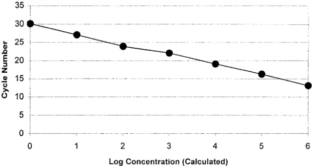
Sensitivity and specificity of PCR in vitro assay.
The
sensi-tivity of the PCR in vitro assay was optimized to four genomic
equivalents (10 fg) of purified
S. pneumoniae
DNA. Real-time
PCR in vitro assay sensitivity optimization using the
S.
pneu-moniae
type strain (ATCC 33400) resulted in a linear
regres-sion curve across 4.4e6 to 4.4e0 genomic equivalents (10 ng to
10 fg of DNA) with an error of
⬍
1% and a correlation
coef-ficient at
⫺
1.00 (Fig. 1). Agarose gel electrophoresis band
intensity correlated with the calculated concentrations of
am-plicon. Human DNA and NTC displayed no detectable
fluo-rescence above background, and upon agarose gel
electro-phoresis, the presence of amplicon was not observed.
Specificity of the in vitro assay initially tested using 1.0 ng of
purified DNA from each of 44 cross-reactivity panel bacterial
organisms and 4.4e3 genomic equivalents of human DNA
dis-played no detectable fluorescence (Table 1). The presence of
amplicon was not observed with agarose gel electrophoresis
examination.
In addition, 1.0 ng of genomic DNA from laboratory stock,
as well as DNA purified by the modified capture disk method,
from each of 10
S. pneumoniae
strains (ATCC 6305, ATCC
49619, ATCC 33400, ATCC 51915, 1301, 1346, 1518, 1661,
1830, and 2113) were correctly identified by the PCR assay.
The presence of the 101-bp amplicon was verified by agarose
gel electrophoresis.
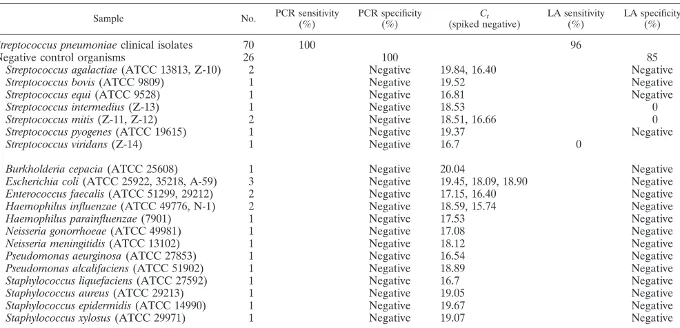
Sensitivity and specificity of PCR-based testing of clinical
isolates.
Double blind PCR-based testing results were
[image:3.587.132.452.74.244.2]concor-dant, and spiked negative data verified the absence of PCR
inhibition (Table 2). Genomic DNA extracted from all
S.
pneu-moniae
samples fluoresced upon exposure to the real-time
PCR assay, and all negative control organism DNAs failed to
report fluorescence. PCR-based testing of 96 specimens from
18-h isolates by real-time fluorescence PCR demonstrated a
sensitivity of 100% (70 of 70) and a specificity of 100% (26 of
26). Sensitivity of the PCR test was 8.8e6 (220 ng) to 8.8e1
genomic equivalents (220 fg) of purified
S. pneumoniae
. There
was no indication of inhibition of the PCR in any of the
neg-ative control DNA samples. All negneg-ative control genomic
DNA samples when spiked with 8.8e4 genomic equivalents of
FIG. 1. Serial dilutions of
S. pneumoniae
(ATCC 33400) DNA isolated by a standardized method and quantitated spectrophotometrically to
4.4e6 through 4.4e0 genomic equivalents were used for determination of real-time PCR assay detection limits or in vitro sensitivity testing. Cycle
number plotted against the log of calculated concentration values resulted in a standard curve with an error of 0.592 and correlation coefficient
at unity. Human genomic DNA at 4,500 genomic equivalents and NTC samples did not fluoresce above background signal. The detection limits
of the PCR assay demonstrated similar results when the dilution series panel was run in testing of all cross-reaction panel and unknown organisms.
TABLE 1. Cross-reactivity panel: negative-control organisms
Genus Species or serovar(s) (subtypes)
Homo
H. sapiens
Streptococcus
S. agalactiae, S. bovis, S. equi, S. equisimilis, S.
pyogenes, S. sanguis
(type II)
Campylobacter
C. coli, C. lari, C. jejuni
Citrobacter
C. freundii
Escherichia
E. coli
H1, O28, O55, O111, 112, 0126, 0128,
O157:H7
Klebsiella
K. pneumoniae
Leclercia
L. adecarboxylata
Neisseria
N. lactamica
Proteus
P. vulgaris
Pseudomonas
P. aeruginosa
Salmonella
Bovis-morbificus, Choleraesuis, Cubana, enteritidis,
Heidelberg, Infantis, Javiana, Lanka, Kovka,
Montevideo, Newport, Paratyphi-A, Poona
Typhi-1 (1078), Typhimurium (528)
Shigella
S. flexneri, S. boydii
(type I)
S. dysenteriae
(type III)
Staphylococcus
S. aureus
Mycoplasma
M. pneumoniae, M. hominis
on May 15, 2020 by guest
http://jcm.asm.org/
[image:3.587.301.541.533.728.2]positive control DNA fluoresced. In spiked negative control
experiments, 8.8e4 genomic equivalents of positive control
DNA had an average
C
tof
⬇
19 (
n
⫽
8; mean
⫽
18.77; range,
17.63 to 19.72). Of all negative control organism DNAs spiked
with 8.8e4 genomic equivalents of positive control DNA, the
average
C
twas
⬇
18 (
n
⫽
26; mean
⫽
18.43; range, 15.74 to
20.04). Upon agarose gel electrophoresis of spiked negative
control DNA, amplicon band intensity correlated with
ampli-con band intensity of 8.8e4 genomic equivalents of positive
control DNA. Nonspiked negative control DNA, tested in
par-allel with the spiked samples, displayed no detectable
fluores-cence when exposed to the PCR assay. The presence of
am-plicon was not observed with agarose gel electrophoresis.
For isolated colonies, the manufacturer has reported that
the Pneumoslide test sensitivity is 98.4% (303 of 308) and that
the test has a specificity of 93% (179 of 192) (BBL
Pneumo-slide test for Streptococcus pneumoniae package insert,
re-vised October 1984). In this study, Pneumoslide tests of 96
fresh clinical isolates had a sensitivity of 96% (67 of 70) and
specificity of 85% (22 of 26) (Table 2). Four false positives
were identified by PCR and upon biochemical analyses proved
to be two
Streptococcus mitis
strains,
Streptococcus intermedius
,
and
Streptococcus viridans
. These data correlate with
perfor-mance characteristics and limitations reported by the
Pneumo-slide manufacturer.
DISCUSSION
This study shows that real-time fluorescence PCR, using
primers and a TaqMan probe complementary to sequences
within the
lytA
gene, is a sensitive and specific assay for the
rapid identification of
S. pneumoniae
from isolates; however,
this method’s inherent value is in its potential for adaptation to
direct detection from clinical specimens. Since the inception of
the PCR over 15 years ago, PCR methods for the detection of
infectious organisms have been recognized as increasingly
valuable clinical diagnostic tools. The development of highly
sensitive and specific PCR assays has alleviated problems
typ-ically associated with microorganisms that are found in low
densities in tissue (or tissue fluids), difficult to culture, or
serologically similar. To prevent false-negative results, the
de-velopment of efficient DNA (RNA) purification methods is
necessary to isolate genetic material from cellular substances
found to inhibit DNA polymerase activity during the PCR (15).
The efficiency of the DNA isolation protocol and level of in
vitro sensitivity, test sensitivity, and specificity make the
method a valid candidate for adaptation to direct detection
from patient specimens and in this embodies our ultimate goal.
Our DNA purification protocol resulted in significant time
,
cost, and labor savings. Thirty-two samples were processed,
and the PCR assay was completed in less than 2 h from time of
isolate collection to completion of the report. Microbial DNA
capture and PCR assay achieved a level of efficiency that
al-lowed complete concordance in the results of double blind
testing in two separate PCR laboratories. The in vitro assay
sensitivity was 4 genomic equivalents, with a test sensitivity of
100% (70 of 70) and a specificity of 100% (26 of 26). In regard
to quantitation, although we have developed an exquisitely
sensitive assay for
S. pneumoniae
in vitro, we have not yet
tested the capture disk protocol for its inherent limit of
detec-tion on clinical specimens. Therefore, until we test a battery of
clinical specimens, we will not know whether this approach is
in itself quantitative and over what sensitivity range. Clearly,
any sample preparation method will impact a quantitative
method. It remains to be seen what the impact of the capture
disk protocol will be on our ability to quantitate. Rather, we
have demonstrated an in vitro sensitivity with a minimum
num-ber of genomic equivalents detectable by the PCR assay that is
very low and that test sensitivity was completely concordant. In
specificity testing, 44 cross-reaction panel organisms
represent-TABLE 2. Results of double blind PCR-based testing of clinical isolates
Sample No. PCR sensitivity(%) PCR specificity(%) Ct
(spiked negative) LA sensitivity(%) LA specificity(%)
Streptococcus pneumoniae
clinical isolates
70
100
96
Negative control organisms
26
100
85
Streptococcus agalactiae
(ATCC 13813, Z-10)
2
Negative
19.84, 16.40
Negative
Streptococcus bovis
(ATCC 9809)
1
Negative
19.52
Negative
Streptococcus equi
(ATCC 9528)
1
Negative
16.81
Negative
Streptococcus intermedius
(Z-13)
1
Negative
18.53
0
Streptococcus mitis
(Z-11, Z-12)
2
Negative
18.51, 16.66
0
Streptococcus pyogenes
(ATCC 19615)
1
Negative
19.37
Negative
Streptococcus viridans
(Z-14)
1
Negative
16.7
0
Burkholderia cepacia
(ATCC 25608)
1
Negative
20.04
Negative
Escherichia coli
(ATCC 25922, 35218, A-59)
3
Negative
19.45, 18.09, 18.90
Negative
Enterococcus faecalis
(ATCC 51299, 29212)
2
Negative
17.15, 16.40
Negative
Haemophilus influenzae
(ATCC 49776, N-1)
2
Negative
18.59, 15.74
Negative
Haemophilus parainfluenzae
(7901)
1
Negative
17.53
Negative
Neisseria gonorrhoeae
(ATCC 49981)
1
Negative
17.08
Negative
Neisseria meningitidis
(ATCC 13102)
1
Negative
18.12
Negative
Pseudomonas aeurginosa
(ATCC 27853)
1
Negative
16.54
Negative
Pseudomonas alcalifaciens
(ATCC 51902)
1
Negative
18.89
Negative
Staphylococcus liquefaciens
(ATCC 27592)
1
Negative
16.7
Negative
Staphylococcus aureus
(ATCC 29213)
1
Negative
19.05
Negative
Staphylococcus epidermidis
(ATCC 14990)
1
Negative
19.67
Negative
Staphylococcus xylosus
(ATCC 29971)
1
Negative
19.07
Negative
on May 15, 2020 by guest
http://jcm.asm.org/
[image:4.587.47.540.82.320.2]ing diverse genera and 26 negative control organisms,
includ-ing seven streptococcal species, did not react. Our method
correctly detected three false-negative specimens and four
false-positive organisms identified as
S. mitis
(two strains),
S.
intermedius
, and
S. viridans
by the Pneumoslide test. As the
method evolves to the next phase of development, testing with
clinical specimens, additional specificity testing will be
con-ducted as more strains become available to us, including typical
and atypical oral streptococci (36), to more fully validate the
PCR assay.
In its current format, our method can be used as a simple
confirmatory assay due to increased sensitivity and specificity
over standardized methods but is not practical for routine
testing due to existing standard biological assays for
S.
pneu-moniae
that are effective, simple to apply, and inexpensive. We
have implemented the method as a part of confirmatory testing
to help us identify isolates that proved difficult to identify using
our laboratory’s routine
S. pneumoniae
assay, the
Pneumo-slide. While bile solubility and optochin tests are commonly
used in many clinical laboratories for the detection of
S.
pneu-moniae,
the Pneumoslide is the assay of preference in our
laboratory because it has been our experience that bile
solu-bility, when done on a blood plate, can be difficult to interpret
and, when done in a tube, requires a large amount of inoculum.
We have found that difficulty with optochin disks may arise, as
some viridans streptococci and aerococci may also periodically
show small zones of inhibition, causing the organism to be
falsely identified as
S. pneumoniae
. If alpha-hemolytic
strepto-cocci are Pneumoslide negative, we routinely identify the
or-ganism using the battery of tests described in the Materials and
Methods section of this paper. As real-time fluorescence PCR
technologies become more widely used, applications will
evolve to increasingly simplified protocols more practical for
routine clinical laboratory diagnosis.
Due to our success with the capture disk DNA isolation
protocol and high degree of sensitivity, specificity, and rapidity
achieved by the PCR assay described in this paper, we have
begun collecting body fluid specimens to develop the clinical
application of the method. We have compared our capture disk
PCR assay to direct PCR of boiled
S. pneumoniae
isolates. The
capture disk procedure performed as described, while the
boiled lysate preparations were negative. Additionally, direct
application of saliva to the capture disk produced sufficient
DNA to conduct a PCR-based assay designed to screen for
transgenic mice markers (16). In our laboratory, the modified
capture disk protocol with a TaqMan PCR assay appears
promising in isolating and detecting
Campylobacter jejuni
DNA
directly from stool specimens (unpublished data).
In this study we have shown that real-time fluorescence
PCR, using primers and a TaqMan probe complementary to
sequences within the
lytA
gene, is a sensitive and specific assay
for the rapid identification of
S. pneumoniae
and that with the
capture disk DNA isolation protocol it provides potential for
clinical applications.
ACKNOWLEDGMENTS
We thank Melisa Gaiser for technical assistance and Rebecca
Me-dina and Andrew J. Rohrer, United States Air Force Academy, for
help in preparation of the manuscript. We thank J. Peter Pelletier,
Wilford Hall Medical Center, Lackland AFB, Tex., for kindly
review-ing the manuscript.
REFERENCES
1.Berry, A. M., R. A. Lock, D. Hansman, and J. C. Paton.1989. Contribution of autolysin to virulence ofStreptococcus pneumoniae. Infect. Immun.57:
2324–2330.
2.Brown, P. D., and S. A. Lerner.1998. Community-acquired pneumonia. Lancet352:1295–1302.
3.Cherian, T., M. K. Lalitha, A. Manoharan, K. Thomas, R. H. Yolken, and M. C. Steinhoff.1998. PCR-enzyme immunoassay for detection of Strepto-coccus pneumoniaeDNA in cerebrospinal fluid samples from patients with culture-negative meningitis. J. Clin. Microbiol.36:3605–3608.
4.Doern, G. V., A. B. Brueggemann, H. Huynh, and E. Wingert.1999. Anti-microbial resistance withStreptococcus pneumoniaein the United States, 1997–98. Emerg. Infect. Dis.5:757–765.
5.du Plessis, M., A. M. Smith, and K. P. Klugman.1999. Application ofpbp1A
PCR in identification of penicillin-resistant Streptococcus pneumoniae. J. Clin. Microbiol.37:628–632.
6.du Plessis, M., A. M. Smith, and K. P. Klugman.1998. Rapid detection of penicillin-resistant Streptococcus pneumoniaein cerebrospinal fluid by a seminested-PCR strategy. J. Clin. Microbiol.36:453–457.
7.Garcia, A., B. Roson, J. L. Perez, R. Verdaguer, J. Dorca, J. Carratala, A. Casanova, F. Manresa, and R. Gudiol.1999. Usefulness of PCR and antigen latex agglutination test samples obtained by transthoracic needle aspiration for diagnosis of pneumococcal pneumonia. J. Clin. Microbiol.37:709–714. 8.Gillespie, S. H.1999. The role of the molecular laboratory in the
investiga-tion ofStreptococcus pneumoniaeinfections. Semin. Respiratory Infect.14:
269–275.
9.Gillespie, S. H., T. D. McHugh, H. Ayres, A. Dickens, A. Efstratiou, and G. C. Whiting.1997. Allelic variation inStreptococcus pneumoniaeautolysin (N -acetyl muramoyl-L-alanine amidase). Infect. Immun.65:3936–3938. 10.Gillespie, S. H., C. Ullman, M. D. Smith, and V. Emery.1994. Detection of
Streptococcus pneumoniaein sputum samples by PCR. J. Clin. Microbiol.
32:1308–1311.
11.Hall, L. M. C.1998. Application of molecular typing the epidemiology of
Streptococcus pneumoniae. J. Clin. Pathol.51:270–274.
12.Hall, L. M., B. Duke, and G. Urwin.1995. An approach to the identification of the pathogens of bacterial meningitis by the polymerase chain reaction. Eur. J. Clin. Microbiol. Infect. Dis.14:1090–1094.
13.Hassan-King, M., I. Baldeh, O. Secka, A. Falade, and B. Greenwood.1994. Detection ofStreptococcus pneumoniaeDNA in blood cultures by PCR. J. Clin. Microbiol.32:1721–1724.
14.Hendolin, P. H., A. Markkanen, J. Ylikoski, and J. J. Wahlfors.1997. Use of multiplex PCR for simultaneous detection of four bacterial species in middle ear effusions. J. Clin. Microbiol.35:2854–2858.
15.Higuchi, R. 1989. Simple and rapid preparation of samples for PCR, p. 31–38.InH. A. Erlich (ed.), PCR technology: principles and application for DNA amplification. Stockton Press, New York, N.Y.
16.Irwin, M. H., R. J. Moffatt, and C. A. Pinkert.1999. Identification of trans-genic mice by PCR analysis of saliva. Nat. Biotechnol.14:1146–1148. 17.Isaacman, D. J., Y. Zhang, E. A. Reynolds, and G. D. Ehrlich.1998. Accuracy
of a polymerase chain reaction-based assay for detection of pneumococcal bacteremia in children. Pediatrics101:813–816.
18.Jacoby, G. A.1996. Antimicrobial-resistant pathogens in the 1990s. Annu. Rev. Med.47:169–179.
19.Kawamura, Y., R. A. Whiley, S. E. Shu, T. Ezaki, and J. M. Hardie.1999. Genetic approaches to the identification of themitisgroup within the genus
Streptococcus. Microbiology145:2605–2613.
20.Kearns, A. M., R. Freeman, O. M. Murphy, P. R. Seiders, M. Steward, and J. Wheeler.1999. Rapid PCR-based detection ofStreptococcus pneumoniae
DNA in cerebrospinal fluid. J. Clin. Microbiol.37:3434.
21.Lu, J.-J., C.-L. Perng, S.-Y. Lee, and C.-C. Wan.2000. Use of PCR with universal primers and restriction endonuclease digestions for detection and identification of common bacterial pathogens in cerebrospinal fluid. J. Clin. Microbiol.38:2076–2080.
22.Morrison, K. E., D. Lake, J. Crook, G. M. Carlone, E. Ades, R. Facklam, and J. S. Sampson.2000. Confirmation ofpsaAin all 90 serotypes of Streptococ-cus pneumoniaeby PCR and potential of this assay for identification and diagnosis. J. Clin. Microbiol.38:434–437.
23.Olive, D. M., and P. Bean.1999. Principles and applications of methods for DNA-based typing of microbial organisms. J. Clin. Microbiol.37:1661–1669. 24.O’Neill, A. M., S. H. Gillespie, and G. C. Whiting.1999. Detection of penicillin susceptibility inStreptococcus pneumoniaebypbp2b PCR-restric-tion fragment length polymorphism analysis. J. Clin. Microbiol.37:157–160. 25.Pozzi, G., M. R. Oggioni, and A. Tomasz.1989. DNA probe for identification
ofStreptococcus pneumoniae. J. Clin. Microbiol.27:370–372.
26.Rudolph, K. M., A. J. Parkinson, C. M. Black, and L. W. Mayer.1993. Evaluation of polymerase chain reaction for diagnosis of pneumococcal pneumonia. J. Clin. Microbiol.31:2661–2666.
27.Salo, P., K. Laitinen, and M. Leinonen.1999. Detection of pneumococcus from whole blood, buffy coat and serum samples by PCR during bacteremia in mice. APMIS107:601–605.
28.Salo, P., A. Ortqvist, and M. Leinonen.1995. Diagnosis of bacteremic
on May 15, 2020 by guest
http://jcm.asm.org/
mococcal pneumonia by amplification of pneumolysin gene fragment in serum. J. Infect. Dis.171:479–482.
29.Smith, A. M., and K. P. Klugman.1998. Alterations in PBP 1A essential for high-level penicillin resistance in Streptococcus pneumoniae. Antimicrob. Agents Chemother.42:1329–1333.
30.Toikka, P., S. Nikkari, O. Ruuskanen, M. Leinonen, and J. Mertsola.1999. Pneumolysin PCR-based diagnosis of invasive pneumococcal infection in children. J. Clin. Microbiol.37:633–637.
31.Ubukata, K., Y. Asahi, A. Yamane, and M. Konno.1996. Combinational detection of autolysin and penicillin-binding protein 2B genes of Streptococ-cus pneumoniaeby PCR. J. Clin. Microbiol.34:592–596.
32.Van Belkum, A., M. Sluijter, R. De Groot, H. Verbrugh, and P. W. M. Hermans.1996. Novel BOX repeat PCR assay for high-resolution typing of
Streptococcus pneumoniaestrains. J. Clin. Microbiol.34:1176–1179. 33.Watson, D. A., V. Kapur, D. M. Musher, J. W. Jacobson, and J. M. Musser.
1995. Identification, cloning and sequencing of DNA essential for encapsu-lation ofStreptococcus pneumoniae. Curr. Microbiol.31:251–259. 34.Whatmore, A. M., S. J. King, N. C. Doherty, D. Sturgeon, N. Chanter, and
C. G. Dowson.1999. Molecular characterization of equine isolates of Strep-tococcus pneumoniae: natural disruption of genes encoding the virulence factors pneumolysin and autolysin. Infect. Immun.67:2776–2782. 35.Whatmore, A. M., and C. G. Dowson.1999. The autolysin-encoding gene
(lytA) ofStreptococcus pneumoniaedisplays restricted allelic variation despite localized recombination events with genes of pneumococcal bacteriophage encoding cell wall lytic enzymes. Infect. Immun.67:4551–4556.
36.Whatmore, A. M., A. Efstratiou, A. P. Pickerill, K. Broughton, G. Woodard, D. Sturgeon, R. George, and C. G. Dowson.2000. Genetic relationships between clinical isolates ofStreptococcus pneumoniae, Streptococcus oralis,
andStreptococcus mitis: characterization of “atypical” pneumococci and or-ganisms allied toS. mitisharboringS. pneumoniaevirulence factor-encoding genes. Infect. Immun.68:1374–1382.
37.Wheeler, J., R. Freeman, M. Steward, K. Henderson, M. J. Lee, N. H. Piggott, G. J. Eltringham, and A. Galloway.1999. Detection of pneumolysin in sputum. J. Med. Microbiol.48:863–866.
38.Wittwer, C. T., M. G. Herrmann, A. A. Moss, and R. P. Rasmussen.1997. Continuous fluorescence monitoring of rapid cycle DNA amplification. Bio-Techniques22:130–138.
39.Wittwer, C. T., K. M. Ririe, R. V. Andrew, D. A. David, R. A. Gundry, and U. J. Balis.1997. The Lightcycler™: a microvolume multisample fluorimeter with rapid temperature control. BioTechniques22:176–181.
40.Zhang, Y., D. J. Isaacman, R. M. Wadowsky, J. Rydquist-White, J. C. Post, and G. D. Ehrlich.1995. Detection ofStreptococcus pneumoniaein whole blood by PCR. J. Clin. Microbiol.33:596–601.