Emerging Zoonotic Pathogen
Streptococcus suis
Matthew T. G. Holden1*, Heidi Hauser1, Mandy Sanders1, Thi Hoa Ngo2, Inna Cherevach1, Ann Cronin1, Ian Goodhead1¤a, Karen Mungall1, Michael A. Quail1, Claire Price1, Ester Rabbinowitsch1, Sarah Sharp1, Nicholas J. Croucher1, Tran Bich Chieu2, Nguyen Thi Hoang Mai3, To Song Diep3, Nguyen Tran Chinh3, Michael Kehoe4, James A. Leigh5, Philip N. Ward6, Christopher G. Dowson7, Adrian M. Whatmore7¤b, Neil Chanter8, Pernille Iversen9, Marcelo Gottschalk10, Josh D. Slater11, Hilde E. Smith12, Brian G. Spratt13, Jianguo Xu14, Changyun Ye14, Stephen Bentley1, Barclay G. Barrell1, Constance Schultsz2,15, Duncan J. Maskell16, Julian Parkhill1
1The Wellcome Trust Sanger Institute, Wellcome Trust Genome Campus, Hinxton, Cambridge, United Kingdom,2Oxford University Clinical Research Unit, Hospital for Tropical Diseases, Ho Chi Minh City, Viet Nam,3Hospital for Tropical Diseases, Ho Chi Minh City, Viet Nam,4Institute for Cell and Molecular Biosciences, The Medical School, University of Newcastle upon Tyne, Newcastle upon Tyne, United Kingdom,5The School of Veterinary Medicine and Science, The University of Nottingham, Sutton Bonington Campus, Sutton Bonington, United Kingdom, 6Nuffield Department of Clinical Laboratory Sciences, Oxford University, John Radcliffe Hospital, Headington, United Kingdom,7Department of Biological Sciences, University of Warwick, Coventry, United Kingdom,8Centre for Preventative Medicine, Animal Health Trust, Newmarket, United Kingdom,9Department of Biology, University of Copenhagen, Copenhagen, Denmark,10Groupe de recherche sur les maladies infectieuses du porc (GREMIP), Universite´ de Montre´al, Montre´al, Que´bec, Canada,11Royal Veterinary College, Hatfield, United Kingdom,12Animal Sciences Group (ASG), Wageningen UR, UR, Lelystad, The Netherlands,13Department of Infectious Disease Epidemiology, Imperial College, London, United Kingdom,14State Key Laboratory for Infectious Disease Prevention and Control, National Institute for Communicable Disease Control and Prevention, China CDC, Beijing, China,15Academic Medical Center-Center for Poverty-Related Communicable Diseases, University of Amsterdam, Amsterdam, The Netherlands,16Department of Veterinary Medicine, University of Cambridge, Cambridge, United Kingdom
Abstract
Background:Streptococcus suisis a zoonotic pathogen that infects pigs and can occasionally cause serious infections in humans.S. suisinfections occur sporadically in human Europe and North America, but a recent major outbreak has been described in China with high levels of mortality. The mechanisms ofS. suispathogenesis in humans and pigs are poorly understood.
Methodology/Principal Findings: The sequencing of whole genomes of S. suis isolates provides opportunities to investigate the genetic basis of infection. Here we describe whole genome sequences of threeS. suisstrains from the same lineage: one from European pigs, and two from human cases from China and Vietnam. Comparative genomic analysis was used to investigate the variability of these strains.S. suisis phylogenetically distinct from otherStreptococcusspecies for which genome sequences are currently available. Accordingly,,40% of the,2 Mb genome is unique in comparison to otherStreptococcusspecies. Finer genomic comparisons within the species showed a high level of sequence conservation; virtually all of the genome is common to theS. suisstrains. The only exceptions are three,90 kb regions, present in the two isolates from humans, composed of integrative conjugative elements and transposons. Carried in these regions are coding sequences associated with drug resistance. In addition, small-scale sequence variation has generated pseudogenes in putative virulence and colonization factors.
Conclusions/Significance:The genomic inventories of genetically relatedS. suisstrains, isolated from distinct hosts and diseases, exhibit high levels of conservation. However, the genomes provide evidence that horizontal gene transfer has contributed to the evolution of drug resistance.
Citation:Holden MTG, Hauser H, Sanders M, Ngo TH, Cherevach I, et al. (2009) Rapid Evolution of Virulence and Drug Resistance in the Emerging Zoonotic PathogenStreptococcus suis. PLoS ONE 4(7): e6072. doi:10.1371/journal.pone.0006072
Editor:Adam J. Ratner, Columbia University, United States of America
ReceivedMarch 11, 2009;AcceptedApril 22, 2009;PublishedJuly 15, 2009
Copyright:ß2009 Holden et al. This is an open-access article distributed under the terms of the Creative Commons Attribution License, which permits unrestricted use, distribution, and reproduction in any medium, provided the original author and source are credited.
Funding:This work was supported by the Wellcome Trust. The funders had no role in study design, data collection and analysis, decision to publish, or preparation of the manuscript.
Competing Interests:The authors have declared that no competing interests exist.
* E-mail: [email protected]
Introduction
Streptococcus suis is a Gram positive coccus that colonises pigs. While it is generally carried asymptomatically in adult pigs, it can cause severe systemic disease in piglets, manifested as a rapidly fatal sepsis associated with meningitis, polyarthritis and pneumo-nia. Why adult pigs carry the causative bacteria asymptomatically while piglets develop acute disease is unknown. The main site of carriage in the adult are the tonsils, but bacteria have also been isolated from the nasal cavities, the gastrointestinal tract and genital tract. The carriage rate in adult pigs can approach 100% and this organism has been a worldwide problem for the pig industry for a number of years.S. suishas also been isolated from a range of other mammalian and avian species [1].
S. suisis an important zoonotic agent. The first case in humans was described in Denmark in 1968 [2]. Human infection withS. suis occurs sporadically in Europe and North-America and case reports suggest that it is almost exclusively related to occupational exposure to pigs or pork products. Incidences of human infection withS. suisare greater in S.E. Asia and China. Meningitis is the most common presentation in humans, but septicaemia and endocarditis are also seen. In areas of Vietnam,S. suisis the main cause of acute bacterial meningitis in adults [3] and it is the third most common cause of meningitis in Hong Kong. The incidence of S. suisinfection in humans is almost certainly under-reported. Most cases have been described as being caused by serotype 2 isolates, but other serotypes can cause human disease [4].
Recently two outbreaks of severe acute disease in humans with high morbidity and mortality in humans have been reported in China [5], in both cases due to serotype 2 strains. An outbreak in 1998 killed 14 of 25 patients, and an outbreak in 2005 affected 204 people, killing 38 of them (19%), a mortality rate around two to four times that previously reported. A high proportion of people in these outbreaks had a toxic shock-like syndrome, and most of the deaths occurred in this group rather than in those suffering meningitis. Streptococcal toxic shock syndrome (STSS) was previously associated withStreptococcus pyogenes, so the emergence of STSS due to presumptive zoonotic S. suis infection is of considerable concern [6]. One isolate from each of these outbreaks has been sequenced [7] and this has identified a proposed pathogenicity island (PI) that may be involved in this particular clinical manifestation of S. suis infection, although this remains speculative at this stage. The PI was identified through comparison of the Chinese strains to the unfinished, unannotated genome sequence of strain P1/7 from the Sanger Institute. In this paper we present the completed fully annotated genome sequence of strain P1/7. In addition we have sequenced the genomes of two otherS. suis strains, SC84 and BM407, which are human isolates from China and Vietnam respectively. Strain SC84 is a representative of the of 2005 outbreak in China [8].
Materials and Methods
Bacterial strains, growth and DNA isolation
S. suis strain P1/7 was isolated from an ante-mortem blood culture from a pig dying with meningitis [9], and is ST1 by MLST [10].S. suisstrain BM407 is also ST1, and was isolated from CSF from a human case of meningitis in Ho Chi Minh City, Vietnam in 2004 [3].S. suisstrain SC84 is ST7, which is closely related to ST1, and was isolated from a case of streptococcal toxic shock-like syndrome in Sichuan Province, China in 2005 [8]. Strain P1/7 is resistant to gentamycin, streptomycin, neomycin, nalidixic acid, and sulfamethoxazole, and sensitive to penicillin, ampicillin, cephalotin, erythromycin, tulathromycin, clarythromycin,
linco-mycin, clindalinco-mycin, pirlimicin, tetracycline, trimethoprim-sulfa, ciprofloxacin, and chloramphenicol. Strain BM407 is resistant to trimethoprim-sulfamethoxazole, tetracycline, erythromycin, azith-romycin and chloramphenicol and susceptible to penicillin, ceftriaxone and vancomycin. Strain SC84 is resistant to tetracy-cline, and susceptible to penicillin, ampicillin, cefotaxime, ceftriaxone, cefepime, meropenem, levofloxacin, chloramphenicol, erythromycin, azithromycin, clindamycin, and vancomycin [11].
Bacteria were cultured in Todd-Hewitt-broth at 37uC for 18 h and pelleted at 10,0006g. The cells were resuspended in 30 ml of lysis solution (10 mM NaCl, 20 mM Tris HCl pH 8, 1 mM EDTA, 0.5% SDS) and incubated at 50uC overnight. Three ml of 5 M sodium perchlorate was added and incubated for 1 h at ambient temperature. After phenol chloroform extraction the DNA was precipitated with ethanol, spooled into deionised water and stored at220uC. DNA was also extracted using a genomic DNA extraction kit (G-500, Qiagen).
Whole genome sequencing
The genome of S. suis strain P1/7 was sequenced to approximately 8-fold coverage, from pUC18 (insert size 2.8– 3.3 kb), and pMAQ1b_SmaI (insert size 3.0–3.3 kb) genomic shotgun libraries using big-dye terminator chemistry on ABI3730 automated sequencers. End sequences from large insert BAC libraries in pBACehr (insert size 10–25 kb) and pBACe3.6 (insert size 12–15 kb) were used as a scaffold. All repeat regions were bridged by read-pairs or end-sequenced polymerase chain reaction (PCR) products.
S. suis SC84 and BM407 genomes were sequenced using multiple sequencing technologies to generate contiguous drafts. The bulk of the sequencing for both projects was generated using 454/Roche GS20 and Solexa sequencing platforms. The 454 sequencing of strain SC84 generated 399,145 reads with an average length of 102.7 bp, which assembledde novointo 281 non-redundant contigs using the 454/Roche Newbler assembly program. The 454 sequencing of strain BM407 generated 223,282 reads with an average length of 103.1 bp, which assembledde novointo 1939 non-redundant contigs. The Solexa sequencing of strain SC84 generated 32,016,513 reads with a length of 37 bp, and of strain BM407 generated 1,769,043 reads with a length of 37 bp which were each assembled by alignment to a reference (strain P1/7) sequence using the ssaha2 [12] alignment program. Data from the 2 technologies, were merged to produce 39 non-redundant contigs in SC84, and 390 non-redundant contigs in BM407.
The merging process involved using the random in silico sheering of the 454de novoassembly consensus contigs to create 500 bp consensus reads with 250 bp overlaps. This generated a fasta tiling path representing each of the 454 de novo assembly contigs. These were then assembled together with all the capillary shotgun reads using phrap2gap [13] to merge the two technologies The contigs were reordered based on BLAST [14] alignments with strain P1/7. The gaps between these contigs were closed by directed PCR and the products sequenced with big dye terminator chemistry on ABI3730 capillary sequencers. Aligned Solexa sequencing was merged to confirm assembly and also confirm sequence in 454 only regions. For the SC84 project capillary coverage was 56, 454 coverage was 196and Solexa coverage was 1906, and for the BM407 project capillary coverage was 56, 454 coverage was 116and Solexa coverage was 206.
The sequence was annotated using Artemis software [15]. Initial coding sequence (CDS) predictions were performed using Orpheus [16], Glimmer2 [17], and EasyGene [18] software. These predictions were amalgamated, and codon usage, positional base preference methods and comparisons to the non redundant protein databases using BLAST [14] and FASTA [19] software were used to refine the predictions. The entire DNA sequence was also compared in all six potential reading frames against UniProt, using BLASTX [14] to identify any possible coding sequences previously missed. Protein motifs were identified using Pfam [20] and Prosite [21], transmembrane domains were identified with TMHMM [22], and signal sequences were identified with SignalP version 2.0 [23]. rRNAs were identified using BLASTN [14] alignment to defined rRNAs from the EMBL nucleotide database; tRNAs were identified using tRNAscan-SE [24]; stable RNAs were identified using Rfam [25].
Comparative genomics
Comparison of the genome sequences was facilitated by using the Artemis Comparison Tool (ACT) [26] which enabled the visualization of BLASTN and TBLASTX comparisons [14] between the genomes. Orthologous proteins were identified as reciprocal best matches using FASTA [19] with subsequent manual curation. Pseudogenes had one or more mutations that would prevent correct translation; each of the inactivating mutations was subsequently checked against the original sequenc-ing data. Small scale variation includsequenc-ing: SNPs and insertions and deletions, were identified by using the SNP detection pipeline of MUMmer [27]. Clonalframe v1.1 was used to identify regions of theS. suischromosomes associated with recombination [28].
Streptococcus sequences used for comparative genomic analysis were:S. suis05ZYH33 (accession number CP000407) [7],S. suis 98HAH33 (accession number CP000408) [7],S. pyogenesManfredo (accession number AM295007) [29], Streptococcus equi 4047 (accession number FM204883) [30], Streptococcus uberis 0140J (accession number AM946015) [31], Streptococcus thermophilus CNRZ1066 (accession number CP000024) [32], Streptococcus pneumoniaeTIGR4 (accession number AE005672) [33],Streptococcus sanguinis SK36 (accession number CP000387) [34], Streptococcus mutans UA159 (accession number AE014133) [35], Streptococcus agalactiae NEM316 (accession number AL732656) [36], and Streptococcus gordonii str. Challis substr. CH1 (accession number CP000725) [37]. The sequences were also compared wth Lactococcus lactis subsp. lactis IL1403 (accession number AE005176) [38].
Phylogenetic analysis
Sequences alignment were made using R-coffee [39] and the alignments inspected and edited using Seaview (v4.0) [40]. Bayesian analysis was performed using Mr Bayes [41,42]. The model of sequence evolution used was the generalized time-reversible (GTR) model with gamma-distributed rate variation. The analysis was run with 26105generations, and the first 56104 were discarded as burn-in. The tree drawn with FigTree (v1.2.2; http://tree.bio.ed.ac.uk/software/figtree/).
Results
The genome ofS. suisstrain P1/7, a pig disease isolate
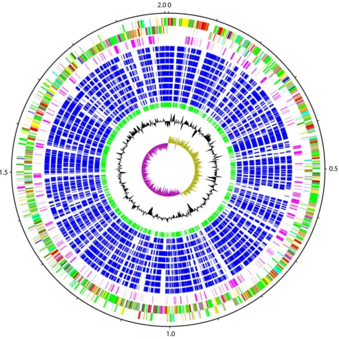
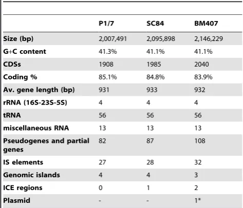
The genome ofStreptococcus suisP1/7 consists of a single circular chromosome of 2,007,491 bp (Figure 1; accession number AM946016) containing 1,908 predicted protein coding sequences (CDSs; Table 1), 82 of which are pseudogenes or gene fragments (Table S1). S. suis is phylogenetically distinct among the
streptococci for which genome sequences are available (Figure 2). Pairwise genomic comparisons reveal very little conservation of gene order with other species (data not shown). Orthologue comparisons against all of the other available streptococcal genomes identified that approximately 60% of the P1/7 genome is orthologous to CDSs in the genomes of otherStreptococcusspecies (Table 2). The highest number of orthologous matches was identified inS. sanguinis(66.1%), followed byS. gordonii(65.6%).
Analysis of the functions of the P1/7 orthologues present in other streptococcal species identified that some functional groups are more widely conserved than others (Figure 3), and that some exhibit a wide range of distribution across the species compared. Many of the core housekeeping functions such as fatty acid metabolism, chaperones, macromolecule biosynthesis, and nucle-otide biosynthesis exhibit the highest conservation. For these classes, .85% of the CDSs of these classes have orthologous matches in all the other streptococci compared. In some cases, for example ribosomal proteins, a lower level of orthologous matches in a strain is due to differences in gene prediction, rather than the absence of a CDSper se. In contrast, some functional groups such as cofactor biosynthesis and amino acid biosynthesis exhibit a lower level of conservation and probably reflect the available nutrients in the niches occupied by the individual streptococcal species. Many of the more variably distributed orthologous functional groups are associated with the acquisition and uptake of nutrients, for example macromolecule and small molecule degradation, as well as responses to environmental stresses and stimuli.
Excluding CDSs in mobile genetic elements (MGE), 283 CDSs (14.9%) in the P1/7 genome do not have orthologous matches (have been) in any of the other Streptococcus species (Table S2). These have been designated S. suis orphans and include: a transporter cluster (SSU0039 to SSU0044), a putative endopep-tidase (SSU0152), a glucosamine-6-phosphate isomerase (SSU0206), a putative lipase (SSU0304), a putative deoxyguano-sinetriphosphate triphosphohydrolase (SSU0327), capsule associ-ated CDSs (SSU0520 to SSU0529), a UDP-galactopyranose mutase (SSU0563), a MATE family transporter (SSU0573), EPS associated CDSs (SSU1115, SSU1125 and SSU1128), a putative oligopeptidase (SSU1679), and a putative choline binding protein (SSU1911) (Table S2).
Virulence genes in the P1/7 genome
The mechanisms by whichS. suiscause disease in humans and pigs are poorly understood. Many putative virulence functions have been identified, however no single function is crucial for the pathogenesis of S. suis. Many putative virulence genes are only present, or expressed, in some S. suis isolates, suggesting that combinations of virulence factors may be more important than individual functions. Additionally, several of the functions may be important for colonization and carriage, rather than disease. Analysis of theS. suisgenomes reveals an array of CDSs encoding potentially important functions.
mediate prostaglandin E2 (PGE2) and matrix metalloproteinase 9 (MMP-9) production by human macrophages [50] suggesting a role in pathogenesis of disease.
The P1/7 genome also contains a cluster of genes that appears to encode the components required for the biosynthesis of a putative exopolysaccharide (SSU1111 to SSU1133; Figure S1a). Included in this are CDSs encoding enzymes for synthesis of the
exopolysaccharide building blocks, for processing and for export. Analysis of the cluster suggests that it is involved in the synthesis of a rhamnose-glucose-like polysaccharide (RGP), and is similar to components of RGP biosynthesis identified inS. mutans (Figure S1b). Four loci have been identified in S. mutans that encode proteins necessary for RGP synthesis: RmlA, RmlC, RmlB, and RmlD direct the synthesis of dTDP-L-rhamnose from D-glucose-Figure 1. Schematic circular diagram of theS. suisP1/7 genome.Key for the circular diagram (outer to inner): scale (in Mb); annotated CDSs coloured according to predicted function are shown on a pair of concentric circles, representing both coding strands;Streptococcus suisorphan CDSs, purple; orthologue matches shared with the Streptococcal species,S. mutansUA159,S. gordoniiChallis CH1,S. sanguinisSK36,S. pyogenesManfredo,
S. equi4047, S. agalactiae NEM316, S. uberis0140J, S. pneumoniaeTIGR4, S. thermophilus CNRZ1066, blue; orthologue matches shared with
Lactococcus lactissubsp.lactis, green; G+C% content plot; G+C deviation plot (.0% olive,,0% purple). Colour coding for P1/7 CDS functions: dark blue, pathogenicity/adaptation; black, energy metabolism; red, information transfer; dark green, surface associated; cyan, degradation of large molecules; magenta, degradation of small molecules; yellow, central/intermediary metabolism; pale green, unknown; pale blue, regulators; orange, conserved hypothetical; brown, pseudogenes; pink, phage and IS elements; grey, miscellaneous.
[image:4.612.61.554.60.553.2]1-phosphate [51,52]; GluA produces UDP-D-glucose [53], RgpG initiates RGP synthesis by transfer of N-acetylglucosamine-1-phosphate to a lipid carrier [54], and proteins from an operon encoding six proteins (rgpABCDEFHI) are required for the transport and assembly of the rhamnan repeat unit backbone, and glucose side chain formation [55,56].
The putative exopolysaccharide biosynthetic cluster between CDSs SSU1111 and SSU1133, contains homologues of rmlACB and rmlD (SSU1129, SSU1130, SSU1132 and SSU1133), and rgpABCD and F (SSU1124, SSU1123, SSU1122, SSU1121 and SSU1120) (Figure S1a). The two clusters of rml and rgp homologues are separated by four CDSs encoding a putative sulphatase (SSU1125), a mannosyl-glycoprotein endo-beta-acetylglucosaminidase gene fragment (SSU1126), a putative N-acetylmuramoyl-L-alanine amidase (SSU1127), and a surface-anchored protein containing an LPXTS motif (SSU1128). In addition to the homologues of theS. mutans rpggenes, there is also a flippase (SSU1119), and six glycosyl transferases (SSU1117, SSU1116, SSU1115, SSU1114, SSU1113 and SSU1111). Homo-logues ofgluA(SSU1828) andrgpG(SSU1672) are located at two other loci on the P1/7 genome. Unlike theS. mutansRGP cluster, the P1/7 cluster does not contain homologues ofrmlE,rmlHand rmlI. These genes encode proteins that are involved in glucose side chain formation [56], therefore it is likely that the polysaccharide produced by P1/7 has a different repeat unit structure to that produced byS. mutans.
The cluster does not appear to encode an obvious repeat unit polymerase. Downstream of the flippase is a membrane protein (SSU1118) that has weak similarity matches to proteins of unknown function. Due to the sequence diversity of repeat unit polymerases, it is not always possible to annotate them by sequence similarity. Hydrophobicity plot profile comparison has been used to identify tentatively these polymerases [57]. A comparison of the hydrophobicity plot of SSU1118 with those of known polymerases identified a similar profile (data not shown).
RGPs constitute serospecific-antigens that form the basis of some streptococcal serotyping. These molecules are associated with modulating host immune cell interactions, and may have a role in eliciting sepsis. Streptococcal RGPs stimulate human
monocytes in vivo to release inflammatory cytokines, including tumor necrosis factor alpha and interleukin-1b [58], induce NO synthase activity [59], and confer resistance to phagocytosis by human polymorphonuclear leukocytes [60]. Elevated NO pro-duction has been observed in animal models of bacterial meningitis and also in human patients with the disease [61,62]. In a mouse model ofS. suisinfection, high levels of inflammatory cytokines were observed both systemically and in the central nervous system [63]. Distinct from the type 2 capsule, it is possible that the exopolysaccharide encoded by this cluster may have an important role in virulence and may contribute to the inflamma-tory response duringS. suisinfection.
Protein secretion and secreted proteins. In the publication by Chenet al.describing the genomes of two ChineseS. suisisolates [7], they presented possible virulence-associated factors or pathways inS. suis, including strain P1/7. This analysis identified thirteen components of a Type II secretion system and one component of a Type III secretion system in the genome of strain P1/7. Our analysis of the genome failed to identify components of either secretion system, although components of Sec and SRP pathways were identified. Neither Type II, nor Type III secretion systems have been identified in Gram-positive bacteria, and it is therefore very unlikely that S. suissecretes proteins via these pathways. It would appear that the description of these secretion system components in S. suis is a case of mistaken identity due to sub-optimal transitive annotation. TheS. suisgenome contains CDSs that have weak similarity to components of these Gram-negative protein secretion systems, but these genes inS. suisencode proteins that are involved in the transport of other molecules, or are components of functionally distinct systems. The observed similarity owes more to distant evolutionary relationships rather than conservation of function [64].
Suilysin (SSU1231) is a secreted thiol-activated cytolysin [65] produced by some strains ofS. suis[66]. This protein is part of a family of cytolysins that play a role in disease caused by several bacterial pathogens [67]. The role that suilysin plays in S. suis disease is unclear. Immunization of pigs with suilysin provided partial protection fromS. suis infection [68]. However, while a suilysin mutant was avirulent in a mouse infection model, it was only slightly attenuated in a model of infection in pigs [69].
Surface proteins. Several of the previously characterizedS. suis virulence factors are cell wall-anchored proteins. These sortase-processed proteins contain a C-terminal LPXTG-type processing motif. The genome of strain P1/7 contains a large number (28) of proteins of this type: all but five contained C-terminal LPXTG motifs followed by non-polar and basic amino acid motifs. The other five putative surface proteins (SSU1128, SSU1885, SSU1186, SSU1888 and SSU1889) contained LPXTS, IPXTG, IPXTG, YPXTG and LYXTG motifs respectively. Previous studies have identified five sortase homologues inS. suis (srtA-E) by PCR screening with degenerate primers [70]. An additional sortase CDS was identified (SSU0428) in the P1/7 genome [71].
[image:5.612.61.298.87.290.2]In addition, the S. suis P/7 genome contains a protein (SSU0879) with an atypical N-terminal LPXTG-type motif. This protein is similar to the large surface zinc metalloproteinases ofS. pneumoniae; 39.9% identical to the zinc metalloprotease precursor ZmpC [72], 33.7% identical to Immunoglobulin A (IgA)1 protease precursor Iga [73], and 28.3% identical to the zinc metallopro-tease precursor ZmpB [74]. InS. pneumoniaethese proteins interact directly with the host immune system, and play an inferred role in virulence. ZmpC mutants inS. pneumoniaeled to reduced mortality in an experimental mouse model of intranasal challenge and sepsis [72]. ZmpC cleaves human matrix metalloproteinase 9 (MMP-9),
Table 1.General properties of the genomes ofS. suisstrains P1/7, SC84 and BM407.
P1/7 SC84 BM407
Size (bp) 2,007,491 2,095,898 2,146,229
G+C content 41.3% 41.1% 41.1%
CDSs 1908 1985 2040
Coding % 85.1% 84.8% 83.9%
Av. gene length (bp) 931 933 932
rRNA (16S-23S-5S) 4 4 4
tRNA 56 56 56
miscellaneous RNA 13 13 13
Pseudogenes and partial genes
82 87 108
IS elements 27 28 32
Genomic islands 4 4 3
ICE regions 0 1 2
Plasmid - - 1*
thought to lead to the proteolytic activation of this matrix remodelling protein [72]. Matrix metalloproteinases can contrib-ute to blood–brain barrier (BBB) disruption during bacterial meningitis [75,76]. MMP-9 produced by leukocytes during meningitis is essential to enable them to migrate through the BBB. A consequence of the increase in the permeability of the BBB mediated by MMP-9, is that bacterial cells are able to migrate to the central nervous system and cause further tissue damage. S. suis capsule induces MMP-9 production in macro-phages in anin vivomodel [50]: it is possible that theS. suiszinc metalloproteinase may also play a role in increasing MMP-9 activity.
IgA1 protease mediates the site-specific cleavage of human IgA1 in the hinge region [73]. IgA is the major class of immunoglobulin in mucosal secretions of the upper respiratory tract, and functions by interfering with microbial attachment to host tissues. The activity of IgA1 protease may help pathogens to subvert the
antigen specificity of the humoral immune response, and facilitate adhesion and persistence at the mucosal surface [77].
Analysis of extracellular proteins produced byS. suisidentified two proteins, muramidase-released protein (MRP) [78] and extracellular protein factor (EF) [79], associated with disease and mortality in pigs (MRP+, EF+) [79]. Variation in the expression of these proteins has been observed inS. suisisolates. Although MRP and EF were originally identified as markers of disease, mostS. suis serotype 2 strains isolated from diseased pigs in Canada are phenotypically negative for MRP and EF [80]. Additionally, mutation of both proteins had no measurable effect on pathogenicity [81]. P1/7 is phenotypically positive for MRP and EF (MRP+, EF+). In the genome of P1/7, EF appears to be encoded in the first ORF of a larger mutated CDS (SSU0171). The entire CDS encodes a putative surface-anchored protein, and is split into three reading frames by a frameshift and nonsense mutations; the 5-prime reading frame encoding EF, contains an N-Figure 2. Phylogenetic relationships ofS. suisto the other genome sequenced streptococci.Unrooted majority-rule tree of Bayesian analysis of combined 16S rRNA and RNase P RNA gene datasets. The was tree built from 16S rRNA andrnpBgene sequences from the genomes of the
Streptococcusspecies:S. suisP1/7,S. mutansUA159,S. gordoniiChallis CH1,S. sanguinisSK36,S. pyogenesManfredo,S. equi4047,S. agalactiae
NEM316,S. uberis0140J,S. pneumoniaeTIGR4, andS. thermophilusCNRZ1066.Lactococcus lactissubsp.lactisIL1403 was included as an outgroup. The numbers at the branches are posterior probabilities indicating the support for the branch. The bar indicates the number of substitutions per site (4 per 100 sites).
[image:6.612.65.550.57.453.2]terminal signal sequence, and the 3-prime reading frame contains a LPXTG sortase-processing motif. The EF+phenotype of P1/7 is probably due to the expression of the 5-prime reading frame.
Unlike MRF and EF, opacity factor (OFS) is a large surface-associated protein that may play a role in the virulence ofS. suis [82]. The N-terminus of OFS is homologous to the N-terminal regions of the fibronectin-binding protein A (FnBA) ofStreptococcus dysgalactiaeand the serum opacity factor ofS. pyogenes. Mutational studies have suggested a role for OFS in virulence, but not colonization. However in strain P1/7 OFS (SSU1474) is mutated, containing two frameshift mutations.
[image:7.612.61.298.85.302.2]Two of the LPXTG proteins were found in a cluster similar to the pilus islands identified inS. agalactiae[83], and comprise the main pilus subunit (SSU0427) and ancillary subunit of this pilus (SSU0425). In addition the cluster contains a signal peptidase (SSU0424) and a sortase (SSU0428). The genome contains a second pilus cluster, containing 3 sortases, srtD, srtC, srtB (SSU1881, SSU1882, SSU1883), ancillary protein 1 (SSU1885), the main pilus subunit (SSU1886), ancillary protein 2 (SSU1888) and ancillary protein 3 (SSU1889).In silicoanalysis of the P1/7 genome suggests that this strain does not express complete pili, as both of the clusters contain pseudogenes; SSU0425 contains a nonsense mutation, and SSU1886 and SSU1888 contain frame-shift mutations. To this end it is worth noting that orthologous CDS in the other sequencedS. suisstrains are also pseudogenes. The genome also contains an incomplete pilus cluster that contains a signal peptidase (SSU0450) and sortase SrtE (SSU0453), but lacks pilus subunits and ancillary subunits. Between the signal peptidase and sortase there are a putative exported protein (SSU0451) and a transposase fragment (SSU0452). It is possible that the putative exported protein represents the N-terminal region of a pilus protein that has been truncated by a deletion event that generated the partial transposase sequence. Evidence for the potential role of pili inS. suispathogenesis has come fromin
Figure 3. Functional distribution of Streptococcal orthologues in theS. suisP1/7 genome.Functional groups are displayed clockwise on the web graph, in decreasing range of orthologue matches for a single category.
[image:7.612.50.555.408.698.2]doi:10.1371/journal.pone.0006072.g003
Table 2.Orthologues ofS. suisP1/7 CDSs in other streptococci.
% of CDSs with orthologous matches
S. sanguinisSK36 66.1
S. gordoniiCH1 65.6
S. agalactiaenem316 64.4
S. agalactiae2603v 64.0
S. agalactiaeA909 63.6
S. uberis0140J 63.9
S. pneumoniaeD39 64.2
S. pneumoniaeTIGR4 63.8
S. pneumoniaeR6 63.6
S. mutansUA159 61.2
S. equi4047 60.1
S. pyogenesManfredo 57.9
S. thermophilusCNRZ1066 55.6
S. thermophilusLMG 18311 55.1
Orthologues matches are displayed as a percentage of the total CDSs in the P1/ 7 genome.
vitrowork looking for preferentially expressed genes inS. suisupon interaction with porcine brain microvascular endothelial cells [71]. The signal peptidase (SSU0424) of theS. agalactiae-like cluster is upregulated, as is srtE, the sortase belonging to the incomplete pilus cluster.
Comparison of cell wall-anchored proteins among streptococcal species shows that they are one of the most variable components of the genome; 13 out of the 29 proteins in P1/7 were unique toS. suis(Table S2), the rest being intermittently distributed. Interest-ingly, orthologues of several of the S. suis cell wall-anchored proteins, or proteins associated with their processing, were conserved within the meningitis-associated streptococci (Table 2). Six of the cell wall-anchored proteins, and four of the sortases were found inS. agalactiaeand/orS. pneumoniae, and were absent in other streptococci [84].
In addition to LPXTG-type cell wall-anchored proteins, the P1/7 genome also contains other surface-exposed proteins that may play an important role in modulating host-cell interactions. The P1/7 genome encodes a protein, SSU0496, which contains a Mac 1 domain (PF09028), an N-terminal signal sequence and a C-terminal transmembrane domain. The Mac 1 domain is found in a small number of streptococcal proteins that modulate the immune response. Mac, also known as IdeS, is a cysteine protease secreted by S. pyogenes [85,86]. Two allelic families of Mac have been identified inS. pyogenes; Mac I and Mac II [87] exhibit sequence divergence in the middle third of the proteins (,50% amino acid identity in this region). Mac was originally named because it had limited sequence homology to thea-subunit of the leukocyteb2 integrin, Mac-1 [85].S. pyogenes Mac proteins block polymorpho-nuclear leukocyte opsonophagocytosis and inhibit the production of reactive oxygen species [87]. The mechanisms of action of theS. pyogenesMac allelic families differ; Mac I binds to, and cleaves, the hinge region of human IgG [88] whereas Mac-2 exhibits lower levels of endopeptidase activity, and does not bind IgG, but instead to the IgG receptor, Fcc [89]. Homologues of Mac have been identified in Streptococcus equi subsp. equi (349 amino acids), the causative agent of strangles, and the related pathogenStreptococcus equi subsp. zooepidemicus [90]. In S. equi the Mac homologue is expressed during infection, and reduces the antiphagocytic activity of equine neutrophils [91].
SSU0496 is similar (,29% sequence identity) in the N-terminal region to theS. pyogenesMac I and II proteins. In comparison, the S. suis protein is considerably larger than the S. pyogenes Mac proteins, comprising 1141 amino acids as opposed to,340 amino acids. The enzymatic activities of S. pyogenes Mac proteins have been characterized, and they belong to a novel family of cysteine proteases [88]. Protein alignment of theS. suisprotein with theS. pyogenesproteins shows that the amino acids of the catalytic triad (Cys94, His262 and Asp284) are conserved. The S. suis protein however, is missing an Arg-Gly-Asp (RGD) integrin-binding motif present in the other streptococcal proteins, suggesting that while it may possess the peptidase activity of its homologues it may lack the ability to bind integrin.
Comparative genomics of streptococci that can cause meningitis
Several species of Streptococcus have been associated with meningitis in humans: S. pneumoniae is an important cause of bacterial meningitis [92], and S. agalactiae is important in this regard [93]. Comparative genomic analysis was used to identify components of theS. suisgenome shared withS. pneumoniaeandS. agalactiaethat were absent in other streptococci. Three strains ofS. agalactiae, NEM316 [94], 2603V/R [95] and A909 [96] and three strains ofS. pneumoniae, R6 [97], TIGR4 [33] and D39 [98], have
been sequenced thus far. WhilstS. pneumoniaestrain TIGR4 is from a case of invasive disease, and S. agalactiae strains NEM316, 2603V/R and A909 are all clinical isolates, none of the strains appears to come from a meningitis case. 71 CDSs were identified that were present in at least one of the strains ofS. pneumoniaeorS. agalactiae, but were absent in otherStreptococcusspecies (Table 3). Of these 12 were found in MGEs in the P1/7 genome. The distribution of orthologue matches is varied between species and between strains; only 5 CDSs (SSU0685, SSU0686, SSU0687, SSU1050 and SSU1403) have orthologue matches in all of the strains for both species. Amongst these common CDSs are 3 CDS that form part of an operon and encode a phosphomethylpyr-imidine kinase, a hydroxyethylthiazole kinase and a thiamine-phosphate pyrophosphorylase. These 3 CDSs catalyse the final steps in the biosynthesis of thiamine monophosphate from pyrimidine and thiazole molecules. The other CDSs in this operon, SSU0684, encodes a putative phosphatase which does not have any orthologue matches. Another of the CDSs common toS. suis, S. agalactiae and S. pneumoniae encodes a PadR family regulatory protein
The final common CDS encodes a hyaluronidase. Hyaluron-idase has been described as a virulence factor ofS. pneumoniae; the addition of hyaluronidase to an intra-nasal inoculum was necessary to cause invasive disease in an infant mouse model of meningitis [99]. Hyaluronidase mutants do not appear to be attenuated in a murine model of meningitis after intracerebral infection [100], suggesting that this degradative enzyme may be important for the bacteria to cross the mucosa or the BBB during the early stages of pathogenesis. Notably in all fiveS. suisstrains sequenced, including the human meningitis strains this CDS is present as a pseudogene. A study investigating the distribution of hyaluronidases in a diverse collection ofS. suisisolates identified fewer than 30% exhibited hyaluronate lyase activity in vitro, although all the isolates tested (n. 309) contained the hyaluronidase gene (hly) [101]. In many cases mutations were identified that disrupted the expression ofhly. Notably hyaluronate lyase activity was absent in the majority of isolates associated with invasive disease (meningitis and septicaemia), whereas activity was detected more often in isolates associated with pneumonia.
Given the unknown potential to cause meningitis of the S. agalactiaeandS. pneumoniaestrains sequenced, variably distributed matches should not be ruled out as having important roles in causing meningitis. Some of these CDSs encode functions that are important for pathogenesis, such as the zinc metalloproteinase previously mentioned. Orthologues of this protein are present in all of theS. pneumoniaestrains compared but absent inS. agalactiae (Table 3). Components of the two pilus clusters are amongst the differentially distributed matches.
Comparative genomics of two geographically distinct human disease strains SC84 and BM407
Table 3.CDSs in the genome ofS. suisstrain P1/7 that have orthologues inS. pneumoniaeand/orS. agalactiae, but not other streptococci.
ID Product S. agalactiae S. pneumoniae
2603V/R A909 NEM316 TIGR4 D39 R6
SSU0003 diacylglycerol kinase protein + + + 2 2 2
SSU0103* replication initiation factor + + 2 2 2 2
SSU0104* conserved hypothetical protein + + 2 2 2 2
SSU0106* FtsK/SpoIIIE family protein + + 2 2 2 2
SSU0110 conserved hypothetical protein 2 2 + 2 2 2
SSU0173 putative membrane protein + + + 2 2 2
SSU0174 ABC transporter ATP-binding protein + + + 2 2 2
SSU0200 conserved hypothetical protein 2 2 2 + + +
SSU0226 putative transcriptional regulator 2 2 2 + + +
SSU0228 putative membrane protein 2 2 2 + + +
SSU0415 Fic protein family 2 2 2 + + +
SSU0417 conserved hypothetical protein 2 2 2 + + +
SSU0418 putative DNA-binding protein 2 2 2 + + +
SSU0423 hypothetical protein 2 + 2 2 2 2
SSU0424 putative signal peptidase I 2 2 + 2 2 2 2
SSU0428 sortase 2 + 2 2 2 2
SSU0440 acetyltransferase (GNAT) family protein 2 2 2 + + +
SSU0452* putative transposase (fragment) 2 2 2 + 2 2
SSU0520 putative rhamnosyl transferase 2 2 2 2 + 2
SSU0524 oligosaccharide repeat unit polymerase + 2 2 2 2 2
SSU0533 putative lipooligosaccharide sialyltransferase + + + 2 2 2
SSU0535 putative N-acetylneuraminic acid synthase + + + 2 2 2
SSU0537 putative transferase + + + 2 + +
SSU0538 N-acylneuraminate cytidylyltransferase + + + 2 2 2
SSU0541* putative transposase 2 2 2 2 + 2
SSU0544* putative transposase 2 2 2 + 2 +
SSU0549* putative transposase (fragment) 2 + 2 2 2 2
SSU0566* putative transposase 2 2 2 2 2 +
SSU0601 conserved hypothetical protein + + + 2 2 2
SSU0685 phosphomethylpyrimidine kinase + + + + + +
SSU0686 hydroxyethylthiazole kinase + + + + + +
SSU0687 thiamine-phosphate pyrophosphorylase + + + + + +
SSU0706 muramidase-released protein precursor 2 2 + 2 2 2
SSU0722 putative L-threonine aldolase + + + 2 2 2
SSU0829 plasmid addiction system, toxin protein 2 2 2 + + +
SSU0830 conserved hypothetical protein 2 2 2 + + +
SSU0879 zinc metalloproteinase 2 2 2 + + +
SSU0885 acetyltransferase (GNAT) family protein 2 2 2 2 + +
SSU1036* putative transposase + 2 2 2 2 2
SSU1050 hyaluronidase precursor (pseudogene) + + + + + +
SSU1054 putative membrane protein 2 2 2 + + +
SSU1067 conserved hypothetical protein + + + 2 2 2
SSU1074 putative membrane protein + 2 2 2 2 2
SSU1126 mannosyl-glycoprotein
endo-beta-N-acetylglucosaminidase (fragment)
+ 2 + 2 2 2
SSU1163 conserved hypothetical protein 2 2 2 + + +
SSU1201 putative surface-anchored protein 2 2 2 2 + +
accounted for by differences in gene prediction. The only large scale region of difference is associated with the 89 kb putative PI that is present in the two sequenced ST7 strains [7], and absent in P1/7.
The genome ofS. suisstrain SC84 consists of a single circular chromosome of 2,095,898 bp (accession number FM252031) and containing 1,985 CDSs (Table 1), of which 87 are pseudogenes or gene fragments (Table S1). Strain BM407 was isolated from a case of human meningitis in Vietnam in 2004 and, like strain P1/7, has MLST sequence type 1. The genome of S. suis strain BM407 consists of a single circular chromosome of 2,146,229 bp (accession number FM252032) containing 2,040 CDSs (Table 1) with 108 pseudogenes or gene fragments, and a plasmid (pBM407) of 24,579 bp, (accession number FM252033) which contains 18 CDSs with 3 pseudogenes or gene fragments (Table S1).
Genome structure
The chromosomes of strains P1/7 and SC84 have a conserved structure and are collinear along their length (Figure 4). The BM407 chromosome is collinear, with the exception of a large inversion (nucleotide 600371 to 1416050). The rearrangement is due to recombination events between identical IS3elements on opposite replichores. Strains P1/7 and SC84 have asymmetric GC
skews (Figure 4), whereas BM407 does not. Whilst the asymmetric conformation is present in two of the three strains sequenced, the BM407 conformation is likely to be the more common form of the S. suis chromosome as it maintains the replichore symmetry, placing the origin and terminus of replication at opposite ends of the circular chromosome, a common feature of bacterial circular chromosomes [102]. Screening a collection of VietnameseS. suis ST1 strains [3] using PCR showed that the chromosome is found in alternative orientations in closely related strains, suggesting that the IS3-mediated recombination generating the chromosomal rearrangement is an ongoing process (data not shown).
Comparison of the threeS. suisgenomes reveals discrete regions of difference that distinguish these closely related strains. For strains P1/ 7 and SC84, the regions of difference are exclusively associated with MGEs; all of the genes identified in strain P1/7 outside MGEs have orthologues in strain SC84. Strain BM407 also contains variation associated with MGEs, but additionally there are two regions that appear to be deleted, resulting in a reduction in the number of genes on the chromosome of this strain in comparison to the other two. The first is a small region of 268 bp, flanked by 30 bp direct repeats, that encodes a putative membrane protein (in P1/7, SSU1245). The second absent region is 3.3 kb and contains 2 complete CDSs encoding subunits of an ATP-dependent exonuclease RexAB and the
ID Product S. agalactiae S. pneumoniae
2603V/R A909 NEM316 TIGR4 D39 R6
SSU1272 type I restriction-modification system S protein 2 2 2 + 2 2
SSU1273 type I restriction-modification system M protein 2 2 2 + + +
SSU1274 type I restriction-modification system R protein 2 2 2 + + +
SSU1383 putative acyl carrier protein phosphodiesterase + + + 2 2 2
SSU1402 putative membrane protein 2 + + + + +
SSU1403 PadR-like family regulator protein + + + + + +
SSU1405* putative transposase 2 2 2 + 2 2
SSU1422* putative transposase 2 2 2 + 2 2
SSU1546 putative membrane protein 2 2 2 + + +
SSU1631* putative transposase 2 2 + 2 2 2
SSU1718 putative membrane protein + + + 2 2 2
SSU1773 putative surface-anchored serine protease 2 2 2 + + +
SSU1795 toxin-antitoxin system, toxin protein + 2 2 + + +
SSU1796 toxin-antitoxin system, antitoxin protein + 2 2 + + +
SSU1852 putative amino-acid ABC transporter permease protein + + + 2 2 2
SSU1866 metal cation ABC transporter membrane protein + + + 2 2 2
SSU1872 CAAX amino terminal protease family protein + + + 2 2 2
SSU1881 sortase SrtD 2 2 2 + 2 2
SSU1882 sortase SrtC + + + + 2 2
SSU1883 sortase SrtB + 2 + + 2 2
SSU1885 putative surface-anchored protein + + + 2 2 2
SSU1886 putative surface-anchored protein (pseudogene) + + + + 2 2
SSU1901 putative nucleotidase 2 + 2 2 2 2
SSU1934 putative exported protein + + + 2 2 2
Orthologues identified using reciprocal Fasta, against theS. pneumoniaestrains R6, TIGR4 and D39, andS. agalactiaestrains NEM316, 2603V/R and A909. Orthologues with matches toS. mutansUA159,S. gordoniiChallis CH1,S. sanguinisSK36,S. pyogenesManfredo,S. equi4047,S. uberis0140J, orS. thermophilusCNRZ1066 were not included in the list. * contained on a putative MGE.
[image:10.612.64.555.84.430.2]doi:10.1371/journal.pone.0006072.t003
C-terminal region of Mrp (SSUBM407_1126; equivalent to bases 733785 to 743796 in strain P1/7). Immediately downstream ofmrpis a partial IS1216 transposase. The 3-prime region of mrp (pSSUBM407_006) is found on the plasmid pBM407, along with rexAB(pSSUBM407_015 and pSSUBM407_016), the genes for the missing chromosomal ATP-dependent exonuclease. The plasmid also contains three copies of IS1216, and it is likely that the chromosomalmrpandrexABlocus was deleted by IS1216-mediated recombination.
Small-scale variation. The three strains also exhibited small-scale variation at the nucleotide level including SNPs, insertions and deletions. Of the three strains BM407 appears to be the most diverse: comparison of strain BM407 to P1/7 identified 977 SNPs, 201 insertions, and 122 deletions in the core chromosome (excluding ICE regions); and comparison to SC84 identified 1017 SNPs, 232 insertions, and 133 deletions, whereas a comparison of strain P1/7 to SC84 identified 85 SNPs, 10 insertions, and 15 deletions.
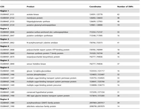
The relative levels of variation are, on the face of it, at odds with the relatedness of the strains as defined by MLST: P1/7 belongs to the same sequence type as BM407, whereas SC84 is a single locus variant of P1/7. MLST is based on variation at only seven loci in the genome however, so it should not be surprising that it does not necessarily reflect the overall diversity at the whole genome level. Analysis of the distribution and density of SNPs in the genomes suggest that recombination has generated allelic variation at certain loci in the BM407 chromosome, thereby increasing the apparent level of diversity. Eight loci in the BM407 chromosome contained a high density of SNPs and were predicted by Clonalframe to be regions that have undergone recombination (Table 4). In total the SNPs in these regions account for,60% of the SNPs in the comparison with P1/7 and SC84.
[image:11.612.64.552.64.468.2]Several of the putatively recombined loci in BM407 encode functions or proteins that potentially have a role in virulence or survival in the host. Two of the loci encode components of transport systems, that include an ABC-type glycine betaine Figure 4. Chromosomal comparisons ofS. suisstrains.Pairwise comparisons of the chromosomes ofS. suisstrains P1/7, SC84 and BM407 displayed using the Artemis Comparison Tool (ACT) [26]. The sequences have been aligned from the predicted replication origins (oriC; right). The coloured bars separating sequences (red and blue) represent similarity matches identified by BLASTN analysis [15], with a score cut off of 100. Red lines link matches in the same orientation; blue lines link matches in the reverse orientation. The green coloured boxes on the horizontal grey lines mark up the extent of genomic islands identified on the chromosomes; pink boxes mark up the extent ICE regions.
transport system protein (SSUBM407_1490) homologous (56.1% identity at the amino acid level) to a bile exclusion system protein BilEB fromListeria monocytogenesshown to have a role in virulence [103]; and a binding protein-dependent transport system (SSUBM407_1447 to SSUBM407_1449) orthologous to a system fromS. mutansresponsible for the uptake of melibiose, raffinose, and isomaltotriose and the metabolism of melibiose, sucrose, and isomaltosaccharides [104]. One of the loci is composed of three CDSs that are part of the putative exopolysaccharide biosynthesis cluster previously described. Two of the loci contain cell wall anchored surface proteins, encoding a zinc carboxypeptidase (SSUBM407_0646) and a N-acetylmuramoyl-L-alanine amidase (SSUBM407_0662). A single regulatory CDS (SSUBM407_0840) is found in the loci that appear to have undergone recombination. This CDS encodes a sensor kinase which is part of a two component regulatory system operon, with the SNPs being found in the N-terminal region of the sensor kinase protein. SSUBM407_0840 is homologous tociaHofS. pneumoniae(49.3% identity at the amino acid level) which plays a role in antibiotics resistance and transformation competence [105]. Mutations within this regulatory protein increase resistance to the cephalosporin cefotaxime.
There are fewer SNPs in the comparisons between P1/7 and SC84 than were described in the publication by Chenet al.(2007) which compared 98HAH33, 05ZYH33 and P1/7. Interestingly, a comparison of SC84 with 05ZYH33, both strains from the same outbreak, identified 2602 SNPs, 596 insertions, and 304 deletions. This is in marked contrast to the low number of SNPs identified in the comparison between P1/7 and SC84, two strains isolated from different hosts, on different continents over 20 years apart.
[image:12.612.63.559.76.460.2]Strains from the Sichuan outbreak have been found to be ST7 [8]. Examination of the sequence at MLST loci of 05ZYH33 revealed that they do not match the expected alleles for those of ST7. In comparison to SC84, which itself belongs to ST7, SNPs were identified at two of the seven MLST loci in the 05ZYH33 sequence: the aroA locus has 29 SNPs, and gki has 2 SNPs. Although, it is possible that 05ZYH33 is actually a different sequence type, albeit a double locus variant of ST7. The predicted sequence type of 98HAH33 is more divergent still, being a triple locus variant of ST7; thearoAhas 6 SNPs,cpnhas 1 single base deletion, and dpr has 1 single base deletion. The most likely explanation for these observations is that the sequence data for the 05ZYH33 and 98HAH33 strains is incorrect.
Table 4.Putative regions of recombination identified in the BM407 genome.
CDS Product Coordinates Number of SNPs
Region 1
SSUBM407_0131 acetate kinase 124591..125778 62
SSUBM407_0132 membrane protein 126092..126643 48
SSUBM407_0133 folypolyglutamate synthase 126699..127955 48
SSUBM407_0134 putative glutamyl-aminopeptidase 129061..128000 19
Region 2
SSUBM407_0646 putative surface-anchored zinc carboxypeptidase 715332..712147 33
SSUBM407_0647 putative cardiolipin synthetase 715548..717095 16
Region 3
SSUBM407_0662 N-acetylmuramoyl-L-alanine amidase 730766..733573 41
Region 4
SSUBM407_0668 polysaccharide export system ATP-binding protein 739785..740999 18
SSUBM407_0669 rhamnan synthesis protein F family protein 741020..742744 40
SSUBM407_0670 exopolysaccharide biosynthesis protein 742777..744036 16
Region 5
SSUBM407_0840 sensor histidine kinase 742777..744036 37
Region 6
SSUBM407_1445 glucan 1,6-alpha-glucosidase 1533333..1531717 16
SSUBM407_1446 sucrose phosphorylase 1534855..1533407 32
SSUBM407_1447 multiple sugar-binding transport system permease protein 1535755..1534925 33
SSUBM407_1448 multiple sugar-binding transport system permease protein 1536641..1535766 23
SSUBM407_1449 multiple sugar-binding protein precursor 1538008..1536773 16
Region 7
SSUBM407_1489 conserved hypothetical protein 1572295..1571534 31
SSUBM407_1490 ABC-type glycine betaine transport system protein 1573816..1572305 38
Region 8
SSUBM407_1989 acetyltransferase (GNAT) family protein 2097869..2097417 39
SSUBM407_1990 aldo/keto reductase family protein 2098790..2097879 14
Mobile genetic elements. Four regions of the P1/7 genome were identified that had properties indicative of recent acquisition, which include 1 prophage-like region and 3 miscellaneous islands, which have been designated genomic islands. None of these genomic islands carried characterized or putative virulence factors. Equivalent genomic islands are present in the SC84 genome. BM407 contains three of the genomic islands present in P1/7, but is lacking genomic island 3 (in strain P1/7 nucleotides 835135 to 846313; Figure 4). In contrast to some streptococcal species such asS. pyogenes[106], the proportion of the genome comprised by MGEs is relatively small, at 1.8% of the P1/7 genome, suggesting that the pan-genome of this species is smaller than other streptococci [96,107], and that there may be some limitations or restrictions to the amount of recent horizontal gene transfer in this species.
Previous sequencing of the twoS. suis strains associated with meningitis outbreaks in China in 1998 and 2005 identified highly similar 89 kb regions present in both genomes which were designated a candidate PI [7]. This region is absent in strain P1/7, but strain SC84 contains an almost identical region. This island has a composite structure, contains several regions that appear to be integrated MGEs and has a structure similar to integrative conjugative elements (ICE) found in other streptococci and other bacteria [108]. Accordingly this region has been designated ICESsuSC84.
Integrative conjugative elements. ICE are commonly found in streptococcal genomes. The most common are Tn916-type elements, which almost invariably carry the tetracycline resistance determinant tetM, often accompanied by other drug resistance genes, and Tn5252-type elements, which are larger and more variable in sequence. In many cases, Tn5252transposons are carrying Tn916elements; varying arrangements of the two are observed in different genomes, suggesting this association has arisen independently on a number of occasions. Analogous with
the bacterial chromosome as a whole, the ICE have core and accessory components to their gene complement. The conjugative machinery appears to be relatively well conserved between the ICE, with all of the streptococcal Tn5252-like elements sharing a large recombination protein, as well as VirB4-type and VirD4-type proteins. Furthermore, a genus-wide comparison of streptococcal ICE reveals a surprising level of similarity in their complement of cargo genes. The pezATaddiction toxin system, first identified in the chromosome of S. pneumoniae, is found on elements present in S. suis, S. pneumoniae and S. agalactiae, presumably aiding fixation of the transposon following integration. Bacteriocins, either alone or as part of a gene cluster encoding the accompanying processing machinery, are present on all the sequencedS. suisandS. pneumoniae[109] elements. Many of the other cargo coding sequences appear to be involved in increasing the stress tolerance of the host:abigenes are common, and the ICE of S. dysgalactiae subsp. equisimilis and S. agalactiae 2603V/R encode multiple heavy metal resistance genes. Of greater clinical importance, sequenced streptococcal ICE can carry genes for resistance to tetracycline, erythromycin, chloramphenicol, trimethoprim, aminoglycosides and streptomycin.
ICESsuSC84is integrated into the SC84S. suisgenome in the
3-prime region of the gene encoding the 50S ribosomal protein L7/ L12 (SSUSC84_0891). ICESsuSC84 has extended similarity to a
[image:13.612.66.550.58.323.2]Tn2424region (Figure 5) found inS. agalactiaestrain NEM316 [94], with three regions of difference. The first of these contains a putative bacteriocin biosynthesis cluster that is disrupted by a putative integron carrying five CDSs including an aminoglycoside resistance gene (aminoglycoside 6-adenylyltansferase; SSUSC84_0863). The second contains MGE-associated CDSs, including a Tn916 insertion that carries a tetracycline resistance gene (tetM; SSUSC84_0827). The third region contains genes associated with putative lantibiotic export/resistance and an asparagine synthetase. The only component of the 89kb island that is an obvious candidate Figure 5. Comparisons of the ICE regions fromS. suisstrains SC84 andS. agalactiaeNEM315.The results of a TBLASTX comparison of the ICESsuSC84from strain SC84 andS. agalactiaeNEM315 Tn2424is displayed using the Artemis Comparison Tool (ACT) [26].
for a pathogenicity determinant is a surface-anchored protein that contains a LPXTG motif that is annotated as an agglutinin receptor (SSUSC84_0873). This protein is found in theS. agalactiaeelement (and other streptococci) and contains a glucan-binding domain.
The BM407 genome contains two regions with extended similarity to ICESsuSC84. The first of these, ICESsuBM4071, is
inserted into a luciferase-like monooxygenase (SSUBM407_0455), resulting in the disruption of this CDS. The second, ICES-suBM4072, is found at the same locus as ICESsuSC84; the element is
inserted into the 3-prime region of the gene encoding the 50S ribosomal protein L7/L12 (SSUBM407_0933). Although ICES-suBM4072 and ICESsuSC84are inserted at the same attachment site
it is likely that they have arisen in these two strains by independent insertion events. Not only do the two elements contain regions of difference, the conserved DNA in the ICEs is more diverse than the core chromosome DNA (the ICEs exhibit,95298% identity, in contrast to ,992100% identity for chromosomal DNA). ICESsuBM4072 and ICESsuSC84have conserved tyrosine family
site-specific integrases (SSUBM407_1003 and SSUSC84_0807; Figure 6), which is probably the explanation for why these elements integrated at the same site in the respective chromo-somes, whereas ICESsuBM4071 contains a serine family
recombi-nase (SSUBM407_0524) that directs the insertion of this element elsewhere on the BM407 chromosome.
Comparison of the BM407 and SC84 ICEs shows that CDSs associated with conjugation are conserved (Figure 6). The ICEs contain regions of sequence divergence with ICESsuBM4071
appearing to be most diverse, containing a greater proportion of unique sequence, and an inverted region. All three elements contain variants of Tn916; ICESsuBM4071 and ICESsuSC84 have
Tn916with the same gene content, whilst ICESsuBM4072 contains
additional genes encoding the tetracycline resistance protein TetL (SSUBM407_0984) and chloramphenicol acetyltransferase (SSUBM407_0980). In addition to the drug resistance genes associated with Tn916, ICESsuBM4072 also carries genes encoding
the tetracycline resistance protein TetO (SSUBM407_0954), erythromycin ribosome methylase ErmB (SSUBM407_0952) and dihydrofolate reductase (SSUBM407_0957), in a region of difference located elsewhere on the element.
Unlike ICESsuBM4072, ICESsuBM4071 does not carry as many
CDSs associated with drug resistance. Amongst its cargo are a lactose utilization operon (SSUBM407_0510 to SSUBM407_0519), includ-ing a glucokinase-like protein, a galactose-6-phosphate isomerase, a tagatose-phosphate kinase, a tagatose 1,diphosphate aldolase, a 6-phospho-beta-galactosidase, and a PTS system lactose-specific transporter, which are absent in ICESsuBM4072 and ICESsuSC84.
The chromosome of BM407 (and P1/7 and SC84) already contains homologues of these CDSs at two loci (SSUBM407_0879 to SSUBM407_0889 and SSUBM407_1060). Interestingly the chro-mosomal copy of the tagatose-6-phosphate kinase lacC (SSUBM407_0883) is mutated in BM407, therefore the provision of an additional copy of this CDS on ICESsuSC84complements the
[image:14.612.60.555.61.404.2]mutation. Complete lactose utilization operons have been found on other ICEs, including theS. agalactiaeNEM315 ICE.
Figure 6. Comparisons of the ICE regions fromS. suisstrains BM407 and SC84.The results of a TBLASTX comparison of the ICESsuBM4071
and ICESsuBM4072 regions from strain BM407, and ICESsuSC84from strain SC84 is displayed using the Artemis Comparison Tool (ACT) [26].
Contained on ICESsuSC84is a two component regulator,salK/
salR (SSUSC84_0849 and SSUSC84_0850), that has been implicated in the virulence of the Chinese isolates in an animal model [110]. These regulatory components are part of a larger operon encoding lantibiotic biosynthesis, similar to the salivaricin cluster of Streptococcus salivarius [111]. Neither of the ICEs in BM407 contains this cluster (Figure 6); these elements contain alternative cargo CDSs in equivalent regions on these elements. Upstream of the operon is a CDS encoding a lantibiotic precursor (SSUSC84_0866). In SC84, and also in 05ZYH33 and 98HAH33, the operon has been disrupted by the insertion of a 7.9 kb element containing 9 CDSs, one of which encodes an aminoglycoside 6-adenylyltransferase (SSUSC84_0863), into the homologue of the S. salivariussalivaricin C biosynthesis genesalM(SSUSC84_0856). The precise regulatory function of SalK/SalR remains unclear, but their operonic association with the lantibiotic cluster suggests that their primary role is associated with this function. However it is possible that regulatory components of this operon may have been adapted to other functions.S. pyogeneswith a mutation insalY, a gene encoding a putative ABC transporter permease of a lantibiotic operon, was attenuated for virulence in a zebrafish invasive-disease model [112]. Analysis of theS. pyogeneslantibiotic cluster showed that it contained a homologue of salM that was mutated, suggesting that like the lantibiotic cluster inS. suisSC84, this cluster also no longer produced a functional lantibiotic, but it was still was essential for full virulence.
Screening of 91 VietnameseS. suisserotype 2 strains of MLST ST1 isolated from patients with meningitis, for the presence of ICESsuSC84using PCR targeted at the insertion sites [7], indicated
that this ICE is not absolutely required for virulence in humans as 16 (17.6%) strains lacked the element (manuscript in preparation). In addition, only 41 (45%) carried sequences homologous to the salK/salRgenes, suggesting that the presence of these genes cannot be used as a marker of virulence. Similar to strains BM407 and SC84, all strains containing ICE-associated sequences were resistant to tetracycline whereas those that were negative in the screening were sensitive, indicating the common presence of ICE carryingtetgenes in these isolates.
Discussion
The sequencing and detailed annotation of the S. suis P1/7, SC84 and BM407 genomes provides valuable data for studying the evolutionary events that are shaping the virulence and drug resistance in this emerging zoonotic pathogen.
There is strong evidence that genetic background influences the ability of S. suis to cause zoonotic infections as human disease isolates are almost always serotype 2 and are very closely related by MLST. Of the 121 STs in theS. suisMLST data base (http:// ssuis.mlst.net/), only five STs have representative isolated from humans, and four (ST1, ST6, ST7 and ST84) out of the five are all part of the same clonal complex. Two of the strains in this study (P1/7 and BM407) introduce a greater degree of diversity into the comparisons, insofar as that they are from different geographical areas, and belong to different STs.
A major feature of the previously sequenced S. suis strains’ genomes hypothesised to be important for pathogenicity, is an 89 kb ICE region designated as a candidate PI [7]. BM407 also contains an almost identical ICE with some notable differences that encompass drug resistance genes, and a two-component regulatory system associated with a bacteriocin cluster. Sequence variation in the BM407 ICE suggest that although it is present at an orthologous site, it has been independently acquired.
The comparative analysis suggests that ICEs have been the major contributor to the evolution of drug resistance in the strains of S. suis examined. Although it is not possible to pinpoint the origins of theS. suis ICEs, due to the limited number of these elements in the databases, it is apparent from the analyses that they are closely related to ICEs found in phylogenetically distant streptococci. It is worth noting that some of these species occupy the same niches as S. suis, suggesting that inter-species genetic exchange is taking place. This has implications for defining the pan-genomes of some streptococcal species, since it may not be possible to define their boundaries if the pool of MGEs they exchange is effectively communal.
Aside from the ICEs, the virulence determinant inventories of P1/7, SC84 and BM407 strains are identical. Small nucleotide differences resulting from point mutation and recombination were observed in the genome comparisons. This variation may have subtle effects on the expression and function of these genes, and as a corollary, generate differences in the virulence phenotypes. To this extent it is possible that some of the small nucleotide differences observed in the SC84 and BM407 genomes in comparison to P1/7 may influence the ability of these strains to cause disease. However, as the capacity of P1/7 to cause human disease is unknown, it is not possible to speculate on the impact of genomic differences identified in this study on host-association. Future studies intending to increase the depth of sequencing in this zoonotic lineage will help unravel the pathogenic significance of sequence variation.
Supporting Information
Table S1 Pseudogenes in the genome of S. suis strains P1/7, SC84 and BM407.
Found at: doi:10.1371/journal.pone.0006072.s001 (0.15 MB DOC)
Table S2 Streptococcus suis orphan CDSs in the in the genome of S. suis strain P1/7.
Found at: doi:10.1371/journal.pone.0006072.s002 (0.24 MB DOC)
Figure S1 Rhamnose-based polysaccharide cluster of strain P1/ 7 A) Structure and functional organisation of the Rhamnose-based polysaccharide cluster of strain P1/7. B) Comparison of the Rhamnose-based polysaccharide cluster from S. suis P1/7 with the RGP rml and rgp gene clusters of Streptococcus mutans UA159. The comparison of the RGP clusters from the S. suis P1/7 (top) and S. mutans UA159 and (bottom) is displayed using the Artemis Comparison Tool (ACT) [26]. The red bars separating each genome represent similarity matches identified by TBLASTX analysis [14].
Found at: doi:10.1371/journal.pone.0006072.s003 (0.35 MB PDF)
Acknowledgments
We would like to thank the Sanger Institute’s Pathogen Production Group for shotgun and finishing sequencing, and the core Informatics Group for support.
Author Contributions


![Figure 4. Chromosomal comparisons of S. suisdisplayed using the Artemis Comparison Tool (ACT) [26]](https://thumb-us.123doks.com/thumbv2/123dok_us/8686898.379171/11.612.64.552.64.468/figure-chromosomal-comparisons-suisdisplayed-using-artemis-comparison-tool.webp)

![Figure 6. Comparisons of the ICE regions from S. suisand ICE strains BM407 and SC84. The results of a TBLASTX comparison of the ICESsuBM4071SsuBM4072 regions from strain BM407, and ICESsuSC84 from strain SC84 is displayed using the Artemis Comparison Tool (ACT) [26].doi:10.1371/journal.pone.0006072.g006](https://thumb-us.123doks.com/thumbv2/123dok_us/8686898.379171/14.612.60.555.61.404/comparisons-tblastx-comparison-icessubm-icessusc-displayed-artemis-comparison.webp)
