ORIGINAL RESEARCH
ADULT BRAIN
Characteristics of Diffusional Kurtosis in Chronic Ischemia of
Adult Moyamoya Disease: Comparing Diffusional Kurtosis
and Diffusion Tensor Imaging
X K. Kazumata, XK.K. Tha, X H. Narita, XY.M. Ito, XH. Shichinohe, XM. Ito, XH. Uchino, and X T. Abumiya
ABSTRACT
BACKGROUND AND PURPOSE: Detecting microstructural changes due to chronic ischemia potentially enables early identification of patients at risk of cognitive impairment. In this study, diffusional kurtosis imaging and diffusion tensor imaging were used to investigate whether the former provides additional information regarding microstructural changes in the gray and white matter of adult patients with Moyamoya disease.
MATERIALS AND METHODS: MR imaging (diffusional kurtosis imaging and DTI) was performed in 23 adult patients with Moyamoya disease and 23 age-matched controls. Three parameters were extracted from diffusional kurtosis imaging (mean kurtosis, axial kurtosis, and radial kurtosis), and 4, from DTI (fractional anisotropy, radial diffusivity, mean diffusivity, and axial diffusivity). Voxelwise analysis for these parameters was performed in the normal-appearing brain parenchyma. The association of these parameters with neuropsychological performance was also evaluated.
RESULTS:Voxelwise analysis revealed the greatest differences in fractional anisotropy, followed, in order, by radial diffusivity, mean diffusivity, and mean kurtosis. In patients, diffusional kurtosis imaging parameters were decreased in the dorsal deep white matter such as the corona radiata and superior longitudinal fasciculus (P⬍.01), including areas without DTI abnormality. Superior longitudinal fasciculus fiber-crossing areas showed weak correlations between diffusional kurtosis imaging and DTI parameters compared with tissues with a single-fiber direction (eg, the corpus callosum). Diffusional kurtosis imaging parameters were associated with general intelligence and frontal lobe performance.
CONCLUSIONS: Although DTI revealed extensive white matter changes, diffusional kurtosis imaging additionally demonstrated micro-structural changes in ischemia-prone deep white matter with abundant fiber crossings. Thus, diffusional kurtosis imaging may be a useful adjunct for detecting subtle chronic ischemic injuries.
ABBREVIATIONS:AD⫽axial diffusivity; AK⫽axial kurtosis; CPT⫽continuous performance task; DKI⫽diffusional kurtosis imaging; FA⫽fractional anisotropy; IQ⫽intelligence quotient; MK⫽mean kurtosis; MD⫽mean diffusivity; MMD⫽Moyamoya disease; RD⫽radial diffusivity; RK⫽radial kurtosis; RST⫽Reading Span Test; SLF⫽superior longitudinal fasciculus; TMT⫽Trail-Making Test
M
oyamoya disease (MMD) is characterized by compensatory development of enlarged and weak basal perforating arter-ies (Moyamoya vessels) due to bilateral occlusive changes in the internal carotid system.1In addition to cerebral ischemia andin-tracranial hemorrhage, patients with MMD demonstrate neuro-cognitive issues, such as executive dysfunction, attention deficits,
and working-memory disturbances.2,3Brain atrophy may
ex-plain cognitive impairment in the absence of infarction, but detection of these changes has been hampered by the limited sensitivity of conventional neuroimaging methods. Diffusion tensor imaging is useful for determining white matter integrity and providing parameters sensitive to changes in axons, my-elin, and organelle structures.4,5Indeed, DTI analysis has
re-vealed a widespread decline in white matter integrity in the normal-appearing brain with MMD.3 Thus, DTI can detect Received August 26, 2015; accepted after revision January 7, 2016.
From the Departments of Neurosurgery (K.K., H.S., M.I., H.U., T.A.), Radiobiology and Medical Engineering (K.K.T.), Psychiatry (H.N.), and Biostatistics (Y.M.I.), Hok-kaido University Graduate School of Medicine, Sapporo, Japan
The authors have no personal or institutional financial interest in the drugs and imaging modalities described herein.
This study was supported by a grant from the Research Committee on Moyamoya Disease, sponsored by the Ministry of Health, Labor, and Welfare of Japan and the Creation of Innovation Centers for Advanced Interdisciplinary Research Areas Pro-grams, Ministry of Education, Culture, Sports, Science, and Technology, Japan.
Please address correspondence to Ken Kazumata, MD, Department of Neurosur-gery, Hokkaido University Graduate School of Medicine, North 15 West 7, Kita, Sapporo 060-8638, Japan; e-mail: [email protected]
Indicates open access to non-subscribers at www.ajnr.org
Indicates article with supplemental on-line appendix and table.
early-stage ischemic injury, which potentially predicts future cognitive outcomes. However, DTI is constrained by technical insufficiencies: It is based on the assumption that water mole-cules diffuse freely and that diffusion can be characterized by a Gaussian distribution.5
In addition, the tensor model is based on the observation that in many tissues, water diffusion is anisotropic (ie, the diffusion is more liberal in some directions and more restricted in others). This anisotropic diffusion can be geometrically de-picted as an ellipsoid, described by eigenvectors and eigenval-ues. This model performs well in regions where fibers are aligned along a single axis. However, it fails in regions with several fiber populations aligned along intersecting axes be-cause it cannot simultaneously map several diffusion maxima.6
Furthermore, because hypoxic-ischemic injury induces neuro-degeneration and regression of dendrite arborization in gray matter, the diffusion properties of gray matter may also reveal the early stages of ischemic injury.7,8Nevertheless, analyzing
isotropic or near-isotropic tissue such as gray matter by DTI may not be valid because its major parameter, fractional an-isotropy (FA), reflects structure only if it is spatially oriented.6
A more recent method called diffusional kurtosis imaging (DKI) quantifies the deviation of water molecule diffusion from the Gaussian distribution without assuming any specific diffusion model.6,9Its parameters are thought to represent the
complexity of tissue microstructure.6 Previous studies have
suggested that DKI is sufficiently sensitive to detect age-related alterations in white matter microstructure.10,11Furthermore,
measurements of diffusion anisotropy by DKI can reveal sex-related and pathologic changes in gray matter.12,13Thus, using
DKI to evaluate the diffusion properties of gray matter and white matter in patients with MMD may be useful for detecting subtle microstructural changes due to ischemia.
Diffusional kurtosis has been investigated to explore tissue reversibility in acute cerebral infarction.14-16However, there
is a paucity of information regarding the microstructural properties measured by DKI in chronic ischemia in living hu-mans. To expand on our prior DTI study, we investigated whether adults with MMD and no overt cerebral infarctions have altered diffusional kurtosis in the entire cerebrum. An exploratory voxel-based whole-brain analysis was performed to map regional DKI parameters and to compare DKI and DTI parameters. We also explored correlations of diffusion param-eters with measures of neurocognitive impairment in an ROI analysis.
MATERIALS AND METHODS
Participants
This prospective study was approved by the Research Ethics Committee of Hok-kaido University Hospital, and written informed consent was obtained from all participants. Participants in the present study are the same as those of our previ-ous study analyzing the relationship be-tween DTI parameters and neuropsy-chological test scores.3 The selection
period was 25 months (April 2012 through April 2014). Twenty-three patients (6 men and 17 women; 21–58 years of age; mean age, 40.9⫾9.5 years) were enrolled. The control group also con-sisted of 23 subjects (10 men and 13 women; 25–56 years of age; mean age, 39.0⫾8.1 years). A brief summary of patient charac-teristics is provided inTable 1.
Neuropsychological Assessment
Neuropsychological examinations consisted of the Wechsler Adult Intelligent Scale-III, Wisconsin Card Sorting Test, Trail-Making Test (TMT; parts A and B), continuous performance task (CPT), Stroop test, and Reading Span Test (RST). The details of neuropsychological examinations and the results are provided in the On-line Appendix.3
MR Image Acquisition
MR imaging was performed with a 3T scanner (Achieva TX; Philips Healthcare, Best, the Netherlands). 3D magnetization-prepared rapid acquistion of gradient echo T1-weighted imaging and axial single-shot spin-echo echo-planar DKI were acquired to evaluate subtle gray and white matter alterations, respectively. The scan parameters for DKI were as follows: TR⫽5051 ms, TE⫽85 ms, flip angle⫽90°, FOV⫽224⫻224 mm2, matrix
size⫽128⫻128, b-values⫽0, 1000, and 2000 s/mm2, number of
diffusion gradient directions⫽32, section thickness⫽3 mm, intersection gap⫽0 mm, number of sections⫽43, and NEX⫽1. The 3D-MPRAGE imaging was performed with TR⫽6.8 ms, TE⫽3.1 ms, flip angle⫽8 °, and TI⫽1100 ms.
Image Processing
Registration between the echo-planar images with no diffusion weighting (b⫽0 s/mm2) and the corresponding DKI data and
correction for eddy current distortion were performed at the MR imaging operator console. The DKI data were processed by using Matlab R2012b (MathWorks, Natick, Massachusetts) and Diffu-sional Kurtosis Estimator (Version 2.5.1; http://nitrc.org/projects/ dke).17Seven DKI and DTI parameters were extracted from the
Diffusional Kurtosis Estimator: mean kurtosis (MK), radial kur-tosis (RK), axial kurkur-tosis (AK), FA, mean diffusivity (MD), radial diffusivity (RD), and axial diffusivity (AD). The DTI parameters (FA, MD, RD, and AD) were calculated from a portion of the DKI data by using a monoexponential model that assumes a Gaussian probability diffusion function by using data from b-values of 0 and 1000 s/mm2.17
[image:2.594.55.380.58.141.2]Following calculation of DKI and DTI parameters, theb⫽0 echo-planar images were warped to the standardized T2 template
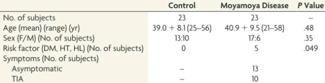
Table 1: Characteristics of the study participants
Control Moyamoya Disease PValue
No. of subjects 23 23 –
Age (mean) (range) (yr) 39.0⫹8.1 (25–56) 40.9⫹9.5 (21–58) .48
Sex (F/M) (No. of subjects) 13:10 17:6 .35
Risk factor (DM, HT, HL) (No. of subjects) 0 5 .049
Symptoms (No. of subjects)
Asymptomatic – 13
TIA – 10
of SPM8 (http://www.fil.ion.ucl.ac.uk/spm/software/spm12). This transformation matrix was applied to the DKI/DTI parame-ter map of each patient. The warped DKI/DTI maps were aver-aged and smoothed with a 6-mm full width at half maximum Gaussian kernel to form customized DKI/DTI templates. Native DKI/DTI maps of all patients and control subjects were then warped to the customized, respective DKI/DTI templates. Indi-vidual maps were then smoothed with a 6-mm full width at half-maximum Gaussian kernel. The warped and smoothed DKI/DTI maps were used for group comparisons between the controls and patients with MMD. To investigate the pathology underlying DKI parameters and the influence of fiber crossings on DTI parame-ters, we investigated the correlations of DKI and DTI parameters in white matter tracts consisting of either a single fiber direction or crossing fibers (ie, multiple directions).18 The ROIs were
placed on the genu of the corpus callosum (ie, a structure with a single fiber direction) and deep white matter tracts corresponding to the bilateral superior longitudinal fasciculus (SLF) (multiple fiber directions) by using the JHU white matter atlas available in FSL (http://fsl.fmrib.ox.ac.uk/fsl/fslwiki/).19
Data Analysis
Whole-brain diffusion parameters were compared voxel by voxel between the controls and patients with MMD by using SPM8, which implemented the general linear model. We used the 2-sam-plettest model, and age was considered a covariate. An explicit mask generated by averaging the normalized CSF space of all par-ticipants was applied. To explore group differences across both gray matter and white matter, we set statistical significance atP⬍
.01 without correction for family-wise error and clusters of 50 voxels or more. The number of voxels demonstrating a significant difference between the controls and patients with MMD was ex-tracted. The Pearson product-moment correlation coefficient was calculated to investigate the correlation between the DKI and DTI parameters by using DKI/DTI values extracted from the ROI analysis.
We also investigated whether the significant changes in DKI/ DTI parameters were associated with neuropsychological exami-nation scores. A thresholdTvalue of 2.42, corresponding toP⬍
.01 without correction for family-wise error, was applied to theT
contrast map of DKI/DTI parameters obtained by comparing controls and patients, and binary mask images containing voxels above the threshold value were generated. The DKI/DTI param-eter values included in the masks were extracted from DKI/DTI of the patients with MMD. The Pearson product-moment correla-tion coefficients were used for analyses involving the Wechsler Adult Intelligent Scale-III, TMT (parts A and B and the difference in score between TMT-A and TMT-B [B–A]). Patients were fur-ther subgrouped into 2, according to their performance scores, error numbers, and reaction time on the Wisconsin Card Sorting Test, Stroop test, CPT, and RST; and the DKI/DTI parameters were compared between these 2 subgroups by usingttests (On-line Appendix). For all correlations and comparisons, aPvalue⬍ .05 was considered statistically significant to explore the possible relationship between neuropsychological scores and diffusion parameters.
RESULTS
Spatial Distribution of DTI/DKI Differences on Voxel-Based Analysis
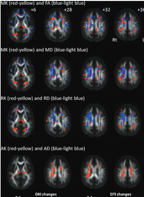
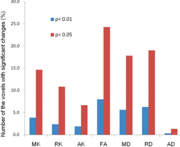
Areas with decreased MK included the right frontal white matter, bilateral thalami, portions of the occipital white matter, corona radiata, corpus callosum, and portions of frontal and parietal white matter corresponding to the posterior segment of the supe-rior longitudinal fasciculus (Fig 1). The decrease in FA was most extensive within the white matter (161,625 voxels), with its de-crease amounting to 12,833 voxels (7.9%) (Fig 2). Significant MK, RK, and AK decreases were observed in 6180 voxels (3.8%), 3828 voxels (2.4%), and 3043 voxels (1.9%), respectively. In contrast, 9028 voxels (5.6%) showed a significant increase in MD; 10,062 (6.2%), in RD; and 548 (0.3%), in AD compared with controls. Figure 1shows DKI/DTI overlap maps, with MK/RK/AK/FA de-creases and MD/RD inde-creases. Areas with FA decrease were more extensive than those with MK decrease; however, the posterior segment of the SLF showed a decrease in only MK/RK. The in-crease in MD/RD was remarkable in the corona radiata; however, the posterior segment of the SLF showed a decrease in MK/RK without an increase in MD/RD. Decreased AK was observed with-out changes in AD for the bilateral thalami, corona radiata, and portions of the temporo-occipital white matter. There was no cortical gray matter with altered DKI/DTI parameters in patients.
Correlation of DTI and DKI Parameters
Correlations between DKI and DTI parameters were examined in both the genu of the corpus callosum and the bilateral SLF (Table 2). In the corpus callosum of both controls and patients with MMD, MK correlated positively with FA (r⬎0.78,P⬍.01), while inverse correlations with MK were found for MD/RD/AD (r⬍ ⫺0.82,P⬍.001). In the corpus callosum, RK correlated inversely with RD (r⬍ ⫺0.84,P⬍.001), and AK inversely cor-related with AD of the corpus callosum (r⬍ ⫺0.64,P⬍.001). A significant correlation between MK and FA was observed in the left SLF of controls, but this was to a lesser degree compared with that in the corpus callosum. No correlation was found between MK and FA in the right SLF of controls. In both controls and patients, correlations between MK and MD/RD/AD were found in the right and left SLFs, albeit to a lesser degree than corpus callosum correlations. The results of correlation between DKI and DTI parameter values are summarized inTable 2.
Association of DKI/DTI Parameters with Neuropsychological Performance Tests
DISCUSSION
We explored the added benefit of diffusional kurtosis measure-ments in the assessment of ischemic burden due to chronic isch-emia in adult MMD. This study confirmed our prior finding that chronic ischemia in MMD preferentially affects the microstruc-ture of normal-appearing white matter.3Detecting
microstruc-tural changes due to chronic ischemia potentially enables early identification of patients at risk of cognitive impairment.3,20
However, detecting microstructural changes has been hampered by the limited sensitivity of DTI in fiber-crossing areas.7In this
study, DKI demonstrated microstructural changes, predomi-nantly in the dorsal part of the deep white matter, where conven-tional MR imaging frequently demonstrates ischemic lesions.21
The significant changes in DKI parameters were associated with neuropsychological scores reflecting general intelligence, execu-tive function, attention, and working memory. Thus, DKI is con-sidered a useful adjunct to conventional DTI, particularly for its ability to detect nascent microstructural changes in areas with abundant fiber crossings.
In this study, we demonstrated that DTI showed widespread regions with alterations in FA, MD, and RD. The finding is con-sistent with previous studies, in which DTI showed more exten-sive white matter changes compared with DKI parameters. Other studies have demonstrated superior sensitivity of DKI to detect white matter changes compared with DTI.11,22,23 DKI showed
significant decreases in frontoparietal subcortical structures and deep white matter. A decrease in DKI parameters, a shift of diffu-sional kurtosis toward free water diffusion, has commonly been interpreted as a reduction in tissue complexity.24Considering the
location of the DKI alterations, a reduction in complexity is thought to reflect microstructural changes in myelin and/or ax-onal attenuation. In the corona radiata, DTI (MD/RD) demon-strated more significant changes compared with DKI (MK/RK). Deep white matter tracts are vulnerable to chronic ischemia be-cause they are located in the terminal field of the blood supply. Myelin degeneration or increased periventricular extracellular fluid may increase RD, while tissue complexity in a radial direc-tion may be maintained, in some part, by glial proliferadirec-tion.25In a
previous study, a trend toward decreased AD was found via anal-ysis of tract-specific spatial statistics,3whereas the voxel-based
analysis of the present study did not show definitive AD changes in white matter. AD decreases in axonal fragmentation; however, axonal degeneration or reduced axonal attenuation can increase AD. In contrast with AD, AK showed significant changes in the
[image:4.594.53.286.47.365.2]FIG 1. Changes in diffusional kurtosis imaging and diffusion tensor imaging parameters in Moyamoya disease shown in maps of 3 diffu-sional kurtosis parameters (mean kurtosis, radial kurtosis, and axial kurtosis) and 4 diffusion tensor parameters (fractional anisotropy, mean diffusivity, radial diffusivity, and axial diffusivity). Areas with significant changes in a combination of DKI/DTI parameters are as follows: decreased MK (red-yellow)/decreased FA (blue-light blue), decreased MK (red-yellow)/increased MD (blue-light blue), decreased RK (red-yellow)/increased RD (blue-light blue), and decreased AK (red-yellow)/increased and decreased AD (blue-light blue). Values from statistical parametric mapping analysis are projected onto axial sections of the average brain space of FA (z⫽12, 28, 32, 36 mm). MK decrease is observed in the thalamus, a portion of the genu and body of the corpus callosum, corona radiata, frontoparietal subcortical white matter, and superior longitudinal fasciculus. RK decrease is ob-served in part of the frontoparietal subcortical white matter, thala-mus, corona radiata, and occipital white matter. AK decrease is ob-served in the thalamus, temporo-occipital white matter, part of the SLF, and corona radiata. The radiologic convention is adopted, with the left side of the brain on the right side of axial panels. The color scale representsTvalues, with colored regions exceeding the signifi-cance threshold ofP⬍.01 (T⫽2.42) with a minimum cluster size of 50 voxels. Rt indicates right; Lt, left.
Table 2: Correlation between DKI and DTI parametersa
Corpus Callosum Rt. SLF Lt. SLF
CNT MMD CNT MMD CNT MMD
r P r P r P r P r P r P
MK vs FA 0.78 .000 0.80 .000 0.08 .715 0.60 .002 0.42 .047 0.47 .022
MK vs MD ⫺0.90 .000 ⫺0.86 .000 ⫺0.60 .002 ⫺0.68 .000 ⫺0.41 .049 ⫺0.49 .017
MK vs RD ⫺0.90 .000 ⫺0.87 .000 ⫺0.54 .008 ⫺0.73 .000 ⫺0.49 .018 ⫺0.55 .006
MK vs AD ⫺0.87 .000 ⫺0.82 .000 ⫺0.50 .014 ⫺0.46 .028 ⫺0.19 .386 ⫺0.35 .101
RK and RD ⫺0.88 .000 ⫺0.84 .000 ⫺0.72 .000 ⫺0.56 .005 ⫺0.47 .025 ⫺0.38 .072
AK and AD ⫺0.75 .000 ⫺0.64 .000 ⫺0.44 .036 ⫺0.61 .001 ⫺0.47 .025 ⫺0.68 .000
Note:—Rt. indicates right; Lt., left; CNT, controls;r; Pearson product-moment correlation coefficient.
[image:4.594.54.534.610.715.2]corona radiata in the voxel-based analysis. Thus, alterations in AD would be marginal at best in patients, while tissue complexity parallel to the principal diffusion direction could be decreased in the corona radiata of patients. Weak correlations between FA and MK in the SLF of controls could be attributed to the crossing/ kissing of white matter fibers. The SLF contains abundant cross-ing fibers projectcross-ing from the corona radiata and corpus callo-sum.18Several fiber populations aligned along intersecting axes in
1 voxel would diminish anisotropy.26
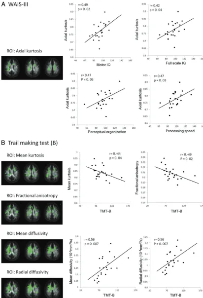
The DKI parameters in patients showed moderate positive correlations with impaired general intelligence and frontal lobe dysfunction. These results are consistent with previous reports showing a relationship between cognitive performance and white matter fiber tracts integrating parietofrontal cortical areas.27-29In
MMD, performance IQ is preferentially affected compared with verbal IQ.1In the present study, AK was significantly correlated
with neuropsychological performance on tests evaluating execu-tive function and working memory (motor IQ, perceptual orga-nization, and processing speed), while no correlation was found with verbal IQ. In an experimental animal model of chronic white matter ischemia, damage to myelin preceded axonal damage, sug-gesting that the change in myelin is the primary pathologic event.30Our observation of a stronger correlation between AK,
rather than AD, and general intellectual ability measured by the Wechsler Adult Intelligent Scale-III may imply that a reduction in axonal density and/or axonal degeneration, a more advanced stage of chronic ischemic injury, is better described by AK than AD. Previous investigations by using DTI and probabilistic trac-tography have shown correlations between neuropsychological examinations and the FA of subcortical white matter. Consistent with previous studies, we observed correlations between DTI pa-rameters and the scores of neuropsychological tests evaluating
frontal lobe function in the present study. A significant correlation of FA/ MD/RD with scores on the Trail-Mak-ing Test suggests that demyelination alone could affect part of the frontal lobe functions.
Transient ischemic attack followed by hyperventilation is a characteristic symptom of childhood MMD, and syn-cope attacks are occasionally observed in both children and adults with MMD.1
The spatial characteristics of nascent brain injury are important for under-standing frontal-dominant neurocogni-tive dysfunction in adult MMD. Pyrami-dal neocortical neurons (layers 3, 5, and 6) in the prefrontal cortex are known to be highly vulnerable to hypoxic-isch-emic insults.31,32 These
hypoxic-isch-emic insults can damage the brain and potentially induce neuronal death or re-gression of dendritic structure.8 We
speculate that microstructural alteration precedes gross volumetric reductions in gray matter. Previous study has revealed gray matter atrophy in the posterior cin-gulate cortex, suggesting that more widespread gray matter changes might be observed in diffusion parameters, particularly in the frontal lobe.3 DKI could reveal early microstructural
changes less constrained by partial volume effects.33Nevertheless,
despite discrete white matter damage, no substantial changes in diffusion parameters were found in the cortical gray matter in MMD. Pathologic tissue changes such as glial proliferation may underlie the lack of significant changes in the diffusion parame-ters of the cortices. A new diffusion MR imaging technique, in-cluding neurite orientation dispersion and density imaging, may detect subtle microstructural changes in the cortex and is poten-tially sensitive to initial ischemic changes before the overt vol-umetric reductions.34,35
The present study revealed microstructural change in the thal-amus, a finding that has never been emphasized with regard to cognitive function in MMD. The mediodorsal thalamus connects to the prefrontal cortex. This is potentially important because innervation of the thalamoprefrontal circuit could modulate pre-frontal neural circuits, which are associated with cognitive as well as affective performance.36
There are several limitations to this study. The statistical power to detect group differences in DKI parameters is signifi-cantly influenced by the number of subjects in a study.37
There-fore, the spatial characteristics of the DKI/DTI alterations found in this study may not represent the topography of ischemic bur-den in adult MMD.37We extracted DKI/DTI values from the
contrast T maps generated from group comparisons between controls and patients to explore the relationship between abnor-mal diffusion parameters in patients and neuropsychological test performance. Although we found an association with neuropsy-chological test scores, voxel-based correlation analysis would
[image:5.594.71.360.49.286.2]mit an objective evaluation of the neural substrates associated with cognitive impairment. Finally, we used a combination of b-values of 0 and 1000 s/mm2for DTI. DTI parameters are
re-ported as dependent on b-values. For DKI, the b-values of 0, 1000, and 2000 s/mm2used in the present study would be feasible for
practical clinical applications. Nevertheless, a different combina-tion of b-values and methods for parameter estimacombina-tion could alter the relationship between DKI and DTI with regard to sensitivity and specificity, which requires further investigation.21
CONCLUSIONS
The results of the present study suggest an additional value of DKI as an adjunct to DTI. DKI parameters can become useful neuro-imaging markers to track ischemic burden in adult MMD.
Disclosures: Ken Kazumata—RELATED:Grant: This study was supported by a grant from the Research Committee on Moyamoya Disease, sponsored by the Ministry of Health, Labor, and Welfare of Japan.* Khin K. Tha—RELATED:Grant: This work was supported (in part) by the Creation of Innovation Centers for Advanced Interdisci-plinary Research Areas Program, Ministry of Education, Culture, Sports, Science, and Technology, Japan.* Masaki Ito—RELATED:Grant: Research Committee on Moya-moya Disease, sponsored by the Ministry of Health, Labor, and Welfare of Japan*;
UNRELATED:Grants/Grants Pending: Masaki Ito was funded by a fellowship (grant) ¥2,000,000 J (approximately US $18,000) for 1 year from SENSHIN Medical Research Foundation donated by the Mitsubishi Corporation, Japan. Funding began April 1, 2015. Takeo Abumiya—RELATED:Grant: grant from the Research Committee on Moyamoya Disease, sponsored by the Ministry of Health, Labor, and Welfare of Japan.* *Money paid to the institution.
REFERENCES
1. Kuroda S, Houkin K.Moyamoya disease: current concepts and fu-ture perspectives.Lancet Neurol2008;7:1056 – 66CrossRef Medline 2. Karzmark P, Zeifert PD, Bell-Stephens TE, et al.Neurocognitive im-pairment in adults with Moyamoya disease without stroke. Neuro-surgery2012;70:634 –38CrossRef Medline
3. Kazumata K, Tha KK, Narita H, et al.Chronic ischemia alters brain microstructural integrity and cognitive performance in adult Moyamoya disease.Stroke2015;46:354 – 60CrossRef Medline 4. Tha KK, Terae S, Nakagawa S, et al.Impaired integrity of the brain
parenchyma in non-geriatric patients with major depressive disor-der revealed by diffusion tensor imaging.Psychiatry Res2013;212: 208 –15CrossRef Medline
5. Basser PJ, Mattiello J, LeBihan D.MR diffusion tensor spectroscopy and imaging.Biophys J1994;66:259 – 67Medline
6. Jensen JH, Helpern JA, Ramani A, et al. Diffusional kurtosis imaging: the quantification of non-gaussian water diffusion by means of magnetic resonance imaging.Magnetic Reson Med2005; 53:1432– 40Medline
7. Back SA.Cerebral white and gray matter injury in newborns: new insights into pathophysiology and management.Clin Perinatal
2014;41:1–24CrossRef Medline
8. Zhang S, Boyd J, Delaney K, et al.Rapid reversible changes in den-dritic spine structure in vivo gated by the degree of ischemia.J Neu-rosci2005;25:5333–38Medline
9. Fieremans E, Jensen JH, Helpern JA.White matter characterization with diffusional kurtosis imaging. Neuroimage 2011;58:177– 88 CrossRef Medline
10. Li X, Gao J, Hou X, et al.Diffusion kurtosis imaging with tract-based spatial statistics reveals white matter alterations in preschool children. Conf Proc IEEE Eng Med Biol Soc2012;2012:2298 –301 CrossRef Medline
11. Coutu JP, Chen JJ, Rosas HD, et al.Non-Gaussian water diffusion in aging white matter. Neurobiol Aging 2014;35:1412–21 CrossRef Medline
12. Gong NJ, Wong CS, Chan CC, et al.Aging in deep gray matter and white matter revealed by diffusional kurtosis imaging.Neurobiol Aging2014;35:2203–16CrossRef Medline
13. Caverzasi E, Henry RG, Vitali P, et al.Application of quantitative DTI metrics in sporadic CJD. Neuroimage Clin 2014;4:426 –35 CrossRef Medline
14. Weber RA, Hui ES, Jensen JH, et al.Diffusional kurtosis and diffusion tensor imaging reveal different time-sensitive stroke-induced microstructural changes. Stroke 2015;46:545–50 CrossRef Medline
15. Grinberg F, Farrher E, Ciobanu L, et al.Non-Gaussian diffusion imaging for enhanced contrast of brain tissue affected by ischemic stroke.PLoS One2014;9:e89225CrossRef Medline
16. Umesh Rudrapatna S, Wieloch T, Beirup K, et al.Can diffusion kurtosis imaging improve the sensitivity and specificity of de-tecting microstructural alterations in brain tissue chronically after experimental stroke? Comparisons with diffusion tensor imaging and histology. Neuroimage 2014;97:363–73 CrossRef Medline
17. Tabesh A, Jensen JH, Ardekani BA, et al.Estimation of tensors and tensor-derived measures in diffusional kurtosis imaging. Magn Reson Med2011;65:823–36CrossRef Medline
18. Reijmer YD, Leemans A, Heringa SM, et al; Vascular Cognitive Impairment Study group.Improved sensitivity to cerebral white matter abnormalities in Alzheimer’s disease with spherical de-convolution based tractography. PLoS One 2012;7:e44074 CrossRef Medline
19. Wakana S, Jiang H, Nagae-Poetscher LM, et al.Fiber tract-based atlas of human white matter anatomy.Radiology2004;230:77– 87 Medline
20. Cheng HL, Lin CJ, Soong BW, et al.Impairments in cognitive func-tion and brain connectivity in severe asymptomatic carotid steno-sis.Stroke2012;43:2567–73Medline
21. Schmidt R, Seiler S, Loitfelder M.Longitudinal change of small-ves-sel disease-related brain abnormalities.J Cereb Blood Flow Metab
2015 Apr 22. [Epub ahead of print]CrossRef Medline
22. Kamagata K, Tomiyama H, Hatano T, et al.A preliminary diffu-sional kurtosis imaging study of Parkinson disease: comparison with conventional diffusion tensor imaging.Neuroradiology2014; 56:251–58CrossRef Medline
23. Fieremans E, Benitez A, Jensen JH, et al.Novel white matter tract integrity metrics sensitive to Alzheimer disease progression.AJNR Am J Neuroradiol2013;34:2105–12CrossRef Medline
24. Steven AJ, Zhuo J, Melhem ER.Diffusion kurtosis imaging: an emerging technique for evaluating the microstructural environ-ment of the brain.AJR Am J Roentgenol2014;202:W26 –33CrossRef Medline
[image:7.594.54.286.64.150.2]25. Cechetti F, Pagnussat AS, Worm PV, et al.Chronic brain hypoper-fusion causes early glial activation and neuronal death, and subse-quent long-term memory impairment. Brain Res Bull 2012;87: 109 –16CrossRef Medline
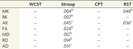
Table 3: Correlations of diffusion parameters with neuropsychological examinationsa
WCST Stroop CPT RST
MK – .004b – .048b
RK – .007b – –
AK – .045c – .036c
FA – .024b – –
MD – .012b – –
RD – .014b – –
AD – .031c – –
Note:—WCST indicates Wisconsin Card Sorting Test, Stroop; Stroop test.
a
Data arePvalues. Correlations of diffusional kurtosis imaging and diffusion tensor imaging parameters with neuropsychological examinations were evaluated in pa-tients with MMD. Three diffusional kurtosis parameters (MK, RK, and AK) and 4 diffu-sion tensor parameters (FA, MD, RD, and AD) were analyzed. Diffudiffu-sion parameters and locations that demonstrated significant relationships with clinical variables follow.
b
Two-tailedttest.
c
26. Jbabdi S, Behrens TE, Smith SM.Crossing fibres in tract-based spa-tial statistics.Neuroimage2010;49:249 –56CrossRef Medline 27. Gla¨scher J, Rudrauf D, Colom R, et al.Distributed neural system for
general intelligence revealed by lesion mapping.Proc Natl Acad Sci U S A2010;107:4705– 09CrossRef Medline
28. Deary IJ, Weiss A, Batty GD.Intelligence and personality as predic-tors of illness and death: how researchers in differential psychology and chronic disease epidemiology are collaborating to understand and address health inequalities.Psychol Sci Public Interest2010;11: 53–79CrossRef Medline
29. Barbey AK, Colom R, Solomon J, et al.An integrative architecture for general intelligence and executive function revealed by lesion mapping.Brain2012;135:1154 – 64CrossRef Medline
30. Kurumatani T, Kudo T, Ikura Y, et al.White matter changes in the gerbil brain under chronic cerebral hypoperfusion.Stroke1998;29: 1058 – 62Medline
31. Lin CS, Polsky K, Nadler JV, et al.Selective neocortical and thalamic cell death in the gerbil after transient ischemia.Neuroscience1990; 35:289 –99Medline
32. Fukuda A, Muramatsu K, Okabe A, et al.NMDA receptor-mediated differential laminar susceptibility to the intracellular Ca2ⴙ
accu-mulation induced by oxygen-glucose deprivation in rat neocortical slices.J Neurophysiol1998;79:430 –38Medline
33. Yang AW, Jensen JH, Hu CC, et al.Effect of cerebral spinal fluid suppression for diffusional kurtosis imaging.J Magn Reson Imaging
2013;37:365–71CrossRef Medline
34. Jelescu IO, Veraart J, Adisetiyo V, et al.One diffusion acquisition and different white matter models: how does microstructure change in human early development based on WMTI and NODDI?
Neuroimage2015;107:242–56CrossRef Medline
35. Zhang H, Schneider T, Wheeler-Kingshott CA, et al. NODDI: practical in vivo neurite orientation dispersion and density im-aging of the human brain.Neuroimage2012;61:1000 –16CrossRef Medline
36. Ferguson BR, Gao WJ.Development of thalamocortical connec-tions between the mediodorsal thalamus and the prefrontal cortex and its implication in cognition.Front Hum Neurosci2014;8:1027 Medline