Evaluation of oxidative and nitrosative stress in
relapsing remitting multiple sclerosis
Štefan Lukáč1*, Terézia Kalnovičová2, Jana Muchová1
1Institute of Medical Chemistry, Biochemistry and Clinical Biochemistry, Faculty of Medicine, Comenius University, Bratislava, Slovakia; *Corresponding Author:
2Department of Neurology, Faculty of Medicine, Comenius University, Bratislava, Slovakia
Received 17 September 2013; revised 28 October 2013; accepted 5 November 2013
Copyright © 2013 Štefan Lukáč et al. This is an open access article distributed under the Creative Commons Attribution License,
which permits unrestricted use, distribution, and reproduction in any medium, provided the original work is properly cited.
ABSTRACT
Multiple sclerosis (MS) is a chronic autoimmune disorder affecting the central nervous system (CNS) through demyelination and neurodegen- eration. Several lines of evidence support a role for oxidative and nitrative stress (OS and NS) in pathogenesis of multiple sclerosis. The mecha- nism of influence of OS and NS on blood-brain- barrier (BBB) has critical importance for evalu- ating antioxidant therapies. As far as we know, markers of oxidative and nitrative stress in MS patients have been investigated independently for their relationship with the state of the blood- brain-barrier. Blood plasma samples of 58 pa- tients with relapse-remitting MS (RRMS) with normal (Group A, n = 48, 36.2 ± 10.5 years) and damaged BBB (Group B, n = 10, 38.2 ± 11.2 years) and of 44 healthy controls (39.2 ± 14.9 years) were analyzed. TAS (total antioxidant plasma status), lipoperoxides, protein carbonyls, 3-ni- trotyrosine and uric acid were evaluated in each group. Our results confirmed decreased TAS (Group A: 1.35 ± 0.55 mmol/l, P < 0.001; Group B: 1.73 ± 0.37 mmol/l vs. 1.9 ± 0.7 mmol/l) and in-creased lipoperoxidation (A: 71.5 ± 42.18 nmol/ml, P < 0.01; B: 127.02 ± 74.67 nmol/ml, P < 0.001 vs. 46.6 ± 27.4 nmol/ml) in RRSM patients. The level of lipoperoxidation positively correlated with the state of BBB (P < 0.05). Elevated concen- trations of protein’s carbonyls (A: 0.48 ± 0.11 nmol/mg protein, P < 0.001; B: 0.43 ± 0.14 nmol/mg protein, P < 0.05 vs. 0.31 ± 0.01 nmol/ mg protein) and 3-nitrotyrosine (A: 107.3 ± 40.7 nmol/l, P < 0.001; B: 89.2 ± 15.9 nmol/l, P < 0.001 vs. 21.6 ± 3.7 nmol/l) indicated free-radical mediated dam- age to plasma proteins, what was confirmed by their positive mutual correlation (P < 0.001).
The level of uric acid was physiological and correlated negatively with protein’s carbonyls (P < 0.05) while there was no significant relationship with 3-nitotyrosine. The results suggest the role of this antioxidant in the protection of the pro- teins against OS what was confirmed by its positive correlation with TAS (P < 0.05). It could be concluded that ROS/RNS in MS patients af- fect a wide range of substances, change their properties and function. High concentration of lipoperoxides indicates a role of lipid peroxida- tion in deterioration of BBB. Considering the body complexity, extremely high levels of dam- aged proteins in blood plasma and abnormal state of BBB should lead us to assumption of changed proteins in CNS that can activate im- mune system and result into autoimmune re- sponse. Therefore, it is necessary to pay atten- tion to ROS/RNS reduction in therapeutic proc- ess to reduce damage to BBB and other adverse effects.
Keywords:Relapsing Remiting Multiple Sclerosis; Oxidative Stress; Nitrative Stress;
Blood-Brain-Barrier; Total Antioxidant Status; 3-Nitrotyrosine; Protein Carbonyls;
Lipoperoxides; Uric Acid
1. INTRODUCTION
(OS) in pathogenesis of demyelination and inflammation [2,3]. In the field of OS and neurodegeneration, perox- initrite is one of the most important free radicals. It has been accepted that the molecules of peroxynitrite are the final molecules responsible for pathological processes in neurodegenerative diseases and MS [3]. Reactive oxygen and nitrogen species are produced by microglia that is activated by T-lymphocytes, especially TH-17 cells in the
reactions of immune response to antigen. Radicals attack different molecules, cause their damage leading to the loss of their function with negative effects on the neural tissue what could enhance progress of the disease. One of the undesired impacts is the deterioration of the blood- brain barrier (BBB) that creates unphysiological state between the CNS and the rest of the body leading to hor- rible consequences. Therefore, it is necessary to evaluate pro- and antioxidative status in these patients due to BBB state.
2. AIM
The aim of our study was to detect total antioxidant status of plasma (TAS) in patients with MS, to analyze various markers of oxidative damage, their mutual cor- relations and correlations with the state of the blood- brain barrier. We also monitored the level of natural an- tioxidant-uric acid (UA), which concentration in plasma of MS-patients is currently leading discussions.
3. MATERIALS AND METHODS
Blood plasma samples from 58 patients with diag- nosed multiple sclerosis according to McDonald’s rules were analyzed. The tested group was divided into group with normal and pathological state of blood-brain barrier according to QA-index. Group A with normal state of BBB consisted of 29 females and 19 males with average age 36.2 ± 10.5 years. Group B with damaged BBB in- cluded 7 females and 3 males with average age 38.2 ± 11.2 years. Every patient had relapsing-remitting form and was out of relapse. According to our knowledge, pa- tients did not suffer from any other disease that would affect markers of oxidative/nitrative stress. Control group consisted of 44 healthy people with average age 39.2 ± 14.8 years (24 males and 19 females). Every participant signed informed consent and agreed with the investiga- tion of mentioned parameters. Statement of ethical com- mittee was not necessary since examination of parame- ters was indicated by the neurologist and was a compo- nent of diagnostic process. None of them had demyeli- nating disease or other diseases associated with an in- crease in oxidative stress. State of BBB of MS-patients was evaluated via QA-index, as ratio between albumin concentration in cerebrospinal fluid and in blood plasma multiplied by 1000; QA index higher than 7.4 indicates
blood-brain barrier deterioration. TAS was analyzed by TEAC method according to Re, et al. [4]. This method
measures the presence of hydrophobic and hydrophilic antioxidants in blood plasma. Oxidative damage to lipids was detected by the rate of lipid peroxidation according to el-Saadani, et al. [5]. To detect protein’s damage OS
and nitrative stress (NS) was evaluated via level of pro- tein’s carbonyls and 3-nitrotyrosine. Carbonyls were de- termined by ELISA method by Buss et al. [6] and the
level of 3-nitrotyrosine was analyzed by commercial ELISA test [Hycult®, Netherlands]. Impact of oxidative damage on metabolism of purine bases was evaluated by measuring the concentration of uric acid by HPLC me- thod (high performance liquid chromatography) with UV detection. After deproteination (with 8% HClO4) and
centrifugation (10,000 g, 20 min, 0˚C - 4˚C) sample was injected into HPLC system. As a standard UA with con- centrations 0.1; 0.25; 0.5; 0.75 µmol/l was used. Mobile phase included: 2% (v/v) methanol in 80 mmol/l KH2PO4,
pH 5.1; fluid speed: 0.8 ml/min, column 250 × 4 mm Nucleosil 120 - 5 C18 (Watrex, Czech Republic). The relationships among studied parameters and with the state of BBB were detected, too. Our results are repre- sented as the mean value ± SD. The data that did not show a deviation from normality were evaluated by one way ANOVA with Bonferroni’s multiple test and Student T-test for uric acid concentration. The Pearson test was used to determine the relationship among the parameters. Statistical evaluation was performed by Stats Direct 2.3.7 (Stats Direct Sales, Sale, Cheshire M33 3UY, United King- dom). P < 0.05 was considered significant.
4. RESULTS
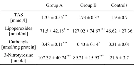
The summary of our results is presented in Table 1.
[image:2.595.309.537.578.688.2]We found out significantly reduced TAS of the patients with MS in both groups (1.35 ± 0.55 mmol/l in Group A and 1.73 ± 0.37 mmol/l in Group B) in comparison to
Table 1. Level of some markers of oxidative stress in patients
with MS and healthy controls.
Group A Group B Controls
TAS
[mmol/l] 1.35 ± 0.55
*** 1.73 ± 0.37 1.9 ± 0.7
Lipoperoxides
[nmol/ml] 71.5 ± 42.18**•• 127.02 ± 74.67***46.62 ± 27.36 Carbonyls
[nmol/mg protein] 0.48 ± 0.11*** 0.43 ± 0.14* 0.31 ± 0.01 3-Nitrotyrosine
[nmol/l] 107.32 ± 40.74*** 89.21 ± 15.93*** 21.6 ± 3.7
Values are expressed as average ± SD. We compared A, B groups to con- trols group: (*P < 0.05); (**P < 0.01); (***P < 0.001) and A versus B group:
(•P < 0.05); (••P < 0.01); (•••P < 0.001). Group A: patients with MS, normal
healthy subjects (1.9 ± 0.7 mmol/L). Statistically signifi- cant difference was confirmed only between group A and the control group, P < 0.001. We did not observe inter- gender differences. Concentration of lipoperoxides in plasma showed a significant difference between MS-pa- tients and the controls. While the concentration of lipop- eroxides in normal subjects was 46.62 ± 27.36 nmol/ml, in MS-patients: Group A 71.5 ± 42.18 nmol/mL, P < 0.01 and Group B 127.02 ± 74.67 nmol/ml, P < 0.001. We have noticed also the difference between patients groups, P < 0.01 and significantly higher rate of lipid peroxida- tion in men than in women, P < 0.05. The average con- centration of 3-nitrotyrosine was significantly increased in both patients’ groups: 107.32 ± 40.74 nmol/l in Group A, P < 0.001, resp. 89.21 ± 15.93 nmol/l in Group B, P < 0.001, in comparison to control group 21.57 ± 3.67 nmol/l. The occurrence of protein carbonyls in patients with MS was: Group A 0.48 ± 0.11 nmol/mg P (protein), P < 0.001, Group B 0.43 ± 0.14 nmol/mg P, P < 0.05, while in plasma of healthy subjects, only 0.31 ± 0.01 nmol/mg P. UA level was analyzed separately for men and women, because there are gender differences of its physiological concentrations. Concentration of uric acid in males was 380.41 ± 86.57 μmol/l and in females 298.86 ± 56.11 μmol/l which is in the physiological range for both genders.
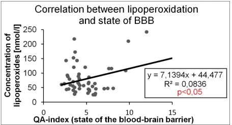
One of significant correlations was between the state of BBB and lipoperoxidation (r = 0.289, n = 58, P < 0.05) (Figure 1). Analyzing the relations between tested pa-
rameters, positive correlation was confirmed between TAS and uric acid concentration (r = 0.308, n = 58, P < 0.05) (Figure 2) and another correlation between protein
damage marker-protein carbonyls and 3-nitrotyrosine (r = 0.436, n = 58, P < 0.001) (Figure 3). There was a
negative relationship between UA concentration and protein carbonyls (r = −0.328, n = 58, P < 0.05) (Figure 4).
5. DISCUSSION
[image:3.595.310.538.84.207.2] [image:3.595.309.537.245.364.2]To present study describes elevated values of various oxidative/nitrative stress markers in multiple sclerosis patients with different state of blood-brain barrier. Oxi- dative resp. and nitrative stress are important factors, which play a role in demyelination-associated changes and neurodegenerative disorders such as MS [3,8]. In accordance with other studies [3,7-9], we found a re- duced TAS in MS patients. Low levels of antioxidants in blood plasma might indirectly point out to their high consumption by free radicals, which could be a proof of prooxidative processes in the body of MS patients [8,9]. Free radicals are not only products of cell metabolism, but also tools of leukocytes in immune intervention. Radi- cals attack molecules of all matters and could cause their damage and loss of function. Lipids are widely spread in
Figure 1. Correlation between lipoperoxidation and state of
[image:3.595.311.536.405.523.2]BBB.
Figure 2. Correlation between total antioxidant status and uric
[image:3.595.309.538.552.676.2]acid concentration.
Figure 3. Correlation between markers of protein damage.
Figure 4. Correlation between uric acid and concentration of
protein carbonyls.
should protect brain and its structures from undesired effects. Elevated lipoperoxidation in both patient groups, positively correlating with the state of BBB (Figure 1),
could indicate an important role of free radicals in the disease progression. Free radicals could attack lipids in membrane of astrocytes, damage phospholipids’ bilayer whereby reduce their function and function of the whole barrier [10].
In MS patients, we also observed a significant increase in another marker of oxidative damage, protein carbonyls. Previous studies [8,16] indicate that plasma proteins are mostly damaged, but when we consider the integrity of the body, deteriorated BBB, we could also think about damaged proteins in the CNS.
3-nitrotyrosine is an indicator of increased formation of peroxynitrite and the most important molecule con- sidered to be in charge of demyelination [3]. In agree- ment with literature data [11], significantly increased plasma levels of 3-nitrotyrosine were reported in our groups of MS patients. It may reflect the significant da- mage to neurons in the CNS when we use similar con- sideration as in protein carbonyls. Also mutual correla- tion between 3-nitrotyrosine and carbonyls (Figure 3)
could show the negative effects of both types of free radicals on proteins. Proteins with changed structure have reduced functions and could also provide epitopse to enhance or induce the immune response which would be another negative effect on disease progression.
In the published literature there were found different levels of uric acid in MS-patients. Some of them describe higher, some lower and some unchanged concentration of uric acid in these patients [11,13]. Our results indicate concentration of uric acid in physiological range, but showed a tendency for higher values. According to our opinion, increased levels of uric acid in MS patients could cause elevated destruction of oligodendrocytes [12] and their nuclei. Human organism could react in this way on the inadequate formation of free radicals since uric acid is a natural antioxidant. Antioxidant function of UA is confirmed by its positive correlation with TAS (Figure 2) and negative correlation with protein carbonyls (Fig- ure 4), although there was no significant correlation with
3-nitrotyrosine. We think that it could be explained by UA’s property to scavenge mainly oxygen reactive spe- cies.
So far, there is no any effective prevention or thera- peutic intervention known that would avoid the patho- logical processes of MS. A certain contribution to this issue also represents our study that confirmed the lack of anti-oxidant capacity of plasma in MS-patients caused by deficiency of hydrophilic and hydrophobic antioxidants. Elevated production of oxygen and nitrogen free radicals results in increased oxidative and nitrative stress with impact on many important molecules in the human body.
This fact should be taken into account in therapeutic practice. In addition, supplementation of both deficient antioxidants and whole-body cryotherapy can be effec- tive to decrease OS [14]. There are new studies describ- ing possibilities to reduce oxidative damage via antioxi- dant pathway action [15], but it is still uncertain whether reactive oxygen and nitrogen species are causes or con- sequences of autoimmune answer. Products of the free radical action could be in future one of the markers of disease progression, as it is actually speculated in some of the studies [8,16]. Oxidative stress is just one of many factors that negatively affect this disease. It is therefore necessary to pay attention to all other pathogenic aspects and their reduction to provide the patients with MS the best possible quality of life with untreatable disease.
REFERENCES
[1] Noseworthy, J.H. (1991) Progress in determining the causes and treatment of multiple sclerosis. Nature, 399, 40-47.
[2] Gonsette, R.E. (2008) Neurodegeneration in multiple scle- rosis: The role of oxidatvie stress and excitotoxicity.
Journal of the Neurological Sciences, 274, 48-53.
[3] Sayre, L.M., Perry, G. and Smith, M.A. (2008) Oxidative stress and neurotoxicity. Chemical Research in Toxicol-
ogy, 21, 172-188
[4] Re, R., Pellegrini, N., Protteggente, A., Pannala, A., Yang, M. and Rice-Evans, C. (1999) Antioxidant activity ap- plying an improved ABTS radical cation decolorization assay. Free Radical Biology & Medicine, 26, 1231-1237. [5] el-Saadani, M., Esterbauer, H., el-Sayed, M., Goher, M.,
Nassar, A.Y. and Jurgens, G. (1989) A spectrophotometric assay for lipid peroxides in serum lipoproteins using a commercially available reagent. Journal of Lipid Research,
30, 627-630.
[6] Buss, H., Chan, T.P., Sluis, K.B., Domigan, N.M. and Win- terbourn, C.C. (1997) Protein carbonyl measurement by a sensitive ELISA method. Free Radical Biology & Medi- cine, 23, 361-366.
[7] Uttara, B., Singh, A.V., Zamboni, P. and Mahajan, R.T.
(2009) Oxidative stress and neurodegenerative diseases: A review of upstream and downstream antioxidant thera- peutic options. Current Neuropharmacology, 7, 65-74.
[8] Besler, H.T. and Comoglu, S. (2003) Lipoprotein oxida- tion, plasma total antioxidant capacity and homocysteine level in patients with multiple sclerosis. Nutritional Neu- roscience, 6, 189-196.
progressive multiple sclerosis. Neurochemical Research, 36, 1012-1016.
[10] Frei, B., Stocker, R. and Ames, B.N. (1988) Antioxidant defences and lipid peroxidation in human blood plasma.
Proceedings of the National Academy of Sciences of USA,
85, 9748-9752
[11] Miller, E., Walczak, A., Saluk, J., Ponczek, M.B. and Majsterek, I. (2012) Oxidative modification of patient’s plasma proteins and its role in pathogenesis of multiple sclerosis. Clinical Biochemistry, 45, 26-30.
[12] Kuračka, Ľ. (2011) Association between degradation pro- ducts of purine nucleotides in cerebrospinal fluid and lev- els of some lipofilic vitamins in plasma of patients with multiple sclerosis. Proceedings of the 50th Faculty Con- ference of Student Science Work, 6th Scientific Confer- ence of Graduants MF CU,14 April 2011, Bratislava, 67-
70.
[13] Amorini, A.M., Petzold, A., Tavazzi, B., Eikelenboom, J.,
Keir, G., Belli, A., Giovannoni, G., Di Pietro, V., Polman, C., D’Urso, S., Vagnozzi, R., Uitdehaag, B. and Lazza- rino, G. (2009) Increase of uric acid and purine com- pounds in biological fluids of multiple sclerosis patients.
Clinical Biochemistry, 42, 1001-1006.
[14] Miller, E., Mrowicka, M., Malinowska, K., Mrowicki, J.,
Saluk-Juszczak, J. and Kedziora, J. (2009) Effects of whole- body cryotherapy on oxidative stress in multiple sclerosis patients. Journal of Thermal Biology, 35, 406-410.
[15] Virgili, N., Mancera, P., Wappenhans, B., Sorrosal, G., Bi- ber, K., Pugliese, M. and Espinosa-Parrilla, J.F. (2013) KATP channel opener diazoxide prevents neurodegenera- tion: A new mechanism of action via antioxidative path- way activation. PLoS One, 11, 8-9.
[16] Sadowska-Bartosz, I., Adamczyk-Sowa, M., Galiniak, S., Mucha, S., Pierzchala, K. and Bartosz, G. (2013) Oxida- tive modification of serum proteins in multiple sclerosis.