CLINICAL MANAGEMENT AND OUTCOMES OF PATIENTS IN THE DUKE CRYPTOCOCCOSIS CLINICAL COHORT, 1996 - 2009
Emily Wenink Bratton
A dissertation submitted to the faculty of the University of North Carolina at Chapel Hill in partial fulfillment of the requirements for the degree of Doctor of Philosophy in the Department of Epidemiology, Gillings School of Global Public Health.
Chapel Hill 2012
Approved by: Charles Poole
Jonathan Juliano
John Perfect
Til Stürmer
© 2012
ABSTRACT
EMILY WENINK BRATTON: Clinical Management and Outcomes of Patients in the Duke Cryptococcosis Clinical Cohort, 1996–2009
(Under the direction of Charles Poole, PhD)
Using this single-center cohort of cryptococcosis patients, we characterized temporal trends among infected, transplant recipients and a third heterogeneous group of HIV-negative, non-transplant patients, over a 14-year period (1996 – 2009). We executed a comparison of clinical management and predictors of poor outcomes with respect to these three groups. Adherence to recommended treatment algorithms was an important research question in our study. The 2010 IDSA Guidelines for treatment of these three groups was created to better inform clinicians, yet supportive evidence from cohort studies is still lacking, particularly in the HIV-negative, non-transplant group.
As recommended, most patients with severe disease received amphotericin B for initial antifungal treatment and the majority of non-severe patients received fluconazole. Receiving a non-recommended antifungal regimen was associated with a higher relative risk of persistent infection at four weeks (RR1.9, 95%CI 0.9 – 4.3). The rate of attributable mortality among those not receiving the recommended dose of initial therapy was higher relative to those receiving recommended dosing (HR 2.3, 95%CI 1.0 – 5.0). Among severe disease patients, flucytosine exposure was associated with a lower overall mortality rate (HR 0.4, 95%CI 0.2 – 0.9) and attributable mortality (HR 0.5, 95%CI 0.2 – 1.2).
ACKNOWLEDGEMENTS
I wish to acknowledge my advisor, Charles Poole and my committee, John R. Perfect, Til Stürmer, David J. Weber and Jonathan J. Juliano for their guidance and knowledge
provided to me to execute this work. Their personal and professional support helped me through the completion of this process.
I extend my thanks to Elizabeth S. Dodds Ashley, PharmD, for assistance with study preparations, such as abstraction tool revisions and database creation, as well as Cody A. Chastain, MD, Michael S. Lee, MD, Mariza Daras, MD, and Lipi Roy, MD, MHS, for assisting with patient chart collection. And also to Nada El Hussieni, MD, who diligently helped with all of the above listed tasks from beginning to end. Her reliable dedication through the data collection and analysis remains extremely appreciated.
TABLE OF CONTENTS
LIST OF TABLES ...x
LIST OF FIGURES ... xi
LIST OF ABBREVIATIONS ... xii
CHAPTERS I. SPECIFIC AIMS ...1
Study rationale ...3
Specific aim 1 ...4
Specific aim 2 ...5
Table ...6
II. BACKGROUND AND SIGNIFICANCE ...7
Disease populations – the three groups ...8
Diagnostics ...13
Treatment ...14
Clinical management ...17
Immune reconstitution inflammatory syndrome ...19
Tables ...21
III. RESEARCH DESIGN AND METHODS ...23
Definitions and data collection ...24
Data analysis ...28
Strengths and limitations of methods ...38
Tables ...41
Figures...45
IV. COMPARISON AND TEMPORAL TRENDS OF THREE GROUPS WITH CRYPTOCOCCOSIS: HIV-POSITIVE, SOLID ORGAN TRANSPLANT, AND HIV-NEGATIVE, NON-TRANSPLANT ...48
Overview ...48
Introduction ...49
Methods...51
Results ...54
Discussion ...60
Tables ...67
Figures...72
V. APPROACHES TO ANTIFUNGAL THERAPY AND THEIR EFFECTIVENESS AMONG PATIENTS WITH CRYPTOCOCCOSIS ...74
Overview ...74
Introduction ...75
Methods...76
Results ...82
Discussion ...90
Tables ...97
Figures...103
Study summary ...105
The three groups ...106
Temporal trends ...107
Persistence and mortality outcomes ...108
Treatment effectiveness ...110
Conclusions ...111
APPENDIX A. PATIENT ABSTRACTION FORM ...114
APPENDIX B. CUMULATIVE ANTIFUNGAL DOSING ...130
LIST OF TABLES
Table 1.1 Indicators of risk for failure ...6
Table 2.1 Primary therapy recommendations (meningeal) ...21
Table 2.2 Primary therapy recommendations (non-meningeal) ...22
Table 3.1 Antifungal treatment response definitions ...41
Table 3.2 Binary variable specification ...42
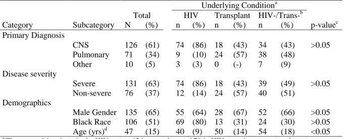
Table 4.1 Patient characteristics at baseline ...67
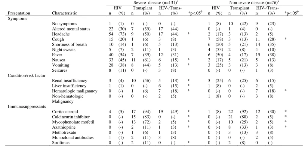
Table 4.2 Presenting symptoms and risk factors ...68
Table 4.3 Differences in mean duration of symptoms ...69
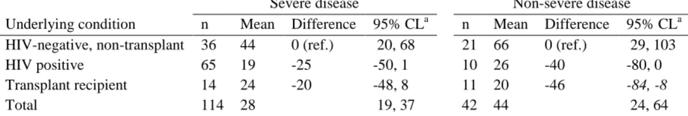
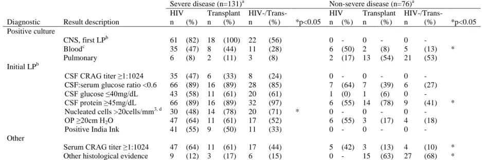
Table 4.4 Diagnostic findings of cryptococcosis disease ...70
Table 4.5 Patient treatment and outcomes ...71
Table 5.1 Baseline covariates prior to starting antifungal therapy ...97
Table 5.2 Patient diagnostics at baseline ...98
Table 5.3 Initial antifungal regimen by baseline severity of disease ...99
Table 5.4 Dosing (mg/kg/day) of initial therapy ...100
Table 5.5 Patient outcomes after antifungal therapy ...101
LIST OF FIGURES
Figure 3.1 Conceptual model showing timing of patient cohort
conditions and events ...45
Figure 3.2 Simplified causal diagram for specific aim 2 (initial therapy) ...46
Figure 3.3 Simplified causal diagram for specific aim 2 (flucytosine) ...47
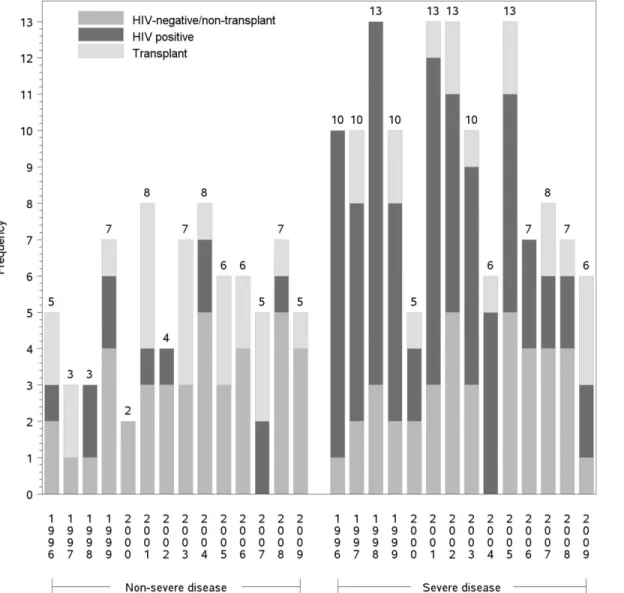
Figure 4.1 Annual cases ...72
Figure 4.2 Use of amphotericin B ...73
Figure 5.1 Diagram of patient flow...103
ABBREVIATIONS
AmpB Amphotericin B
AmpBd Amphotericin B deoxycholate ABLC Amphotericin B Lipid Complex ARDS Acute respiratory distress syndrome ARV Antiretroviral
DCCC Duke Cryptococcosis Clinical Cohort DUMC Duke University Medical Center CFU Colony forming units
CI Confidence Interval
CL Confidence Limits
CM Cryptococcal meningitis CNS Central nervous system CSF Cerebrospinal fluid
GVHD Graft Versus Host Disease
HAART Highly Active Antiretroviral Therapy HIV Human Immunodeficiency Virus
HR Hazard Ratio
IDSA Infectious Disease Society of America LFAmpB Lipid formulation amphotericin B L-AmpB Liposomal amphotericin B
LP Lumbar puncture
RR Risk Ratio
SD Standard deviation SOT Solid organ transplant WBC White Blood Cell
CHAPTER I. SPECIFIC AIMS
The IDSA 2010 Clinical Practice Guidelines for the management of cryptococcosis [1] provide informative categories for analysis by classifying patient groups (HIV positive, solid organ transplant recipients, and HIV-negative/non-transplant) and severity of disease (meningeal, non-meningeal). Taking these categories into consideration, we examined the differences between the three different clinical groups and address the degree to which the various clinical manifestations of cryptococcosis and baseline patient immune status modify the effectiveness of antifungal treatment.
The three-month mortality rate during management of acute cryptococcosis with CNS involvement is still nearly 20% despite medical advances and the advent of highly active antiretroviral therapy (HAART) [2,3,4]. In resource-limited settings, the two-week mortality rates after presenting to a health clinic have been reported as high as 100% [5]. With the high risk of mortality with severe disease, treatment and clinical management strategies are imperative to improving patient survival and preventing relapse or persistence of infection.
Definitions of treatment success varies based on immune status—particularly HIV status. HIV positive patients may be cured with initial therapy, with “cure” meaning the eradication of the fungal organism and elimination of symptoms, but early studies reveal rates of relapse nearing 50% [6,7,8,9]. Thus, commonly with this particular
With the introduction of maintenance therapy, rates of relapse have dropped significantly to under 10% in geographic areas where induction therapy is the standard of care [3,10,11,12]. A study in France comparing cryptococcosis relapse rates among HIV-positive patients in the pre-HAART versus HAART eras found that relapse after initiating maintenance therapy was largely associated with a lower CD4 cell count at baseline, a slower rate of increase of CD4 cells in the first two weeks of HAART, as well as a serum
cryptococcal antigen titer ≥1:512 at any time during follow-up [13]. Close monitoring of the patient for relapse may still be required despite this study showing that HIV patients with a history of cryptococcosis may be taken off maintenance therapy. Relapse rates among non-AIDS patients are still around 15-20% [3,14]. Commonly, HIV-positive patients with cryptococcosis are followed indefinitely with lifelong maintenance therapy and generally non-HIV patients are followed for at least one year [3].
Data is limited on predicting risk factors for failure of cryptococcal treatment. Based on previous studies [13,15,16,17,18,19,20,21], some risk factors that contribute to treatment failure for cryptococcosis, particularly cryptococcal meningitis (the most commonly studied), include: positive cultures (CSF, blood), high serum or CSF antigen titers, headache, and abnormal mental status at admission (Table 1.1). These suggested risk factors are not necessarily exhaustive, nor do they represent a consensus among the medical community.
mortality rate is not necessarily due to cryptococcosis, but can be a result of an underlying condition or co-infection.
StudyRationale
Cryptococcus is an invasive mycosis that causes considerable morbidity and mortality, but there are few larger cohort studies that focus on this disease, many of which are now more than a decade old [13,18,19]. With the global burden of cryptococcal infection reaching 1 million cases per year [23], many questions remain regarding the clinical
management on of HIV-infected individuals, organ transplant recipients and more recently immunocompetent individuals or other non-HIV/non-transplant hosts.
Many HIV positive patients are on lifelong antifungal maintenance therapy with monthly monitoring. Therefore, identifying risk factors and treatment recommendations for HIV patients with cryptococcal disease is important not only for the potential prevention of immune reconstitution inflammatory syndrome, increased intracranial pressure, drug resistance and cryptococcomas, but also to identify the optimal treatment regimens of cryptococcal disease to reduce costs associated with treatment failure. This is particularly true in resource limited settings coping with the HIV epidemic without proper access to diagnostic tools or medications. Due to the absence of new drug development, there is much need for well-supported disease management strategies.
wealth of information on all three clinical groupings defined by the Infectious Disease Society of America (IDSA) [1] that include: 1) HIV-infected individuals; 2) solid organ transplant recipients; 3) non-HIV and non-transplant hosts. The third group is notably heterogeneous, but will most likely form the smallest proportion of patients in this database, although this group could prove of unique interest to clinicians and to serve as a comparison.
Specific Aim 1. How do the following three groups with cryptococcosis differ with respect to clinical presentation and management: positive, transplant recipients, and HIV-negative/non-transplant patients?
Objectives:
1. Examine the changing populations for acquiring Cryptococcus (HIV positive, transplant recipients, HIV-negative/non-transplant)
2. Summarize temporal trends in diagnosis and amphotericin B formulations as initial therapy
3. Describe and assess differences and similarities of underlying three groups with respect to initial presentation, diagnostics and approaches to clinical management
a. Examine prevalence and type of immunosuppression in the three groups— HAART exposure in HIV-infected patients, corticosteroid use and
immunosuppressive agents among transplant and HIV-negative/non-transplant, as well as prevalence of patients appearing
Specific Aim 2. How does initial antifungal therapy for cryptococcosis influence patient outcomes of persistence of infection, attributable mortality and overall mortality?
Objectives:
1. Determine the association of aspects of initial treatment on poor patient outcomes that include the risk of persistent infection and rates mortality due to cryptococcosis and overall 1-year mortality
a. Estimate the effect of initial antifungal treatment type on persistence and mortality
b. Estimate the effect of initial treatment dosing on persistence and mortality c. Estimate the effect of flucytosine exposure among patients with severe
cryptococcosis disease on persistence and mortality
i. Estimate the effect of ≤ 7 days or >7 days of flucytosine on the risk of persistence and mortality rate among patients surviving at least 14 days from the time of cryptococcosis diagnosis.
2. Assess other strong predictors for the above outcomes in (1).
Table
Table 1.1. Indicators of risk for failure (including mortality) of cryptococcosis antifungal treatment.
Non-HIV Infected HIV-infected
Positive India ink High LP OP Low CSF glucose Extra-neural sites CSF antigen titer >1:32 CSF antigen titer ≥1:1024 Steroid therapy
Lymphoreticular malignancy Hematologic malignancy No headache
Organ failure
Abnormal mental status Low CSF leukocyte counta Absent antibodya
Age ≥60 Male gender
Positive culture at 2 weeks Positive blood and urine cultures Treatment without flucytosine High serum or CSF antigen titer Age >30 years
Low CSF glucose Intravenous drug usea High MIC to fluconazoleb Mechanical ventilationa ICU admissiona
Organ failure
a
These risk factors were not measured in our study and will be uncontrolled in our analyses; CSF nucleated host cell count was used as a proxy for CSF leukocyte count
b
CHAPTER TWO - BACKGROUND AND SIGNIFICANCE
Cryptococcus spp., an encapsulated yeast, is an opportunistic human fungal pathogen isolated from the environment worldwide, particularly in urban areas [24,25]. Humans are thought to be exposed by inhaling the fungal basidiospores, which are small enough to establish in the alveoli of the lung [26]. Pigeon guano is a common source for infectious propagules of C. neoformans and is postulated to play a central role in transmission from the environment to humans [3,24,27,28,29,30,31,32,33]. As the spores are inhaled, the lungs are the portal of entry and pneumonia is a common manifestation of cryptococcal diseases, however, the most common manifestation of disease is when the pathogen spreads to the central nervous system (CNS) and causes meningoencephalitis. This is more common among immunocompromised individuals who are unable to contain the infection at the primary site (the lung) and from the advent of the HIV epidemic, C. neoformans has become an increased global concern among those who are HIV infected [34]. Furthermore,
cryptococcal meningitis is uniformly fatal without treatment [3,24,35,36,37].
immunosuppressed, and more often than C. neoformans, immunocompetent humans and animals [38,39]. C.gattii is rare in the Southeastern U.S. with only one identified clinical case in an immunocompromised adult [40], hence this species was not the focus of our study.
Diagnosis without laboratory capabilities is difficult because clinical presentation of human cryptococcosis can be very nonspecific. However, the central nervous system (CNS) and respiratory tract are the most common organs involved in cryptococcal infection [34]. Clinical manifestations have a wide range of severity from an asymptomatic nodule in the lung to sudden death from septic shock with acute respiratory distress syndrome. Clinical signs can be seen in the CNS, lung, skin, eye, genitourinary tract, bone and joints, muscle, heart, gastrointestinal tract, breast, lymph nodes, thyroid, adrenal gland, and head and neck [3].
Cryptococcosis can occur at any age. Being at least 30 years of age is reported as a risk factor for cryptococcosis treatment failure among HIV positive individuals [41,42], while cases that are ≥60 years are reported among HIV-negative patients [21]. Age may have an independent impact on survival or incidence of treatment failure. A male predominance exists in both HIV positive and HIV negative cryptococcosis patients [4,41,43,44].
Disease populations—the three groups
The 2010 IDSA Cryptococcal Guidelines defined three distinct risk groups for
HIV Positive
Cryptococcal infections in HIV-positive patients almost always occur in advanced stages of the disease, and are for the most part, incurable but treatable— individuals who survive initial illness require lifelong maintenance therapy and close monitoring [3].
Symptoms and clinical findings common in AIDS patients include: headaches, fever, shorter duration of symptoms than non-AIDS patients, positive India ink examination, cerebrospinal fluid (CSF) antigen titer ≥1:1024, CSF pleocytosis <20/l, CD4 counts <100 cells/l, serum antigen positive, cryptococcemia and increased intracranial pressure (Table 2.1)
[3,6,7,8,9,45,46]. There is less knowledge on common symptoms among HIV-negative patients, but similar to HIV-positive patients headache, fever and abnormal CSF chemistry (glucose <40, protein >45) are considered most common (Table 2.1) [45].
Parallel with the rise of the AIDS epidemic in 1981, the incidence of cryptococcosis increased [3]. Before the HIV epidemic, cryptococcosis was much more uncommon and occurred predominantly among patients with compromised immunity, such as those with hematologic malignancy or had undergone solid organ transplantation at a rate of 0.2 – 0.8 per 100,000 depending on geographic area [34,47]. In more recent decades, HIV-associated cryptococcal infection is estimated to account for up to 80% of all cases [34,47,48]. It is estimated the current rate of HIV-associated cryptococcosis is approximately 1 million cases per year worldwide [23]. Despite this high estimate of disease, the incidence among
observation was also made in a large retrospective study in France, with a 46% decrease in cryptococcosis incidence between pre-HAART and post-HAART eras [41].
Incidence rates of cryptococcosis remain the highest in areas where HIV disease is high and availability of highly-active antiretroviral therapy (HAART) is limited [1,23]. About 5-10% of patients with AIDS in the United States will develop cryptococcosis (pulmonary and/or meningeal) [1,3]. Prevalence estimates among HIV patients in the U.S. range from 5-13% depending on region and study year (1985-1997) [3,6,7,8,9,49,50,51]. In an eight-year surveillance study in the United States, under one-third of the HIV-infected cryptococcosis cases had been placed on HAART before diagnosis [42], emphasizing the importance of access and adherence to HAART in prevention of opportunistic disease, not to mention being tested for HIV infection. Though progress has been made, the 3-month mortality rates from acute cryptococcal meningitis continue to hover around 20% [2,4].
Solid Organ Transplant
The risk of cryptococcosis has not vanished in developed countries because of the increased use of immunosuppressive therapy and continued progress of transplant medicine [34]. Cryptococcosis is the third most common invasive fungal infection affecting solid organ transplant recipients, with a mortality rate of 10-20% and near 40% with CNS
syndrome (IRIS) [43]. Between 20-60% of cryptococcosis in HIV-negative patients occurs in solid organ transplant patients [3,21,47,55].
Cryptococcosis disease is diagnosed at a median of 21 months after transplantation [56] and most commonly in renal transplant patients; though liver transplant patients are more likely to have disseminated disease [56]. Studies have reported a lifetime
cryptococcosis prevalence of 3-4% post-transplantation [52,57,58,59]. Immunosuppressive drug administration for transplant recipients to prevent organ rejection can place them at increased risk for cryptococcal infection. Corticosteroid use is associated with an increased risk of cryptococcosis [16,43,60]. The use of calcineurin inhibitors as an immunosuppressive agent appears to be associated with lower mortality, but the interaction of this class of drug with amphotericin B deoxycholate (AmpBd) can contribute to nephrotoxicity [3,61].
Therefore, lipid formulation amphotericin B (LFAmpB) is recommended for organ transplant recipients (Table 2.2). A study by Sun et al (2009) demonstrated that LFAmpB was
significantly associated with better survival in transplant patients compared to AmpBd (OR, 0.11; 95% CI, 0.02-0.57) [61].
HIV-negative, non-transplant
The healthy human population is rarely infected by cryptococcosis, but cases with limited frequency have been reported [3,4,21,62]. High risk patients other than HIV-positive individuals and organ transplant recipients are a heterogeneous group. Patients with
or chronic lung diseases may contribute to susceptibility, and simultaneous infection with other fungi cannot be ruled out [3]. There are still many questions surrounding this patient group, particularly with risk factors for cryptococcosis disease given their “healthy” immune status. Therapeutic approaches are primarily based on expert opinion, out-dated explorative and retrospective cohort studies. Lacking a consensus, further study is needed.
It is difficult to consistently treat such a diverse group with mortality rates still as high as 24% [19,22]. More studies to inform the management of the HIV-negative/non-transplant group and to understand how host immunity plays a role in poorer prognosis are needed so as to reduce observed elevated mortality in this group. In an important study by Pappas et al (2001), 306 HIV-negative cryptococcosis patients from 15 different centers in the U.S. were shown to have successful treatment in 74% of cases. Relapse occurred in 4% of patients, all-cause mortality was 30% and death due to cryptococcosis was 12% [21]. Factors influencing mortality among all patients included organ failure, hematologic
malignancy, age ≥60 years, unsuccessful therapy, site of infection other than pulmonary and a positive blood culture for C. neoformans (Table 2.1) [21]. A recent multi-center study of 86 cryptococcal meningitis patients also found the highest mortality in the
Diagnostics
India ink stain (direct observation)
Latex agglutination test (antigen detection) primarily in CSF and serum Culture (24-72 hours up to 5-7 days)
Cryptococcus neoformans can be rapidly diagnosed with an India ink stain of the CSF fluid. It is not as sensitive or specific as serological tests, but serves more as an immediate diagnostic tool that can also reveal a general idea of organism burden. The detection of the cryptococcal polysaccharide antigen in body fluids (latex agglutination test) is another highly sensitive and specific (~95%) method to identify infection and indicate fungal burden. A high antigen titer level (≥1:1024) translates into a higher burden of organisms. Generally, the CSF is tested to rule out meningoencephalitis and the serum is tested for disseminated
disease using the antigen test. Other body fluids can be tested, such as urine or pulmonary fluids. In geographic areas with a high burden of HIV, the serum cryptococcal antigen is now being evaluated as a screening tool for patients at-risk for cryptococcosis. Culturing Cryptococcus neoformans is another diagnostic tool, but takes longer than the other two tests, and CSF cultures for viable organisms may be difficult if there is a case of chronic
Treatment
Before 1950 cryptococcosis was uniformly fatal, however its treatment has improved dramatically in the last 20 years [1,3]. There was a dramatic rise in cryptococcal infections in parallel with the AIDS epidemic in the early 1980’s. The widespread use of fluconazole for antifungal prophylaxis is thought to be a contributing factor of the decline in incidence beginning in the early-mid 1990s [47]. Currently the most common antifungal therapies used to treat cryptococcal disease are: amphotericin B (a polyene antimycotic) and flucytosine, fluconazole, and lipid formulations of amphotericin B. These drugs are used alone or in combination therapy dependent on the underlying disease state [1]. Success of treatment varies due to a lack of randomized clinical trials; selection of a treatment regimen is largely arbitrarily decided by the prescribing physician [1].
Treatments have been more widely studied among HIV-related cryptococcal disease, as many of these patients must receive lifetime antifungal therapy. Side effects of
combination therapy are rather common in this immunocompromised group and despite advances, mortality associated with cryptococcal meningitis can be up to 25% among
cryptococcal meningitis in patients with AIDS, despite the substantial danger of drug-related toxicities and fluconazole resistance [15].
Amphotericin B
Amphotericin B (AmpB) began as cryptococcal therapy in 1956 [71] and to date, is the most potent and effective drug for treating cryptococcal meningitis with success rates ranging between 60-70% [3]. This drug is limited by its poor penetration of the subarachnoid space accompanied risk of nephrotoxicity; therefore it is commonly administered in
combination with flucytosine or fluconazole [3,72]. Amphotericin B is also available in more expensive lipid formulations (LFAmpB) that reduce the risk of renal toxicity and thus allow higher doses to be administered (3-6mg/kg/day for liposomal AmpB [L-AmpB] or amphotericin lipid complex [ABLC] 5mg/kg/day). Lipid formulations are also beneficial in patients with baseline renal concerns [1,3]. Optimal dosing of LFAmpB and effectiveness in combination therapy are still unclear. There is some evidence that LFAmpB at 4mg/kg/day is equal or more fungicidal than AmpBd [1,73,74]. Human studies are still undersized, making clear recommendations for treatment difficult.
Flucytosine (5FC)
AIDS patients it has been demonstrated that antifungal regimens containing flucytosine is an independent predictor of treatment success [13,78]. This drug is not without some serious drawbacks that generally arise between days 4-14 of treatment [79]. Side effects include bone marrow suppression, leucopenia and gastrointestinal disturbances [80,81]. Eliminated primarily by the kidneys, changes in renal function should also be checked [3]. Drug levels should be monitored to avoid bone marrow toxicities, with therapeutic ranges between 30-80 µg/ml (and not exceed 100µg/ml) two hours after first recommended dose of 100 mg/kg/day. This dose was found effective by van der Horst et al. (1997) with only a 3% withdrawal rate [13]. It is still uncertain what drug levels correspond with gastrointestinal side effects and pancytopenia.
Fluconazole
Treatment Recommendations
Treatment recommendations based on the most recent 2010 IDSA Guidelines for effective treatment of cryptococcosis according to each category of presentation of cryptococcosis are in Tables 2.1 and 2.2 [1]. Proper induction therapy for cryptococcal meningoencephalitis was defined using the categories in Table 2.1. For pulmonary or other types of cryptococcosis, appropriate therapy was defined by IDSA recommendations in Table 2.2 [1].
Fluconazole, flucytosine and amphotericin (deoxycholate or lipid preparation) are the antifungal medications that comprise the 2010 IDSA Guidelines for effective treatment of cryptococcosis and were also primary treatments recommended for use in the previously published 2000 guidelines [1,15]. These four drugs are sometimes used in combination or alone (except for flucytosine, which is not given alone), and choice of treatment relies on the severity of disease, immune and HIV status, and other possible underlying conditions. They are the same four treatments used and available to patients dating back to the beginning of our cohort study in 1996; part of the reasoning for our study period range along with the availability of HAART among HIV infected patients. Interestingly and considering their long history of use, there have been very few reports of drug-resistant strains (either azole or polyene) of C. neoformans [86].
Clinical management
inflammatory reactions [34]. Management of patients with severe disease that have elevated intracranial pressures (≥ 20 – 25 cm water) is important in preventing poor outcome, but its requirement in conjunction with symptom development (e.g., increasing headache, mental status changes, new neurologic findings) and a precise opening pressure for treatment has not yet been established [34]. One study of HIV patients with cryptococcal meningitis showed elevated pressures two weeks after starting treatment predicted poor clinical response [15], however another study did not find a significant association between opening pressure at day 14 and mortality at 10 weeks [87]. A recent study found increased intracranial pressure was more common among HIV-infected (49%) and non-immunosuppressed (67%)
cryptococcosis patients compared to immunosuppressed patients. However, HIV-patients were more likely to receive repeat lumbar punctures that other patient groups [62]. Options for managing acute elevated intracranial pressure include: repeated lumbar punctures, lumbar drain insertion, ventriculostomy, or ventriculoperitoneal shunt. In patients who have IRIS, corticosteroids are needed to control symptoms [34].
CSF cryptococcal antigen titers [89]. Both of these measures can serve as a measure of organism load and can be used in follow-up evaluation after cryptococcal treatment. A larger combined cohort study of 262 HIV-infected cryptococcal meningitis cases found an
association between the rate of clearance of infection and survival [90]. The strength of the association in multivariate analysis was stronger with survival at 2 weeks (HR 1.34; 95% CI, 1.06–1.68) than at 10 weeks (HR 1.18; 95% CI, 1.04–1.33) [90].
Immune reconstitution inflammatory syndrome (IRIS)
Rapid changes in immunity, such as with the introduction of HAART among HIV-infected patients, can result in a clinical worsening or radiographic manifestations consistent with an inflammatory process but produces negative results for biomarkers or cultures [34]. Referred to as IRIS, this condition has been reported in up to one-third of HIV patients with cryptococcosis upon initiation of HAART [91]. Timing of onset varies, generally 4 – 6 weeks after starting HAART and is associated with increasing CD4 counts and diminishing viral load [91,92]. There is evidence that IRIS is more common in patients with a higher fungal burden and disseminated infection or fungemia [91,93]. The recommendations on when to begin HAART following cryptococcosis infection vary widely between 2 – 10 weeks [1].
fungal burden, influence the development of IRIS, and graft survival is also reduced in this patient group [95]. Management of IRIS in transplant recipients and other
Tables
Table 2.1. Primary therapy recommendations for cryptococcal meningoencephalitis based on the 2010 IDSA Guidelines [1].
Underlying condition Primary induction therapy Maintenance therapy HIV-positive
AmpBd (0.7 – 1.0 mg/kg/d IV)a plus 5FC (100mg/kg/d po) for at least two weeks
Fluconazole 400mg (6mg/kg) po daily for a minimum of 8 weeks, then fluconazole 200mg po daily ≥1yrb
Organ transplant recipients
Liposomal AmpB 3-6mg/kg/d IV or ABLC 5mg/kg/d IV plus 5FC 100mg/kg/d for at least 2 weeks
Fluconazole 400-800mg po daily for 8 weeks; followed by
fluconazole 200mg po daily for 6-12 months
HIV-uninfected, non-transplant
AmpB deoxycholate ≥0.7 – 1.0mg/kg/d + 5FC (if tolerant) 100mg/kg/d for ≥4 weeks, ≥6 weeks if intolerant to flucytosine
Fluconazole 400mg po daily for 8 weeks; followed by
fluconazole 200mg po daily for 6-12 months
a
Can substitute amphotericin B deoxycholate for lipid formulations AmpB (3-6 mg/kg/d) for at least 2 weeks if patients have or are predisposed to renal dysfunction
b
Table 2.2. Primary therapy recommendations for non-meningeal cryptococcosis (pulmonary or other) adapted from the 2010 IDSA Guidelines [1].
Underlying condition Characteristics of disease Treatment Immunosuppressed
patients
Pneumonia associated with CNS or evidence of
dissemination and/or ARDSa
Treat like CNS disease (Table 2.1) including maintenance therapy; due to risk of
nephrotoxicity, AmpBd is not recommended for first line therapy (LFAmpB should be used)b
Immunocompetent
No evidence of dissemination, mild-to-moderate symptoms, absence of diffuse pulmonary infiltrates, absence of severe immunosuppression
Fluconazole 400mg/d for at least 6-12 months
Severe disease Treat like CNS disease (Table 2.1) including maintenance treatment
Mild to moderate symptoms Fluconazole 400mg daily for 6-12 months
a
ARDS = Acute Respiratory Distress Syndrome b
Recommendation for organ transplant recipients. Immunosuppressive management should include: sequential or step-wise reduction of immunosuppressants – lowering corticosteroids dose first
CHAPTER III. RESEARCH DESIGN AND METHODS
Study Population
The clinical cohort used for this study originates from the Duke University Medical Center (DUMC) in Durham, North Carolina in the Southeastern United States. During the study period from January 1, 1996 through October 31st, 2009 we retrospectively collected demographic and clinical information on all adult patients discharged from DUMC with ICD-9 diagnosis codes of cryptococcosis, cryptococcal meningitis, pulmonary Cryptococcus, and disseminated Cryptococcus. Subjects were considered eligible if they had confirmed cryptococcal disease and received treatment for their cryptococcal infection at DUMC with a sufficient electronic medical record or paper chart available for review. The complete medical record for each patient was reviewed and data regarding different interventions and outcomes were extracted. Investigators recorded all data on a standardized abstraction form that was reviewed by an epidemiologist and clinician prior to and during data entry.
Definitions and Data Collection
A cryptococcosis case was confirmed by having at least one of the following:
positive cerebral spinal fluid (CSF) antigen or fungal culture, direct histological examination of Cryptococcus, positive serum cryptococcal antigen (CRAG) test, or positive culture from blood or pulmonary sites. Positive CSF India ink staining alone was not an acceptable diagnostic tool, but was used for supportive evidence for infection.
Basic demographics, such as age, gender, state of residence, and race/ethnicity were collected. Although race and gender are possibly associated with cryptococcosis disease, HIV status, or organ transplant receipt, there is no strength of evidence that these
demographics are associated with which antifungal induction treatment a patient is given, but other factors such as immunosuppression and severity of cryptococcosis, are more important predictors of what antifungal regimen is chosen.
Upon the first admission for cryptococcosis, patient information regarding presenting symptoms, self-reported duration of symptoms and radiological findings (brain CT, brain MRI, chest CT and chest X-ray) were recorded closest to the date of diagnosis. Abnormal radiology was defined as evidence of hydrocephalus, gyral enhancement, and/or multiple nodules that may be enhancing or non-enhancing [96]. Nodules can be either single or multiple [97]. Demographic information included birth date, race/ethnicity, gender, and country of origin if not the United States. Patient weight (kg) closest to first cryptococcosis diagnosis was recorded to assess accurate antifungal dosing.
were: the date of HIV diagnosis, any documented evidence of non-compliance with antiretroviral (ARV) therapy and/or antifungals, whether cryptococcosis was an AIDS-defining illness, and the use of ARV therapy before, during and after cryptococcosis diagnosis until lost to follow-up or death.
Information on solid organ transplant recipients included: date and type of transplant, if they experienced graft-versus-host disease (GVHD) status ≤6 months after transplant, and type and dose of immune suppression medications at the time of cryptococcosis diagnosis (corticosteroid, calcineurin inhibitor, glucocorticoid, monoclonal antibodies, methotrexate, sirolimus, mycophenolate mofetil, azathioprine, or other). It was also noted if these drugs were changed or stopped due to cryptococcosis disease. There was one patient with a stem cell transplant (not solid organ) that was included in this immunosuppressed transplant group.
Regardless of whether patients had HIV or received and organ donation, other possible causes of immune suppression (hematological or other malignancy, rheumatologic disorder, chronic organ failure, or steroid therapy) were noted. If there was no apparent immunosuppression, patients were classified as “immunocompetent.”
for collection tube #4 were recorded, and if not available, the highest tube number was used for LP data. Co-morbid infections, positive cultures for other organisms and new cancer diagnoses were noted during the time of hospitalization or while on treatment for
cryptococcosis. Creatinine levels and positive culture results were recorded at each
admission, after induction therapy, and at 2 weeks, 10 weeks, 1 year and >1 years of follow-up. Peak creatinine was recorded while the patient was on induction therapy. All available flucytosine serum drug levels were obtained. Hematologic parameters (hemoglobin, hematocrit, WBC count and platelets) at the start date of flucytosine treatment and at the nearest available date with valid measurements 14 days after starting flucytosine were collected. Treatment information (type, dose and date of start and date of stopping) was abstracted from the first admission for cryptococcosis until lost to follow-up, death or until the end of the study period.
Organ failure before, during and after antifungal therapy was an important variable collected in this study, particularly transplanted organ failure during or after antifungal therapy among transplant recipients. Poor clinical response either from treatment failure (persistence of clinical findings or positive microbiology) or treatment toxicity (notably renal failure) was noted along with modifications made to treatment due to these adverse events. Date of death and death due to cryptococcosis were also recorded.
In a paper by Seagal et al (2008), authors proposed guidelines for defining treatment responses and study outcomes to invasive fungal diseases in clinical trials (Table 3.1) [98]. These recommendations informed our study definitions used for persistence of
include success and failure in non-CNS cases. “Relapse” was defined as clinical,
mycological or radiographic evidence of recrudescence after stopping antifungal therapy, if the patient had initially experienced “success” [21].
Figure 3.1 is a conceptual diagram of cohort patient flow from first diagnosis (1) and admission for cryptococcosis through death or lost to follow-up. After diagnosis, patients are typically started on treatment immediately—sometimes even prior to positive culture results if clinical signs indicate cryptococcosis disease (2). Treatment exposure depends on multiple factors and recommendations for treatment are listed in Tables 2.1 and 2.2. Two major considerations for treatment shown in these tables are HIV status or alternate possible immunosuppression and CNS involvement. Patients will then either: die while on treatment (2a), fail therapy due to toxicity or persistence of disease (2b), or experience mycological cure or suppression (3). Patients cured or suppressed on maintenance therapy (3) are followed until they die or are lost to follow-up. Some patients will experience relapse (3a) and have to restart antifungal maintenance therapy or have the dose increased. In some cases, patients will undergo re-induction therapy. Patients experiencing immune
Data Analysis
Specific Aim 1. How do the following three groups with cryptococcosis differ with respect to clinical presentation and management: positive, transplant recipients, and HIV-negative/non-transplant patients?
Exposure definitions
Clinical presentation variables used for comparison included demographics,
symptoms and duration of symptoms of cryptococcosis, diagnostic tools used for diagnosis, and underlying conditions and possible risk factors of disease (Table 3.2).
Demographics – Gender, race/ethnicity, and age at time of first diagnosis of
cryptococcosis (at DUMC) were collected. As this is the first time this study cohort will be presented in the literature, demographics showing patient composition are valuable to readers.
Immune status – HIV, transplant recipients, and HIV-negative/non-transplant patients (3 general categories); we know from prior research that baseline immune status is associated with treatment exposure, treatment failure, and mortality.
is recommended as sufficient. Clinical diagnostics, such as serum and CSF antigen, CSF culture, and radiographic tests will likely contribute to what treatment patients will receive. These clinical and microbiological results can also be indicators of future treatment failure or mortality. Because they are associated with treatment exposure and patient treatment success and mortality, the following are possible confounders and may need to be adjusted for in subsequent analyses.
Outcome definitions
Clinical management and patient outcomes during follow-up were abstracted from patient charts. HAART during and after treatment for cryptococcosis, including regimen and start and stop dates were recorded when available. It was noted whether HAART was
stopped during antifungal induction treatment and if so, when it was re-started (if at all). Similarly, trends of immunosuppressive medications for HIV-negative patients were examined, including dose at the time of cryptococcosis diagnosis and any changes to dose during antifungal treatment.
Management of elevated intracranial pressures using serial LPs, ventricular shunting, lumbar drain, and/or pharmacological therapy was recorded.
to be negative results for cultures or stable/reduced biomarkers for the initial fungal pathogen during diagnostic work-up for the inflammatory process.
Analysis Plan
Objective 1: Examine the changing populations for acquiring Cryptococcus (HIV positive, transplant recipients, HIV-negative/non-transplant)
Variables were examined using descriptive statistics and stratified based on the three groups and/or severity of cryptococcosis as needed. Where appropriate, the Student’s t-test was used to test the difference of two means and the Kruskal-Wallis test was used for the difference between medians for non-parametric continuous data. Chi-square (Χ2) tests were used to examine differences between categorical frequency distributions. The statistical significance level of alpha (α) equal to 0.05 was used for each two-tailed test performed, thus a “significant” result refers to a p-value <0.05.
Objective 2: Summarize temporal trends in diagnosis and amphotericin B formulations as initial therapy
Graph thepercentage of patients who received amphotericin B deoxycholate and lipid formulations for initial therapy each year. Examine any notable trends in use between these two regimens over time.
Objective 3: Describe and assess differences and similarities of underlying three groups with respect to initial presentation, diagnostics and approaches to clinical management
agents among transplant and HIV-negative/non-transplant, as well as prevalence of patients appearing “immunocompetent.”
Specific Aim 2. How does initial antifungal therapy for cryptococcosis influence patient outcomes of persistence of infection, attributable mortality and overall mortality?
Exposure definitions
Initial therapy - antifungal therapy regimen based on criteria provided in Tables 2.1 and 2.2.
Each patient was checked to see if they follow the 2010 IDSA guidelines for the treatment of cryptococcosis. Appropriate initial treatment would be amphotericin B for severe disease and fluconazole for non-severe disease. Induction therapy refers to the entire period of initial therapy, not the initial antifungal drug that was used.
Treatment dose – the patient was given the appropriate dose of initial antifungal therapy.
This study exposure is based on mg/kg units calculated using the daily dose of antifungal therapy given to the patient divided by the patient’s measured weight in kg at the closest date to cryptococcosis diagnosis. For example, if 0.7-1.0 mg/kg/day is the
fluconazole. Rounding to the nearest tenth for AmpBd and the nearest integer for AmpB lipid products were used to categorize appropriate dosing.
Flucytosine-containing regimens – flucytosine was given as a combination regimen with
primary initial amphotericin B therapy among patients with severe disease (1=yes, 0=no). Secondary data analysis will examine relative differences between 0 – 7 days and >7 days of flucytosine in combination with initial therapy. Unique to this exposure variable, we
hypothesized that flucytosine is more frequently paired with amphotericin B deoxycholate than other polyene formulations, so it was listed as a possible confounding factor (Figure 3.3).
Outcome Definitions:
Persistent cryptococcosis,two weeks (severe disease only): Positive cultures (CSF, blood,
pulmonary sites, other) two weeks (14 days) after starting therapy. The patient had to have survived two weeks to be eligible for this outcome because at least two weeks of induction therapy is recommended in this group. We acknowledge that the IDSA Guidelines
Persistent cryptococcosis, four weeks (severe and non-severe disease): Positive cultures (CSF, blood, pulmonary sites, other) four weeks after starting therapy and/or the positive indication of the presence of symptoms (headache, photophobia, fever, etc.) four weeks (30 days) since starting primary therapy. The patient had to have survived four weeks to be eligible for this outcome. Deaths among this patient group in this time period were considered persistent infection as a sensitivity analysis.
Because data were observational, measures for indicating persistent infection were not taken at exactly two weeks and four weeks to test for positive culture, antigen testing, and/or infection-related symptoms. Acceptable values were used if they did not overlap with the preceding measurement (e.g. a baseline culture could not be used for a two week test result), and did not extend beyond the designated time point (e.g. a measure at 3 weeks would not be counted as a week 2 measure, but instead a 4 week measure if there was not an observation at 4 weeks). Persistence measures for two weeks had to have occurred ≥1 week of therapy. Measures beyond the final time point (4 weeks, 30 days) were accepted for that final time point if within 90 days since starting therapy.
Cryptococcal-attributable mortality: A determination made by a panel of experts at DUMC.
All-cause mortality through one year: In order to assess one-year mortality risk for all patients, we obtained data on survival and mortality up to one year after their date of cryptococcosis diagnosis from the Duke Data Support Repository (DSR), which uses the Social Security Administration death index, the Tumor Registry and The Duke Information System for Cardiovascular Care death data to report mortality status.
Secondary outcomes: frequency of re-induction(s) with amphotericin B, IRIS and renal
toxicity during initial therapy, receipt of ≥7 days of flucytosine compared to receipt of 0 – 7 days of flucytosine (severe disease), and the changing initial therapy (interrupted therapy).
Re-induction: Patient had to have finished initial induction therapy for at least three days or have been placed on consolidation or maintenance therapy, then placed back on amphotericin B as part of re-induction status.
IRIS: The definition for IRIS for this study, adapted from Singh and Perfect [94] was the following: A clinical or radiographic manifestations consistent with an inflammatory process such as contrast enhancing lesions on imaging studies (CT/MRI) along with (a) and (b) and at least one of (c) through (f):
a) Symptoms occurred during receipt of appropriate therapy and could not be explained by newly acquired infection
b) Negative results for cultures or stable/reduced biomarkers for the initial fungal pathogen during diagnostic work-up for the inflammatory process
e) Histopathology showing granulomatous lesions f) Unexplained hypercalcemia
Renal toxicity: Creatinine values measured closest to day 0 and day 14 of treatment for patients with severe disease who received induction therapy with amphotericin B were used to determine renal toxicity. Defined as a >50% decrease in Glomeruler Filtration Rate (GFR), also known as estimated creatinine clearance, during initial induction treatment. GFR was calculated using the CKD-EPI (Chronic Kidney Disease Epidemiology
Collaboration) formula [99].
Analysis Plan
Objective 1: Determine the association of aspects of initial treatment on poor patient outcomes that include the risk of persistent infection and rates mortality due to
cryptococcosis and overall 1-year mortality.
1. Estimate the effect of initial antifungal treatment type on persistence and mortality 2. Estimate the effect of initial treatment dosing on persistence and mortality
3. Estimate the effect of flucytosine exposure among patients with severe cryptococcosis disease on persistence and mortality
a. Estimate the effect of ≤ 7 days or >7 days of flucytosine on the rate patient mortality and the risk of persistence among patients surviving at least 14 days from the time starting antifungal therapy
Follow-up begins on the first day of starting treatment. Because the follow-up periods were short (two weeks and four weeks) for persistence outcomes, thus minimizing competing risks, binomial regression was used to estimate the risk ratios (relative risk [RR]) for the separate (main) effects of receipt of recommended initial treatment type,
recommended initial treatment dose and flucytosine combination treatment exposure (severe disease only) on the dichotomized outcomes of persistence at two weeks (severe disease only) and persistence at four weeks, adjusting for important covariates (Figures 3.2 and 3.3). Patients who were untreated with anti-cryptococcal therapy (n=3) were excluded from this analysis.
To evaluate confounding we will assess the bivariate associations between all
covariates and main exposures and outcomes. Minimum adjustment sets will be determined using the Directed Acyclic Graph (DAG) program (v0.21) [100]. Should minimum
adjustment sets exceed what our limited study size could operably model, the minimum adjustment sets for each of our three chosen exposures (with slight variation), based on previous studies that predicted poor outcomes [13,15,16,17,18,19,20,21], variables associated with severe underlying condition and high fungal burden from our minimal adjustment sets will be prioritized and we will proceed with multivariate adjustment using a change-in-estimate approach with a 10% cut-off criterion [101]; eliminating variables chosen by the DAG program that did not confound the association of effect estimates between the main exposures and outcomes. Effect measure modification by confounding variables will be examined through the inclusion of interaction terms in the models and using the spreadsheet by Andersson et al. (2005) to determine the relative excess risk due to interaction
estimate of exposure and outcome association. Changes in the precision of estimates will be examined with the confidence limit ratio (CLR).
Binomial regression will be used to estimate the risk ratio (RR) of the association between treatment exposures and these outcomes. Cox proportional hazards models will be used to estimate hazard ratios (HR) for the association between treatment exposures and mortality outcomes. Assessment of the proportional hazards assumption (PHA) will be performed using graphical methods (ln – ln survival plot) and by adding an interaction between exposure and (log) time.
Abstraction forms were entered into Microsoft Office Access (2007) and data
analyses were performed using SAS v9.2 (SAS Institute, Cary, North Carolina). Investigators recorded all information on a standardized abstraction form developed in collaboration with epidemiologists and clinicians.
Objective 2. Report the frequency of secondary outcomes of Immune Reconstitution Inflammatory Syndrome (IRIS), renal toxicity measured by at least a 50% decline in Glomerular Filtration Rate (GFR), re-induction of antifungal therapy, and changing antifungal therapy during initial induction.
Strengths and limitations of methods
Despite its limitations, this study represents an important insight into how the cryptococcosis patient is being managed and what the outcomes have been. To our knowledge it is the largest single-center cryptococcosis cohort and provides in-depth information on a heterogeneous group of patients experiencing disease (HIV-infected, transplant recipients and other HIV-negative patients). The importance of trends within and between these three groups will help to inform clinicians regarding at-risk populations, and how these groups may be shifting as HAART among HIV-infected patients expands and immunosuppression places other groups at a higher potential risk for cryptococcosis. With careful retrospective chart review we were able to capture the intricacies of patient treatment, which included: halting corticosteroid use, dose changes, switching of initial antifungal therapy, and duration of therapy. Future studies combining our cohort with additional patient groups from other institutions would provide beneficial robustness for treatment
effectiveness analyses and encourage this group cooperation.
not estimable in this study and the underlying source population and referral patterns could shift over time.
Retrospective chart review has the potential for incomplete or incorrect information capture due to loss of paper documentation or lack of entry in electronic medical record. We used both sources to ensure data was as complete as possible and discrepancies were
minimized. Erroneous self-report of symptoms or symptom duration was a possibility, but this is a limitation of many observational clinical studies. Despite our careful abstraction process, missing or incomplete data could lead to bias in categorization of symptoms or derived outcome definitions, such as IRIS. Lumbar puncture opening pressure data was inconsistent and missing in about 40% of initial procedures. However, knowing how infrequent lumbar punctures are performed is an informative fact of real-world clinical practice. Other measures may be needed to identify elevated intracranial pressures and clinically manage patients with severe cryptococcosis.
Being a rare disease, limited numbers of cases prevented robust statistical analyses. Importantly, much of the clinical attention over the last two decades has centered around two groups (HIV-infected and transplant recipients). There has been less focus on treatment of HIV-negative/non-transplant patients and yet this group accounts for over one-third of the total cases.
In order to obtain a reportable picture of various outcomes, we created definitions of severe versus non-severe, persistence of infection (two and four weeks), attributable
Tables
Table 3.1. Antifungal treatment response definitions in patients with cryptococcal disease. Adapted from Seagal et al, 2008 [98].
Success
Complete (or partial)
Survival and resolution (or improvement) of attributable signs and symptoms of disease;
AND documented clearance of pathogen from CSF, blood, other sites; AND improvement or stabilization of positive radiologic findings
Failure
Stable
Progression
Death
Survival and minor or no improvement in attributable signs and symptoms of disease;
AND persistently positive CSF or other cultured specimens Worsening of clinical signs or symptoms;
AND persistently positive CSF or other cultured specimens;
OR new sites of disease or worsening of preexisting radiologic lesions; Death during the period of evaluationa
a
42
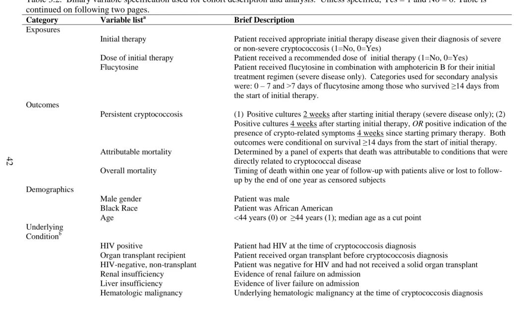
Table 3.2. Binary variable specification used for cohort description and analysis. Unless specified, Yes = 1 and No = 0. Table is continued on following two pages.
Category Variable lista Brief Description Exposures
Initial therapy Patient received appropriate initial therapy disease given their diagnosis of severe or non-severe cryptococcosis (1=No, 0=Yes)
Dose of initial therapy Patient received a recommended dose of initial therapy (1=No, 0=Yes)
Flucytosine Patient received flucytosine in combination with amphotericin B for their initial treatment regimen (severe disease only). Categories used for secondary analysis were: 0 – 7 and >7 days of flucytosine among those who survived ≥14 days from the start of initial therapy.
Outcomes
Persistent cryptococcosis (1) Positive cultures 2 weeks after starting initial therapy (severe disease only); (2) Positive cultures 4 weeks after starting initial therapy, OR positive indication of the presence of crypto-related symptoms 4 weeks since starting primary therapy. Both outcomes were conditional on survival ≥14 days from the start of initial therapy. Attributable mortality Determined by a panel of experts that death was attributable to conditions that were
directly related to cryptococcal disease
Overall mortality Timing of death within one year of up with patients alive or lost to follow-up by the end of one year as censored subjects
Demographics
Male gender Patient was male
Black Race Patient was African American
Age <44 years (0) or ≥44 years (1); median age as a cut point Underlying
Conditionb
HIV positive Patient had HIV at the time of cryptococcosis diagnosis
Organ transplant recipient Patient received organ transplant before cryptococcosis diagnosis
HIV-negative, non-transplant Patient was negative for HIV and had not received a solid organ transplant Renal insufficiency Evidence of renal failure on admission
Liver insufficiency Evidence of liver failure on admission
43
Category Variable lista Brief Description Baseline Disease
Severe disease Patients with disease where induction therapy with amphotericin B is
recommended by IDSA Guidelines (CNS disease, or treat as CNS disease) [1]; non-severe cases were non-CNS disease where fluconazole therapy for primary treatment is recommended
Positive CNS culture Patient had positive fungal culture from CSF Positive blood culture Patient had positive fungal culture from blood
Positive pulmonary culture Patient had positive fungal culture from lung biopsy or bronchoalveolar lavage Other histological evidence Other histological test positive for Cryptococcus
Symptoms
No symptoms Patient reported no disease-attributable symptoms Altered mental status Yes/No
Headache Yes/No
Cough Yes/No
Shortness of breath Yes/No
Night sweats Yes/No
Fever Yes/No
Nausea Yes/No
Vomiting Yes/No
Seizures Yes/No
Initial LPc
CSF CRAGd titer ≥1:1024 High was defined as ≥1:1024 Low CSF:serum glucose ratio CSF:serum glucose ratio was <0.6 Low CSF glucose CSF glucose ≤40mg/dL
High CSF protein CSF protein ≥45mg/dL High nucleated cells Nucleated cells >20cells/mm3
High LP OPe OP ≥20cm H2O
44
Category Variable lista Brief Description Other
High serum CRAG titer Serum CRAG titer ≥1:1024
Corticosteroid exposure Patient on corticosteroid therapy at time of cryptococcosis diagnosis Calcineurin inhibitor Patient taking calcineurin inhibitor
Mycophenolate mofetil Patient taking mycophenolate mofetil Azathioprine Patient taking azathioprine
Methotrexate Patient taking methotrexate
Monoclonal antibodies Patient taking monoclonal antibodies
Sirolimus Patient taking sirolimus
HAART HIV-infected patient reported prior or current exposure to HAART a
Unless unavailable and where applicable, all information was gathered from the procedure or chart entry closest to cryptococcosis diagnosis date. b
Since HIV positive, transplant recipients and HIV-negative/non-transplant groups were exclusive categories, dummy variables indicating the three groups were used for multivariate models.
c
LP = Lumbar puncture; first measured value for each patient d
CRAG = Cryptococcal antigen e
Figures
Figure 3.1. Conceptual model showing timing of patient cohort conditions and events. All patients enter the study at the time of diagnosis and are followed until death, lost to follow-up or until the end of the study period (October 31st, 2009).
1. Patient is diagnosed with cryptococcosis 1a. Died before therapy 2. Antifungal treatment
started 3. Patient cured or suppressed with maintenance therapy 4. Patient dies or lost to follow-up 2a. Patient
dies while on therapy
Change in antifungal therapy
3a. Relapse of cryptococcosis or
IRIS
3b.Patient dies 2c. Patient dies
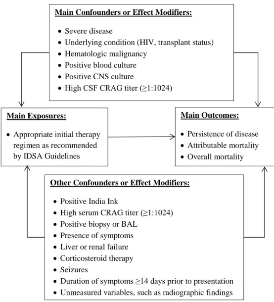
Figure 3.2. Simplified causal diagram for specific aim 2: initial treatment and dose of initial treatment. Covariates listed were determined as the minimum adjustment set for models by the DAG program by Knüppel [100].
Main Exposures:
Appropriate initial therapy regimen as recommended by IDSA Guidelines
Main Outcomes:
Persistence of disease
Attributable mortality
Overall mortality Main Confounders or Effect Modifiers:
Severe disease
Underlying condition (HIV, transplant status)
Hematologic malignancy
Positive blood culture
Positive CNS culture
High CSF CRAG titer (≥1:1024)
Other Confounders or Effect Modifiers:
Positive India Ink
High serum CRAG titer (≥1:1024)
Positive biopsy or BAL
Presence of symptoms
Liver or renal failure
Corticosteroid therapy
Seizures
Duration of symptoms ≥14 days prior to presentation
Figure 3.3. Simplified causal diagram for specific aim 2: flucytosine exposure (severe disease only). Covariates listed were determined as the minimum adjustment set for models by the DAG program by Knüppel [100].
Main Exposures:
Flucytosine combination therapy used (yes/no)
≤7 vs. >7 days of flucytosine
Main Outcomes:
Persistence of disease
Attributable mortality
Overall mortality Main Confounders or Effect Modifiers:
Hematologic malignancy
Positive blood culture
Positive CNS culture
Underlying condition (HIV, transplant status)
High CSF CRAG titer (≥1:1024)
Receipt of AmpBd as initial primary therapy
Other Confounders or Effect Modifiers:
Corticosteroid therapy
High serum CRAG titer (≥1:1024)
Positive India ink
Seizures
Liver or renal failure
Duration of symptoms ≥14 days prior to presentation
Presence of symptoms
CHAPTER IV. COMPARISON AND TEMPORAL TRENDS OF THREE GROUPS WITH CRYPTOCOCCOSIS: HIV-INFECTED, SOLID ORGAN TRANSPLANT
AND HIV-NEGATIVE/NON-TRANSPLANT
Overview
The Infectious Disease Society of America (IDSA) 2010 Clinical Practice Guidelines for the management of cryptococcosis outlined three key populations at risk of disease: (1) HIV-infected, (2) transplant recipient, and (3) HIV-negative/non-transplant. However, direct comparisons of management, severity and outcomes of these groups have not been
HIV-positive and HIV-negative/non-transplant patients accounted for 89% of severe disease cryptococcosis-attributable deaths and 86% of all-cause mortality. In this single-center study, the frequency of cryptococcosis did not change in the last two decades, although the underlying case mix shifted (fewer HIV-positive cases, stable transplant cases, more cases with neither). Cryptococcosis had a relatively uniform and informed treatment strategy, but disease-attributable mortality was still common.
Introduction
Cryptococcus neoformans is an invasive mycoses that can cause meningoencephalitis, particularly among those who are immunocompromised, but in some cases
an active Infectious Disease group at our institution with a particular interest in the pathogenesis and treatment of cryptococcosis.
HIV-positive populations with cryptococcosis have been the most widely studied group over the last two decades [5,13,41,42,47,78,105,106,107,108,109,110,111] and have received greater attention recently due to the recognition that cryptococcosis incidence in this group remains high and paralleled with the AIDS epidemic in sub-Saharan Africa [23]. Starting in the 1960 – 1980’s, use of immunosuppressive medications to treat severe diseases or for solid organ transplantation has increased the pool of patients susceptible to
Cryptococcus and, in the late 1990’s Cryptococcus gattii emerged in Vancouver Island, British Columbia, Canada, resulting in an outbreak of infections in both immunosuppressed and immunocompetent hosts [38,39]. Though clinical isolates were not typed in this study, serotype A (C.neoformans var. grubii) predominates this region [112]; C.gattii is rare in the Southeastern U.S. with only one identified clinical case in an immunocompromised adult [40]. The HIV-negative cryptococcosis patient group had been excluded from clinical review for several decades but has gained more attention recently [21,45,62,68,69,70]. Cryptococcal patients who are HIV-negative, particularly those who have few or no underlying risk factors (i.e., “apparently immunocompetent”), may experience more of a delay in time to presentation and diagnosis than HIV-positive or transplant recipient patients [21]. In particular, recent evidence from a study showed that HIV-negative,
non-immunosuppressed cryptococcal meningitis patients suffered higher mortality rates than HIV-positive patients [62].
opinion and outdated and retrospective cohort studies, with few representing HIV-negative populations and comparatively developed countries [19,21,22,48]. In this relatively large, retrospective single-center study, our goal was to provide an in-depth look at how
cryptococcosis was managed clinically in the positive, transplant recipient and HIV-negative/non-transplant patient groups in order to improve our understanding of this disease.
Methods
Objectives
The goals of this study were to describe trends in cryptococcosis symptoms, diagnosis, treatment and mortality through a 14-year study period (1996–2009) within the context of the three groups defined by the IDSA Guidelines.
Participants
We identified all consecutive adult patients (≥18 years old) discharged from DUMC with International Classification of Diseases, 9th Revision (ICD-9) diagnosis codes of cryptococcosis (117.5), and cryptococcal meningitis (321.0) between January 1, 1996 and October 31, 2009 through electronic medical records. Eligible subjects had confirmed cryptococcal disease and a sufficient medical record (electronic and/or paper chart) available for review. A cryptococcosis case was confirmed by having ≥1 of the following: positive cerebral-spinal fluid (CSF) cryptococcal antigen (CRAG) or fungal culture, direct