CSEIT1831358 | Received : 16 Feb 2018 | Accepted : 28 Feb 2018 | January-February-2018 [(3) 2 : 245-252 ]
International Journal of Scientific Research in Computer Science, Engineering and Information Technology © 2018 IJSRCSEIT | Volume 3 | Issue 2 | ISSN : 2456-3307
245
Image Processing Strategies for Fusion of Multiple Images: A
Comprehensive Analysis
Prabhjit Kaur, Prabhpreet Kaur
MTECH Guru Nanak Dev University, Amritsar, Punjab, India
ABSTRACT
The digital image processing is capable of handling the problems extracted out of several domains. Information collected over the several domains is required to be filtered. The process of extracting information out of several domains is known as image fusion. Application areas of image fusion could be many. This paper highlights the application of image fusion in fields of health care using MRI and CT images etc. The phases associated with image fusion include feature detection, feature matching, transform model estimation and image resampling and transformation. Each of these phases is elaborated for detecting the enhancement parameter for future endeavours. Comparative analysis is presented to determine the optimal technique that can be worked upon to obtain optimal parameter listening.
Keywords: Image processing, image fusion, feature detection, matching, transform model estimation, image resampling and transformation
I.
INTRODUCTION
Image processing becomes catastrophic in modern era for providing the relevant information to the user.[1], [2] The information is represented in digital form and is extracted out in the user understandable format. Bandwidth conservation is always an issue associated with the image processing. In order to tackle this issue image fusion is utilized. Several images are collaborated together to form a single image.[3], [4] to fuse the image, first of all image acquisition is performed. After acquisition is complete registration is performed. After performing registration, images are fused together. Each of these steps is critical and failure in any one of the phases could lead to the failure of image fusion. Image fusion mechanisms are categorised as under
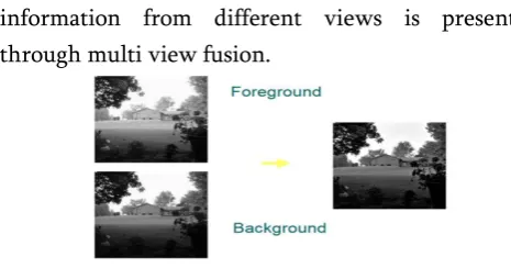
1.1 Multi view Fusion
[5]Images of the same modality are collected and fused together. The images are taken at the same time from distinct places. Complementary
information from different views is presented through multi view fusion.
Figure 1. Multi view Fusion
1.2 Multi Model Fusion
[6] Images of different modality are collaborated together. The images of multi model includes PET, CT, MRI etc. the aim is to decrease the amount of data required to be transmitted and to conserve bandwidth representation.
1.3 Multi temporal Fusion
[7]Images belonging to same scene but taken at different time periods are collaborated to form multi temporal fusion. Multi temporal fusion yield mechanism to detect changes within the images. Subtraction is commonly done to obtain change detection.
Figure 3. Multi temporal fusion
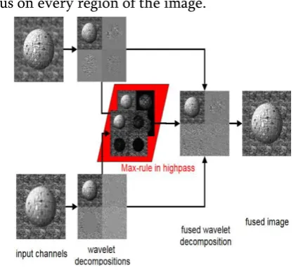
1.4 Multi Focus Fusion
[5], [6]Multi focus fusion is accomplished by dividing the image into regions. Every region is in focus in at least one channel. The aim of this fusion is to set a focus on every region of the image.
Figure 4. Multi Focus Fusion 1.5 Image Fusion for restoration
[8]Image consist of many distinct components. These components may include optimal and degradation components. Removing degradation components from the image forms image fusion for restoration. All of these fusion methods provide the way of conserving the bandwidth and provide a way to highlight main features associated with the images.
The phases associated with image fusion is described as under
1.6 Feature Detection
[9]Image is associated with large number of features. These features are represented with slandered deviation, mean, median kurtosis etc. all of these features are required to be detected and extracted. This is accomplished using this phase.
1.7 Feature Matching
[10] There exist a training set of images. The extracted features are matched against the already extracted features stored within training set. Matched features are retained and rest of the features are neglected.
1.8 Model estimation
[10]Model estimation is used in order to determine the optimal methodology that can be used in order to represent the features extracted from the image. Entropy in case of optimal feature extraction is exceedingly high. Entropy represents degree of relationship between pixels.
1.9 Resampling
[11] This process is used to collaborate the pixels to form a new image. The image sampling then extracts the parameters for comparison. MSE and PSNR and basic parameters used for enhancement of image and to check optimality.
Next section described literature survey of techniques and phases associated with image fusion.
II.
LITERATURE SURVEY
Literature survey describes the existing algorithms used to perform image fusion operation. The algorithms are listed as under
2.1 Simple Average
density. Thus this mechanism is simple way of obtaining an output or desired image with all critical regions in focus. The value of pixel from each region is taken and added together. This sum is divided by total number of pixels obtained within the scene. The average value gives the region of interest.
∑
2.2 Select Maximum
The highest pixel intensity value is selected for determining in focus region. Thus this algorithm selects the pixels with highest intensity pixels from each input image. The highest pixel intensity level pixels replace the corresponding pixels for image fusion.
2.3 Discrete Wavelet Transform
DWT is generally utilized as a part of numerical and utilitarian investigation. In these territories wavelets are thought to be discretely circulated. DWT has advantage that both area and recurrence data is considered. DWT has advantage more than Fourier change since it has transient determination. The idea of wavelet is basic. [12]They are utilized for multistage examination prepare. Depiction of multistage wavelet is portrayed considering the case as
Example 1
The sequence of wavelets are considered using n=23 y={1,1,2,3,1,3,2,2}
Consider vectors P and L computed through algorithm for multistage which can be applied as follows
1.
√
2.
√ ( )
3.
4. If I=0 then stop else move to step 1
The essential thoughts behind wavelets are depicted through the above recorded calculation. The calculation gives essential comprehension of the wavelets or gives smaller structure investigation of put away data.
Discrete wavelet change is additionally depicted as far as vitality. The vitality protection can be depicted considering the accompanying illustration.
Example 2
Grouping of example can be considered in which vitality is put away. The example can be broke down utilizing taking after
|| ||
|| ||
Transform coefficient can be calculated using following equation
|| || (√ ) (√ ) ( √ )
( ) ( √ )
( √ )
|| ||
Since both qualities are equivalent consequently vitality is monitored.
There exists R bundle which can be connected to wavelet change to get the bend as takes after
2.4 Correlation
Confront location is intricate since numerous particular expressions are included. The facilitate examination is directed to decide match or confuse expression. For this reason relationship is valuable. Connection communicates connection between pixels through position values including x and y. Relationship is communicated with the assistance of connection coefficient shown with r. Estimation of r lies in the vicinity of 0 and 1. Relationship coefficient is best portrayed considering the accompanying illustration.
X Y
X1 Y1
X2 Y2
X3 Y3
X4 Y4
X5 Y5
--- --
--- --
Xn Yn
Then correlation coefficient is calculated using following equation.
2.5 Principal Component Analysis
Relationship component is mind boggling and high cost is experienced as intricate pictures are taken care of. So as to determine the issue PCA technique is utilized. PCA is a measurable methodology that utilizations orthogonal change to change over conceivably related qualities into set of non associated direct values known as primary parts. It uses the arrangement of Eigen values that are works from the arrangement of preparing informational collections. From these Eigen values the preparation confront pictures have been figured which are masterminded finding the most difference in picture. After this the Euclidean separation from the info confront has been computed for every Eigen values. This can ordered the picture into parts in light of
Euclidean separation. The weighted total of Eigen faces spoken to by content face pictures anticipated on to the space extended by Eigen faces. The appearances can be distinguished by these weights. [13]
The accompanying is route through which the related qualities have been computed:
∑
∑
2.6 Linear Discriminant Analysis
Linear Discriminant examination is valuable to decide consolidated components that do the partition of the classes. The length and multifaceted nature related with the figuring’s are lessened utilizing LDA approach. The dimensionality diminishment and characterization of face acknowledgment is proficient utilizing slightest time and space multifaceted nature. Twisting inside the picture is normal.[14] This is likewise refined through LDA. Mathematically, a set of n dimensional vectors xi, x2 ,---,xn belongs to l classes of faces.
Max
Where
Sn=∑ ( )
Sw=∑ ∑ ( )( )
U is the mean of training images presented to the simulation. Sw is within the scatter matrix and Sn is between class scatter matrixes.
III. COMPARISON TABLE OF EXISTING LITERATURE
The comparison of existing techniques is presented in order to derive the best possible technique for future enhancement.
Table 1. Qualitative analysis of various image fusion mechanisms Ref
no.
Author’s name
Title of paper Techniques used
Para-meters
Future Work
Merits Demerits
1. S. Avinash between pixels
is not Breast Cancer Lesions[18] Analysis of Face Recognition
Entropy is low
5. M. Satone et.al. (2014)
Feature
Selection Using Genetic
Algorithm for
Face like accuracy and MSE is not
operation can be improved along with accuracy through noise cancellation procedure 8. M. Rahate
et.al.(2013)
Image Fusion to Enhance the
Noise handling mechanism phase results in low PSNR
IV. RESEARCH GAP
The existing techniques analysed describe image fusion in the field of multi modal systems. Multi modal systems includes images from the MRI and CT scan that are collaborated together to achieve fusion.
to improve parameters such as accuracy and Peak signal to noise ratio.
V.
CONCLUSION AND FUTURE SCOPE
This works provide in-depth study of techniques that are applied over the fused image to find abnormality within the image. The comparative analysis also suggest the merits and demerits which can be used to improve the current strategy for abnormality detection. Most of the techniques analysed lacks Noise handling procedures. Also image enhancement procedures are missing. So in future noise handling and enhancement procedure can be merged along with image fusion for better abnormality detection within the fused image.
VI. REFERENCES
[1]. N.Singla, "A Comparative Study of Noising And Denoising Technique In Image Processing," vol.4, no.3, pp.38-42, 2016.
[2]. Y.Ma, D.Lin, B.Zhang, Q.Liu, and J.Gu, "A Novel Algorithm of Image Gaussian Noise Filtering based on PCNN Time Matrix," in 2007 IEEE International Conference on Signal Processing and Communications, 2007, pp.1499-1502.
[3]. P.Yuvarani, "Image Denoising and Enhancement for Lung Cancer Detection using Soft Computing Technique," pp.27-30, 2012. [4]. S.Huda, J.Yearwood, H.F.Jelinek, M.M.Hassan,
and M.Buckland, "A hybrid feature selection with ensemble classification for imbalanced healthcare data : A case study for brain tumor diagnosis," vol.3536, no.c, pp.1-13, 2016. [5]. I.Reducindo, E.R.Arce-santana,
D.U.Campos-delgado, F.Vigueras-g, A.R.Mej, and G.Rizzo, "Non-rigid Multimodal Medical Image Registration Based on the Conditional Statistics of the Joint Intensity Distribution," vol.7, pp.126-133, 2013.
[6]. V.Bhavana and H.K.Krishnappa, "Multi-Modality Medical Image Fusion using Discrete Wavelet Transform," vol.70, pp.625-631, 2015. [7]. I.Kosesoy, M.Cetin, and A.Tepecik, "A Toolbox
for Teaching Image Fusion in Matlab," Procedia - Soc.Behav.Sci., vol.197, no.February, pp.525-530, 2015.
[8]. P.B.Dasgupta, "Analytical Comparison of Noise Reduction Filters for Image Restoration Using SNR Estimation," vol.17, no.3, pp.121-124, 2014.
[9]. R.Kaushik, R.Kumar, and J.Mathew, "On Image Forgery Detection Using Two Dimensional Discrete Cosine Transform and Statistical Moments," vol.70, pp.130-136, 2015.
[10]. I.D.T, B.Goossens, and W.Philips, "MRI Segmentation of the Human Brain : Challenges , Methods , and Applications," vol.2015, 2015. [11]. D.Van De Ville, M.Nachtegael, D.Van Der
Weken, E.E.Kerre, W.Philips, I.Lemahieu, and S.Member, "Noise Reduction by Fuzzy Image Filtering," vol.11, no.4, pp.429-436, 2003. [12]. P.Dave and M.Tech, "Study and Analysis of
Face Recognition system using Principal Component Analysis ( PCA )."
[13]. X.Luan, B.Fang, L.Liu, W.Yang, and J.Qian, "Extracting sparse error of robust PCA for face recognition in the presence of varying illumination and occlusion," Pattern Recognit., vol.47, no.2, pp.495-508, 2014.
[14]. K.H.An, S.H.Park, Y.S.Chung, K.Y.Moon, and M.J.Chung, "Features for Face Detection Based on Ada-LDA," pp.1117-1122, 2009.
[15]. S.Avinash, "An Improved Image Processing Analysis for the Detection of Lung Cancer using Gabor Filters and Watershed Segmentation Technique."
[16]. M.Satone and G.Kharate, "Feature Selection Using Genetic Algorithm for Face Recognition Based on PCA , Wavelet and SVM," vol.6, no.1, pp.39-52, 2014.
[18]. V.Ponomaryov, "Computer-aided detection system based on PCA/SVM for diagnosis of breast cancer lesions," 2015 Chil.Conf.Electr.Electron.Eng.Inf.Commun.Tec hnol., pp.429-436, 2015.
[19]. P.N.Dangat and P.S.D.Joshi, "Efficient Disease Detection Approach Based on MRI and CT Images Fusion Technique," vol.3, no.7, pp.918-922, 2014.