Western University Western University
Scholarship@Western
Scholarship@Western
Electronic Thesis and Dissertation Repository
12-4-2015 12:00 AM
Exploring And Training Spatial Reasoning Via Eye Movements:
Exploring And Training Spatial Reasoning Via Eye Movements:
Implications On Performance
Implications On Performance
Victoria A. RoachThe University of Western Ontario
Supervisor
Dr. Timothy D. Wilson
The University of Western Ontario
Graduate Program in Anatomy and Cell Biology
A thesis submitted in partial fulfillment of the requirements for the degree in Doctor of Philosophy
© Victoria A. Roach 2015
Follow this and additional works at: https://ir.lib.uwo.ca/etd
Part of the Cognition and Perception Commons, Cognitive Neuroscience Commons, Educational Assessment, Evaluation, and Research Commons, Educational Psychology Commons, Experimental Analysis of Behavior Commons, and the Science and Mathematics Education Commons
Recommended Citation Recommended Citation
Roach, Victoria A., "Exploring And Training Spatial Reasoning Via Eye Movements: Implications On Performance" (2015). Electronic Thesis and Dissertation Repository. 3446.
EXPLORING AND TRAINING SPATIAL REASONING VIA EYE MOVEMENTS:
IMPLICATIONS ON PERFORMANCE
Integrated Article
by
Victoria Roach
Graduate Program in Anatomy and Cell Biology
A thesis submitted in partial fulfillment
of the requirements for the degree of
Doctor of Philosophy
The School of Graduate and Postdoctoral Studies
The University of Western Ontario
London, Ontario, Canada
Abstract
This dissertation sought to determine if eye movements could serve as an indicator of success in spatial reasoning, and if eye movements associated with successful completion could be applied to strategically improve spatial reasoning.
Using the line images of Shepard and Metzler, an electronic test of mental rotations ability (EMRT) was designed. Two versions of the test were created, allowing for both a timed (6 seconds per question) and untimed testing environment. Four experiments were designed and completed to relate mental rotation ability (MRA) scores from the EMRT, to patterns in chrononumeric and visual salience data. In each experiment, participants completed the EMRT under a different protocol. These protocols included an untimed EMRT, a timed EMRT, a within-participant crossover study where participants
completed both the timed, and untimed EMRT in series, and a training crossover study where low MRA participants completed the timed EMRT in both a guided and unguided environment.
In the untimed experiment, individuals of high and low MRA were asked to complete the EMRT while their eye movements were observed. As no time limit was imposed, the results allowed for observations based on MRA alone, and served to demonstrate and how individuals of different skill level differ in terms of eye movement.
In the following experiment, the addition of a time limit to the EMRT revealed how individuals of high and low MRA perform when under a time restriction. The results of the Timed experiment confirmed differences between the high and low MRA group in terms of eye movements, and attention to salient regions of test images.
accuracy, and identification of visually salient elements.
The results from the first three experiments were then applied in the Guidance
Experiment to confirm the role that visual salience plays in the context of spatial problem solving. By mapping the apprehension patterns of successful high MRA individuals onto the EMRT, low MRA individuals could be guided to salient areas on the timed EMRT. The results revealed that the application of visual guidance is an effective mechanism for MRA training.
This research attends to a previously unaddressed niche in eye-movement and spatial ability training literature. As a result, it may serve as a foundation to cultivate methods of honing and improving spatial skills in the general population.
Keywords
Co
-
Authorship Statement
The written material in this thesis is the original work of the author. Victoria Roach participated in all aspects of the work contained herein: conception of the hypotheses, conduction of the experiments, and authorship of the manuscripts. The role of the co-authors are detailed below by chapter.
Chapter 3: Eye movements during untimed tests of mental rotation ability.
The manuscript is published in the journal Anatomical Sciences Education. All authors on the manuscript shared in the conception of this research study. V. Roach and J. Kryklywy, using digital images created by Michael Peters, with permission, developed the Electronic Mental Rotations Test (EMRT). The data for this study was collected, analyzed and interpreted by V. Roach and G. Fraser. V. Roach carried out the composition of the manuscript with inputs from Drs. Wilson and Mitchell.
Chapter 4: Eye movements during timed tests of mental rotation ability.
Conception of this research study was shared by V. Roach, T. Wilson, and D. Mitchell. V. Roach and J. Kryklywy carried out the modifications to the Electronic Mental Rotations Test. The data for this study was collected, analyzed and interpreted by V. Roach and G. Fraser. V. Roach carried out preparation of the manuscript with inputs from Drs. Wilson and Mitchell. The manuscript is under review at the journal Cognitive Science.
Chapter 5: Comparison of eye movements during tests of mental rotation in timed and untimed conditions.
from Drs. Wilson and Mitchell. The manuscript is under review at the journal Anatomical Sciences Education.
Chapter 6: The effects of visual guidance on success in tests of mental rotation.
Conception of this research study was shared by V. Roach, T. Wilson, and D. Mitchell. V. Roach, J. Kryklywy and G. Fraser completed the modifications to the Electronic Mental Rotations Test. The data for this study was collected, analyzed and interpreted by V. Roach and G. Fraser. V. Roach carried out preparation of the manuscript with inputs from Drs. Wilson and Mitchell. The manuscript will be submitted to the journal
Acknowledgments
First and foremost I offer my sincerest thanks to my primary supervisor, Dr. Timothy D. Wilson. His support, mentorship and patience have carried me through this degree, and given me countless opportunities to grow into an independent scientist.
I would also like to thank my advisors; Drs. Brian Allman, Derek Mitchell, Mathew Heath and Marjorie Johnson for giving sage advice and helping me sort out the technical details of my work. They were a sounding board for my work, and provided countless insightful comments and constructive criticisms that helped me to focus my ideas and guide me throughout my research.
Further, I must express my sincere thanks to my research colleagues, James Kryklywy and Graham Fraser. Their technical savvy, resilience and patience were instrumental in the execution of each and every experiment in this dissertation, and I am wholly indebted to them.
Moreover, I must thank my many friends in Anatomy and Cell Biology. I am particularly grateful to Drs. Charys Martin and Michele Barbeau and Ngan Nguyen, who set me up for success in my PhD, and to Dr. Stefanie Attardi, Lauren Allen, and Danielle Brewer, for helping me stay positive, and focused. Further, I would like to thank my brother Andrew Roach, and Shannon Lovell, for their tireless support and motivation through the challenging last few years.
Table of Contents
Abstract ... i
Co-Authorship Statement ... iii
Acknowledgments ... v
Table of Contents ... vi
List of Acronyms And Abbreviations ... x
List Of Tables ... xi
List of Figures ... xii
General Introduction ... 1
1.1Spatial Ability ... 1
1.2 Mental Rotation Ability ... 1
1.3 Training Spatial (and Mental Rotation) Ability ... 2
1.4 Gaze-Directed Training ... 3
1.5 Overview of Dissertation ... 4
1.6 References ... 6
Literature Review ... 9
2.1 The Eyes, Sight, and Tracking their Movements ... 9
2.1.1 The Anatomy of the Visual System ... 9
2.1.2 The Eye and Extra-Ocular Muscles ... 9
2.1.3 The Retina ... 11
2.1.4 Movements of the Eyes ... 12
2.1.5 Eye Tracking Technology ... 14
2.1.6 Applications of Eye Tracking ... 16
2.1.7 Summary ... 16
2.2 Spatial Ability ... 17
2.2.1 A Brief History ... 17
2.2.2 Factors Comprising Spatial Ability ... 18
2.2.3 Developmental Research in Spatial Ability ... 19
2.2.4 Links to Academics, STEM, and other disciplines ... 20
2.2.6 Summary ... 22
2.3The Effect of Time Limits in Scholastic Assessment ... 22
2.3.1 General Introduction ... 22
2.3.2 Time Limits and Speededness ... 22
2.3.3 The Validity of Speeded Tests ... 23
2.3.4 Individual Differences in Working Memory ... 24
2.3.5 Individual Differences in Mental Processing Speed ... 25
2.3.6 Individual Differences in Strategy ... 25
2.3.6 Summary ... 27
2.4Neural Underpinnings ... 27
The Visual Processing System ... 27
2.4.1 Brief Summary of the Cortical Areas in the Visual Pathway ... 27
2.4.2 Visual Pathways ... 30
2.4.3 Neural Underpinnings of Mental Rotations Ability ... 32
2.4.4 The Neural Underpinnings of Working Memory ... 33
2.4.5 Summary ... 35
2.5 Training using Expert Eye Modeled Examples ... 36
2.5.1 Gaze-Based Training ... 36
2.5.2 Expert-Data-Driven Approaches to Visual Guidance ... 37
2.5.3 Types of Eye Movement Modeled Example (EMME) Cueing ... 37
2.5.4 EMME Training in Psychomotor Tasks ... 39
2.5.5 EMME Applications in Surgery ... 39
2.5.6 EMME Applications in Perceptual Tasks ... 40
2.5.7 Summary ... 41
2.6 Overview of Empirical Chapters ... 41
2.7 Overall Objectives ... 42
2.8 References ... 43
Chapter 3 ... 56
3 Eye Movements During Untimed Tests of Mental Rotation Ability ... 56
3.1 Introduction ... 56
3.5 References ... 75
Chapter 4 ... 79
4 Eye Movements During Timed Tests of Mental Rotation Ability ... 79
4.1 Introduction ... 79
4.2 Materials & Methods ... 82
4.3 Results ... 86
4.4 Discussion ... 94
4.5 References ... 99
Chapter 5 ... 102
5 A Comparison of Eye Movements During Tests of Mental Rotation in Timed and Untimed Conditions ... 102
5.1 Introduction ... 102
5.2 Materials & Methods ... 106
5.3 Results ... 110
5.4 Discussion ... 126
5.5 References ... 132
Chapter 6 ... 135
6 The Effects Of Visual Guidance On Success In Tests Of Mental Rotation Ability ... 135
6.1 Introduction ... 135
6.2 Materials & Methods ... 137
6.3 Results ... 142
6.4 Discussion ... 146
6.5 References ... 149
Chapter 7 ... 153
7 Discussion ... 153
Limitations ... 161
Future Directions ... 163
References ... 164
Appendices ... 167
A. Copyright Releases ... 167
List of Acronyms And Abbreviations
1. Mental Rotation Ability (MRA) 2. Mental Rotation Test (MRT)
3. Vandenberg and Kuse Mental Rotation Test (VKMRT) 4. Electronic Mental Rotation Test (EMRT)
5. High Mental Rotation Ability (HMRA) 6. Low Mental Rotation Ability (LMRA) 7. Eye Movement Modeled Examples (EMME) 8. Region of Interest (ROI)
9. Analysis of Variance (ANOVA) 10.Standard Deviation (SD)
11.Male (M) /Female (F) 12.Confidence Interval (CI)
13.Science, Technology, Engineering and Mathematics (STEM)
14.Science, Technology, Engineering, Mathematics and Medicine (STEMM). 15.Electro-oculography (EOG)
16.Photo-oculography (POG) 17.Video-oculography (VOG)
18.Congenital adrenal hypoplasia (CAH) 19.Milliseconds (ms)
20.Hertz (Hz)
21.Three-dimensional (3D)
22.Posterior Parietal Cortex (PPC)
List Of Tables
List of Figures
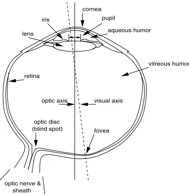
Figure 1: The basic anatomy of the structures of the human eye, adapted from Duchowski (2007) with permission © 2007 Springer Science & Business Media ... 10 Figure 2: Extrinsic (Extra-ocular) muscles of the eye, adapted from Davson (1980) with
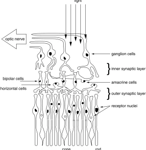
permission © 1980 Academic Press. ... 11 Figure 3: The basic composition of the layers of the human retina, adapted from
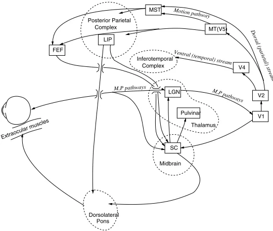
Duchowski (2007) with permission © 2007 Springer Science & Business Media. . 12 Figure 4: Schematic representation of the brain and the visual pathways that are relevant
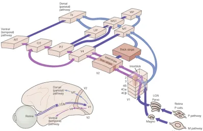
to eye movements and attention, adapted from Duchowski (2007) with permission © 2007 Springer Science & Business Media ... 30 Figure 5: The basic anatomy of the visual pathways of the human brain. ... 31 Figure 6: Four sample questions based on the Shepard and Metzler block pairs used in the EMRT15. Each pair represents one question. Participants used a keyboard to indicate
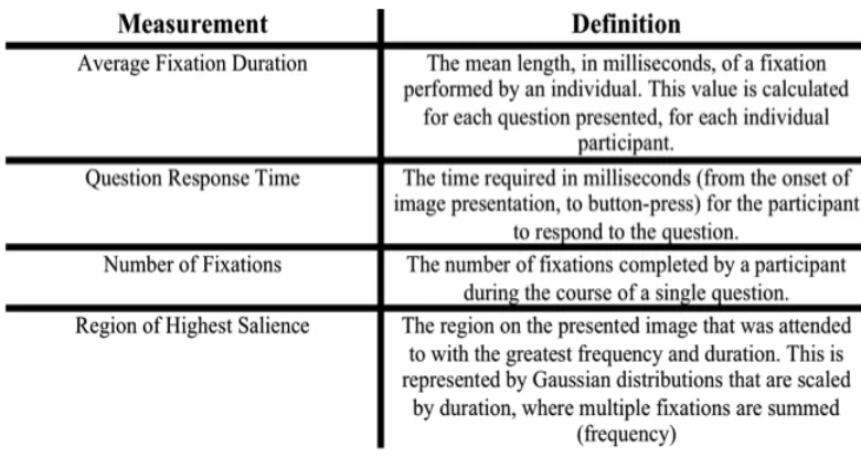
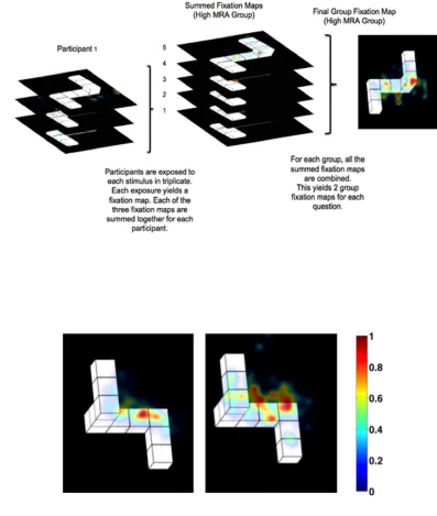
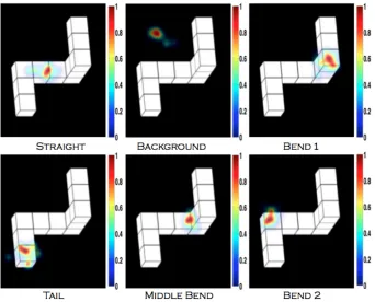
if shapes were the same or different. Answer key: clockwise from the top-left: images are different from each other, different, identical and identical. ... 60 Figure 7: A schematic representing the creation of salience maps. A, A representation of
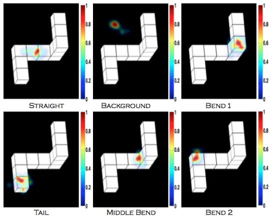
the protocol employed to interpret spatial data employing the summation of individual fixation maps to the development of a group level heat map; B, The dichotomy between high and low MRA group heat maps indicative of the position of highest salience. High MRA, on the left, fixate predominantly on one location and on little else; while Low MRA on the right attend to several points of interest. The color bar indicates the salience of the region, where red (1) represents areas of highest salience, and blue (0) represents areas of lowest salience in arbitrary units.. ... 62 Figure 8: A representation of the six categorizations for the location of highest spatial
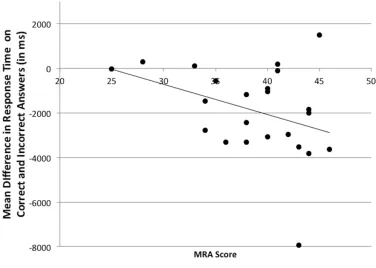
salience, scales to right of diagrams are arbitrary units. ... 63 Figure 9: The relationship between MRA score and the mean difference of response time
Figure 10: The frequency distribution of highest saliency by region, for both high and low groups. No significant difference between the two groups was observed,
suggesting that the groups attend to the regions in the same proportion. ... 68 Figure 11: A diagrammatic representation of the 6 possible categorizations for the
location of highest spatial salience. The areas colored red represent the most attended region of the image, and are indicative of the highest salience across the group. ... 84 Figure 12: The relationship between Timed EMRT Scores, and VKMRT Scores,
confirming the Timed EMRT as a valid measure of MRA. The maximum score is 24 on the VKMRT and 48 on the Timed EMRT. ... 86 Figure 13: High MRA individuals conduct fixations that are quicker than those completed
by LMRA individuals, on average, during spatial problem solving. Error bars
indicate one standard deviation……...……….86 Figure 14: Average Fixation Duration is constant across different levels of accuracy for
both MRA groups. Error bars indicate 95% confidence interval for within-group comparison. ... 88 Figure 15: High and LMRA individuals exhibit equivalent response times on average.
Error bars represent one standard deviation. ... 89 Figure 16: High and LMRA individuals show different response times based on accuracy. Error bars represent 95% Confidence Interval for within –group comparison. ... 90 Figure 17: High and Low MRA Individuals are equivalent in terms of average number of
fixations per question. Error bars represent one standard deviation. ... 91 Figure 18: Correctly answered questions exhibit significantly fewer fixations per question
than incorrectly answered questions. Error bars represent 95% confidence interval for within-group comparison………...………...…..91 Figure 19: The dichotomy between High and Low frequencies across the categories of
salience. ... 93 Figure 20: The two MRA Groups attend to the same salience category in only 34% of
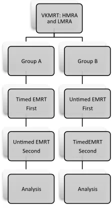
Figure 21: Schematic representation of experimental design and data collection in the experiment………..……….... 105 Figure 22: Diagrammatic representation of the six possible categorizations for the
location of highest salience, derived from Chapter 3: The Untimed Experiment. The areas having the highest salience are presented in red, and indicate the most
attended areas of the image ... 108 Figure 23: A between group comparison of performance (average EMRT score ± SD),
regardless of timing condition. The HMRA group outperforms the LMRA group consistently and significantly F(1.12) = 14.41 (p = 0.003), confirming the significant difference between the groups. ... 111 Figure 24: The performance (average EMRT Score ± 95% Confidence Interval) of both
MRA groups for both timing conditions. Both groups show lower EMRT scores in the timed condition, compared to the untimed condition ... 112 Figure 25: A between-group comparison of average response time (average response time
± SD), regardless of timing condition. There is no difference in average response time between HMRA and LMRA groups ... 113 Figure 26: A within-participant comparison of average response time (average response
time ± 95% Confidence Interval) across timed, and untimed conditions, for both MRA groups. There is a significant difference in average response time between timed and untimed conditions. ... 114 Figure 27: A within-participant comparison of average response time (average response
time ± 95% Confidence Interval) across timed, and untimed conditions, for both MRA groups. There is a significant difference in average response time between timed and untimed conditions. ... 115 Figure 28: The between-group comparison of average fixation duration (average fixation
duration ± Group SD). There is no significant difference in average fixation duration between HMRA and LMRA groups ... 116 Figure 29: The within-participant comparison of average fixation duration (average
Figure 30: The within-participant comparison of average fixation duration (average fixation duration ± 95% Confidence Interval) across both timing conditions, for both MRA groups. No significant difference between average fixation duration was observed according to timed and untimed conditions. ... 118 Figure 31: The between-group comparison of average fixations per question (average
fixations per question ± SD) regardless of timing condition or accuracy of the answer. There was no difference observed in average fixations per question between HMRA and LMRA groups ... 119 Figure 32: The within-group comparison of average fixations per question (average
fixations per question ± 95% Confidence Interval) over correct and incorrectly answered questions, for both MRA groups. There was a significant difference noted in average fixations per question between correct and incorrect answers for both groups ... 120 Figure 33: The within-group comparison of average fixations per question (average
fixations per question ± 95% Confidence Interval) for both MRA groups, on the timed, and untimed conditions. There is a significant difference in average fixations per question between the timed and untimed conditions ... 121 Figure 34: The between-group comparison of salience location frequency, for both timing
conditions. There is a significant difference between the four groups: HMRA:
Timed, HMRA: Untimed, LMRA: Timed and LMRA: Untimed (Fisher Exact Test = 122.18, p <0.001). ... 122
Figure 35: A) The between-group comparison of salience center distribution in the untimed condition. There is no difference between the distribution of salience centers between the HMRA and LMRA groups (Fisher Exact Test: 1.31, p = 0.95). B) The between-group question-by-question agreement for the untimed condition. The groups share salience centers in 75% of questions, showing significant
agreement between the groups during untimed testing (κ = 0.67). ... 123 Figure 36: The between-group comparison of salience center distribution in the timed
groups share salience centers in 42% of questions, showing no agreement between the groups during timed testing (κ = 0.20). ... 124 Figure 37: A) The within-group comparison of salience center distribution in the HMRA
group. There is a significant difference between the distribution of salience centers between the timed and untimed conditions (Fisher Exact Test: 30.33, p <0.001). B) The within-group question-by-question agreement for the HMRA group. The group demonstrates the same salience centers in 38% of questions, showing fair agreement between the testing conditions (κ = 0.25). ... 125 Figure 38: A) The within-group comparison of salience center distribution in the LMRA
group. There is a significant difference between the distribution of salience centers between the timed and untimed conditions (Fisher Exact Test: 76.46, p <0.001). B) The within-group question-by-question agreement for the LMRA group. The group demonstrates the same salience centers in 13% of questions, showing no agreement between the testing conditions (κ = 0.013). ... 126 Figure 39: Exemplar of EMME Guidance Test Question ... 139 Figure 40: Schematic of Experimental Design ... 141 Figure 41: Within-group contrast of EMRT scores ± 95% CI across the two tests, for each test group. A significant interaction was observed between Test and Group, ... 142 Figure 42: Within-Groups contrast of Mean EMRT Scores ± 95% CI across the treatment
groups. ... 143 Figure 43: Between-Group Contrast of Mean Differences between EMRT Tests ± 95%
CI across treatment groups ... 144 Figure 44: The visual salience agreement between EMME exemplars and the LMRA
Chapter 1
General Introduction
1.1
Spatial Ability
Loosely defined as a cluster of cognitive skills that allow an individual to navigate their surroundings, spatial ability has been closely studied by cognitive psychologists for decades1–5. Unfortunately, despite the years of dedicated research, a unified, firm definition of spatial ability has yet to be reached6. The lack of a firm definition is likely the result of the multi-faceted nature of spatial ability. It is accepted that spatial ability is not a singular unitary construct, but more accurately, an assortment of sub-skills which each lend to an individual’s ability to interact with their 3D environment in different ways2.
Research in spatial cognition and spatial ability has served to elaborate on each of the sub-skills of spatial ability. At present, several schemas exist to categorize the sub-skills of spatial ability, including those of Carroll (1993), Lohman (1988), and Linn and Petersen (1985). In the sub-division by Linn and Peterson, three sub-skills of spatial ability were identified as discrete constructs: spatial perception, spatial visualization, and mental rotation2.
1.2
Mental Rotation Ability
Mathematics)disciplines14–18 . The correlates to academic success have resulted in mental
rotations ability catching the attention of the American National Science Board, and led to the publication of “Preparing the next generation of STEM innovators: Identifying and developing our nation's human capital” in which spatial ability features heavily19. In the publication, the authors argue that individuals possessing high spatial ability are an “untapped, pool of talent that are critical to our highly technological society” (p20). This report alludes to individuals possessing high spatial ability, and raises the question of training this cognitive skill19.
1.3 Training Spatial (and Mental
Rotation) Ability
In a recent meta-analysis by Uttal et al. the concept of training spatial ability is explored through a review of current published and unpublished studies attempting to train or elevate individual spatial ability scores18. Uttal et al. was able to discern three different
categories of training efforts: those employing video games as a method of training 20,
those employing an instructional course21and those employing practice, or repeated exposure to spatial tasks as a method of training22,23. The findings of this meta-analysis concluded that spatial training, regardless of technique, yielded an average improvement in spatial scores by almost one-half of a standard deviation on related, untrained spatial tasks18. This suggests that spatial skills are in fact, moderately malleable, and further research is required to discern the most advantageous method of spatial ability training.
In effort to decipher the most advantageous mechanisms to train spatial ability, research has sought to decipher the cognitive underpinnings that govern mental rotations ability. One theory, proposed by Just and Carpenter, suggests that mental rotations ability may manifest in the finite and measureable movements of the eyes. The theory suggests that as an individual attends to spatial task stimuli, their attention is manifest in each fixation of the eye; defined as the act of maintaining visual gaze on a single location for a
duration exceeding 200 milliseconds24. Just and Carpenter further suggest that each fixation of the eye is intimately involved with the ability to visually encode spatially distributed information25,26. With this in mind, patterns in eye movements may represent the initial cognitive stages (i.e., search, transformation and comparison, and
information gathering stage of learning27.
In a foundational study by Just and Carpenter (1985) employing eye-tracking technology and two computer simulation models, significant differences were observed in the visual approaches taken by individuals of high and low MRA during the performance of the Cube Comparison Test28. The significant result obtained by Just and Carpenter gives credence to the theory of a link between cognitive processing and eye movement. The relationship thus provides a sound rationale for the exploration of an eye movement-based, or “gaze- directed”, protocol to formulate and guide the training of mental rotations ability.
1.4 Gaze
-
Directed Training
Gaze-directed training has been employed with varied success in variety of domains; the majority of which pertain to the psychomotor relationship of hand-eye coordination during the completion of physical tasks. Studies within the field of kinesiology have long employed successful gaze-directed training approaches to inform athletes where and when to look for the best results on a variety of athletic maneuvers29–33. The studies of gaze and attention in sport subsequently informed a series of studies in surgical education that applied the gaze-directed approach to training novice laparoscopists. In the studies of several research groups, eye patterns of novice and expert laparoscopists were collected during the performance of standardized technical laparoscopic tasks34–36. These studies exploited differences in the patterns of eye movements between the novices and experts, and elucidated the successful approach employed by the experts34,35. The findings of these two studies yielded a gaze-directed approach employing the eye movements of expert laparoscopists to train novices on specific laparoscopic techniques. When evaluated, the gaze-directed approach based proved to be an effective protocol to guide the attention of novices, and improve their performance on a specific laparoscopic task37.
thesis aims to identify patterns existing in the eye movements of high and low mental rotation ability individuals, as they complete the Electronic Mental Rotation Test (EMRT). The EMRT is based on the line drawings of Shepard and Metzler38,39. These patterns will provide a foundation for the development of a gaze-directed mental rotation training tool to enhance the performance of low spatial individuals on subsequent spatial tests.
1.5
Overview of Dissertation
The purpose of this dissertation is to explore the relationship between eye movements and spatial reasoning in groups of individuals with varying spatial ability, and the implications of this relationship for training mental rotation. Spatial ability, the ability to comprehend three-dimensional structures, is key in how individuals perceive and interact with their surroundings. One subset of spatial ability, mental rotations ability, is linked to success in skill acquisition, and academic success in the STEM disciplines, particularly anatomy, and may serve as a consideration when designing methods for instruction. By using eye tracking, we investigated how eye movements can be recorded and related to underlying cognitive processes associated with spatial test completion. The value of this approach is that it allows us to characterize visual apprehension strategies used by adept individuals during spatial reasoning with and without a time limit, and employ successful visual apprehension strategies to improve mental rotation performance in individuals who typically struggle. In addition to enabling us to improve the performance of low ability individuals, this approach also allows us to begin to assess more effectively how visual cueing strategies can be used to guide instruction in spatially complex disciplines, including anatomy, aeronautics, surgery and STEM.
The remainder this dissertation is divided into 6 chapters:
spatial ability, and briefly refer to working memory as it relates to spatial problem solving. Finally, I discuss the effect of time limitations on accuracy in spatial problem solving, and refer to literature that summarizes the efforts made to train spatial problem solving, including those employing eye movement modeled examples to direct the visual attention of trainees.
Chapters 3 – 6 are the four experiments in this dissertation. “Chapter 3: The Untimed Experiment” examines whether there are observable differences in eye movement patterns between High and Low MRA individuals on an untimed version of the
Electronic Mental Rotations Test. “Chapter 4: The Timed Experiment” examines if there are observable differences in eye movement patterns between High and Low MRA individuals on a timed version of the Electronic Mental Rotations Test. “Chapter 5: The Crossover Experiment” examines the within-group ramifcations of the application of time limits in terms of eye movement patterns, and accuracy on the Electronic Mental
Rotations Test. Finally, Chapter 6: The Guidance Experiment” examines the effect of using expert-based eye-movement-modeled-exemplars (EMME) as cues to guide the visual attention of Low MRA individuals during the Electronic Mental Rotations Test.
1.6 References
1. Lohman D. Individual Differences in Speed and Level of Spatial Ability. Standford University; 1979. p. 1-226.
2. Linn M, Petersen A. Emergence and Characterization of Sex Differences in Spatial Ability: A Meta-Analysis. Child Development. 1985;56:1479–1498.
3. Carroll J. Human cognitive abilities: a survey of factor-analytic studies. Cambridge, New York: Cambridge University Press; 1993. p.1-35.
4. Thurstone L. Primary Mental Abilities. Chicago: Chicago University Press; 1938. 5. Hegarty M, Waller DA. Individual Differences in Spatial Abilities. In: The
Cambridge Handbook of Visuospatial Thinking. Cambridge, UK: Cambridge University Press; 2005. p. 121–169.
6. Hegarty M, Waller D. A dissociation between mental rotation and perspective-taking spatial abilities. Intelligence. 2004;32:175–191.
7. Hoyek N, Collet C, Rastello O, Fargier P, Thiriet P, Guillot A. Enhancement of mental rotation abilities and its effect on anatomy learning. Teaching and learning in medicine. 2009;21:201–6.
8. Wanzel K, Hamstra S, Anastakis D, Matsumoto E, Cusimano M. Effect of visual-spatial ability on learning of visual-spatially-complex surgical skills. Lancet.
2002;359:230–231.
9. Wanzel K, Matsumoto E, Hamstra S, Anastakis D. Teaching technical skills: training on a simple, inexpensive, and portable model. Plastic and Reconstructive Surgery. 2002;109:258–263.
10. Wanzel K, Hamstra S, Caminiti M, Anastakis D, Grober E, Reznick R. Visual-spatial ability correlates with efficiency of hand motion and successful surgical performance. Surgery. 2003;134:750–757.
11. Nguyen N, Mulla A, Nelson A, Wilson T. Visuospatial anatomy comprehension: The role of spatial visualization ability and problem-solving strategies. Anatomical Sciences Education. 2014;7:280–288.
12. Nguyen N, Nelson A, Wilson T. Computer visualizations: factors that influence spatial anatomy comprehension. Anatomical Sciences Education. 2012;5:98–108. 13. Lufler RS, Zumwalt AC, Romney CA, Hoagland TM. Effect of visual–spatial
ability on medical students’ performance in a gross anatomy course. Anatomical sciences education. 2012;5:3–9.
14. Smith IM. Spatial ability: Its educational and social significance. RR Knapp; 1964. 15. Wai J, Lubinski D, Benbow C. Spatial ability for STEM domains: Aligning over
50 years of cumulative psychological knowledge solidifies its importance. Journal of Educational Psychology. 2009;101:817–835.
16. Lubinski D. Spatial ability and STEM: A sleeping giant for talent identification and development. Personality and Individual Differences. 2010;49:344–351. 17. Newcombe NS, Frick A. Early education for spatial intelligence: Why, what, and
how. Mind, Brain, and Education. 2010;4:102–111.
19. NRC. Learning to Think Spatially: GIS as a Support System in the K-12 Curriculum. Geographical Education. 2007;20:79–80.
20. Feng J, Spence I, Pratt J. Playing an action video game reduces gender differences in spatial cognition. Psychological science. 2007;18:850–855.
21. Sorby SA, Drummer T, Hungwe K, Charlesworth P. Developing 3-D Spatial Visualization Skills for Non-Engineering Students. Engineering Design Graphics Journal. 1999;63:21–32.
22. Lohman D, Nichols P. Training spatial abilities: Effects of practice on rotation and synthesis tasks. Learning and Individual Differences. 1990;2:67–93.
23. Wright R, Thompson WL, Ganis G, Newcombe NS, Kosslyn SM. Training generalized spatial skills. Psychonomic bulletin & review. 2008;15:763–771. 24. Carpenter R. Movements of the eyes. 2nd ed. London, UK: Pion Limited; 1988. p.
568 - 769.
25. Shepard R, Cooper L. Mental images and their transformations. The MIT Press; 1986. p. 1-364.
26. Just M, Carpenter P. Eye fixations and cognitive processes. Cognitive Psychology. 1976;8:441–480.
27. Kirschner PA. Cognitive load theory: implications of cognitive load theory on the design of learning. Learning and Instruction. 2002;12:1–10.
28. Just M, Carpenter P. Cognitive coordinate systems: Accounts of mental rotation and individual differences in spatial ability. Psychological Review. 1985;92:137– 172.
29. Vine S, Moore L, Wilson M. Quiet eye training facilitates competitive putting performance in elite golfers. Frontiers in psychology. 2011;2:1–9.
30. Wilson M, Vine S, Wood G. The influence of anxiety on visual attentional control in basketball free throw shooting. Journal of Sport and Exercise Psychology. 2009;31:152–168.
31. Williams AM, Davids K, Burwitz L, Williams JG. Visual search strategies in experienced and inexperienced soccer players. Research quarterly for exercise and sport. 1994;65:127–135.
32. Savelsbergh GJP, Williams AM, Kamp J Van Der, Ward P. Visual search, anticipation and expertise in soccer goalkeepers. Journal of sports sciences. 2002;20:279–287.
33. Harle SK, Vickers JN. Training quiet eye improves accuracy in the basketball free throw. Sport Psychologist. 2001;15:289–305.
34. Wilson M, McGrath J, Vine S, Brewer J, Defriend D, Masters R, Surg E.
Psychomotor control in a virtual laparoscopic surgery training environment: gaze control parameters differentiate novices from experts. Surgical Endoscopy. 2010;24:2458–64.
35. Wilson M, McGrath J, Vine S, Brewer J, Defriend D, Masters R. Perceptual impairment and psychomotor control in virtual laparoscopic surgery. Surgical Endoscopy. 2011;25:2268–2274.
37. Wilson M, Vine S, Bright E, Masters R, Defriend D, McGrath JS. Gaze training enhances laparoscopic technical skill acquisition and multi-tasking performance: a randomized, controlled study. Surgical Endoscopy. 2011;25:3731–3739.
38. Shepard R, Metzler J. Mental Rotation of Three-Dimensional Objects. Science. 1971;171:701–703.
Chapter 2
Literature Review
2.1 The Eyes, Sight, and Tracking their Movements
2.1.1 The Anatomy of the Visual System
The human visual system consists of a series of visual pathways that serve to connect the photosensitive cells of the retina to the visual processing areas of the brain. The pathways that connect multiple brain areas associated with common visual functions are termed "streams". These structures, and their roles in the processing of visual stimuli are discussed herein.
2.1.2 The Eye and Extra-Ocular Muscles
When considering the anatomy and physiology of sight, it is often useful to consider the mechanics of a digital camera. In the human eye, light entering the eye is focused first by the cornea; which acts much like the lens of a camera. The light passing through the cornea then passes through the iris of the eye; which serves much like the aperture blades of the camera. Both structures serve to control the amount of light that reaches the
Figure 1: The basic anatomy of the structures of the human eye, adapted from Duchowski (2007) with permission © 2007 Springer Science & Business Media
Six extra-ocular muscles serve to move the eyes about the orbit, and bring objects of interest into view through coordinated contractions and relaxations. Side to side (lateral) movement is achieved through actions of the medial and lateral recti, while up-and-down movements are largely achieved through the action of the superior and inferior recti. Finally diagonal movements of the eyes are achieved through the efforts of the superior and inferior obliques2. Three cranial motor nuclei provide efferent control of the extra-ocular muscles: the abducens nucleus, the trochlear nucleus and the oculomotor complex. The abducens nucleus transmits its axons along the abducens nerve to provide
innervation to the lateral rectus. The trochlear nucleus sends its axons along the trochlear nerve, which decussates to control the superior oblique of the contralateral eye. Finally, the oculomotor complex houses nuclei that send their axons along the oculomotor nerve, to control of the medial rectus, inferior oblique, inferior rectus and superior rectus (Figure 2)1.
2.2 The Retina 19
retina lens iris cornea pupil aqueous humor vitreous humor fovea visual axis optic axis
optic nerve & sheath
optic disc (blind spot)
Fig. 2.3. The eye. Adapted from Visual Perception, 1st edition, by Cornsweet (1970) © 1970. Reprinted with permission of Wadsworth, a division of Thomson Learning: <www.thomsonrights.com>.
respond to brighter chromatic light (daylight vision). The retina contains approxi-mately 120 million rods and 7 million cones.
Figure 2: Extrinsic (Extra-ocular) muscles of the eye, adapted from Davson (1980) with permission © 1980 Academic Press.
2.1.3 The Retina
As mentioned previously, at the rear of the eye there is a multi-layered structure known as the retina. The retina houses many light-sensitive cells (photoreceptors) that serve to transform light energy into electrical impulses. These electrical impulses are conveyed as neural signals to the deeper centers of the brain responsible for visual information
processing. There are two varieties of photoreceptors: rods that are responsible for detecting achromatic light and provide scotopic vision, and cones that are sensitive to brighter chromatic light and provide photopic vision. Each retina holds approximately 120 million rods, and 7 million cones
The photoreceptors are arranged in parallel to comprise one layer of the retina. One may be surprised to observe that the photoreceptive layer actually resides in the deepest, innermost layer of the retina, farthest away from the incoming light. The three cellular layers of the retina are separated by plexiform or synaptic layers, which provide
42 4 Taxonomy and Models of Eye Movements
Left (view from above):1, superior rectus; 2, levator palbebrae superioris; 3, lateral rectus; 4, medial rectus; 5, superior oblique; 6, reflected tendon of the superior oblique; 7, annulus of Zinn.Right (lateral view):8, inferior rectus; 9, inferior oblique.
2 7 4 5 1 6 3 1 2 9 8 3
Fig. 4.1.Extrinsic muscles of the eye. Adapted from Davson (1980) with permission © 1980 Academic Press.
1. The eye movement system is, to a large extent, a feedback circuit.
2. Signals controlling eye movement emanate from cortical regions that can be functionally categorized as voluntary (occipital cortex), involuntary (superior colliculus), and reflexive (semicircular canals).
The feedbacklike circuitry is utilized mainly in the types of eye movements requir-ing stabilization of the eye. Orbital equilibrium is necessitated for the steady retinal projection of an object, concomitant with the object’s motion and movements of the head. Stability is maintained by a neuronal control system.
4.2 Saccades
move-the ganglion cells in move-the outermost layer of move-the retina. The ganglion cells in move-the outermost layer of the retina receive signals from the photoreceptors, and transmit those signals along their unmyelinated axons. The unmyelinated axons then converge at the structure called the optic disk, and unite to form the myelinated optic nerve. The optic nerve is myelinated to provide insulation that accelerates impulse conduction and facilitate signal transmission. However, as myelin would block the light passing to the photoreceptors, the axons of the ganglia preceding the optic disk are not myelinated (Figure 3)1.
Figure 3: The basic composition of the layers of the human retina, adapted from Duchowski (2007) with permission © 2007 Springer Science & Business Media.
2.1.4 Movements of the Eyes
In an effort to position the fovea in line with a stimulus of interest, the extra-ocular muscles work in coordination to execute movements. The most common of these
20 2 Neurological Substrate of the HVS
}
}
ganglion cells
inner synaptic layer
amacrine cells
outer synaptic layer
receptor nuclei light
cone rod
bipolar cells optic nerve
horizontal cells
Fig. 2.4.Schematic diagram of the neural interconnections among receptors and bipolar, gan-glion, horizontal, and amacrine cells. Adapted from Dowling and Boycott (1966) with permis-sion © 1966 The Royal Society (London).
connections between inner nuclei cells and ganglion cells are formed. The top layer, or the ganglion layer, is composed of ganglion cells.
movements is called a saccade. Saccades are the rapid, ballistic movements of the eyes that are used to position the fovea in line with a stimulus of interest. Saccades can range in amplitude; being very short while an individual is reading, to long and sweeping while gazing around a vast space. When the eyes are open, saccades occur reflexively, but can be commanded voluntarily. Saccadic eye movement begins within 200 milliseconds of stimuli detection in the periphery of the visual field. In the 200 milliseconds prior to saccade, the distance between the current foveal position and the position of the peripheral target stimuli is calculated. This distance is termed the “motor error”. The motor error is then translated to a motor command that signals the extra-ocular muscles to move the eyes to the calculated position. It should be noted that saccades are considered ballistic due to the fact that once a saccade generation signal is fired, any changes to the target position will cause a misalignment in the position of the fovea with respect to the target.That is, if the target moves during the 200 millisecond latency prior to saccade, the saccade will miss the target, and a second saccade will be required to reach the revised target position3.
The period between two saccades, when the fovea is held relatively still and in line with a target of interest, is termed a fixation. Fixations are the maintenance of gaze on a single position in the visual field. This is the time period in which all visual input occurs1. In order to achieve fixation on a stationary stimuli, the eye conducts three types of micro-movements: microsaccades, ocular drift, and microtremor. Consequently, the human eyes are never completely stationary.
Microsaccades, first discovered by Robert Darwin, are small, “jerk-like” involuntary movements of the eye, which are much like voluntary saccades4. Microsaccades occur during prolonged visual fixations (lasting several seconds, and typically, range from in amplitude from 0.03 to 2 degrees of visual angle.
Tremor, often referred to as “physiological nystagmus”, is a wave-like motion of the eyes7. The smallest movement of the eyes, tremor amplitudes are approximately the same
size as the diameter of a retinal cone6,8,9. While difficult to record with accuracy, it is unclear as to tremor’s role in the maintenance of vision. Research suggests that the high frequency of tremor maintains the activity of the visual system, and thus facilitates visual perception5.
2.1.5 Eye Tracking Technology
The primary requirement of analyzing eye movements is the identification of fixations and saccades. It is assumed that these movements provide evidence of voluntary, overt visual attention. Naturally, fixations relate to the desire to maintain one's gaze on a region of interest, while saccades are considered manifestations of the desire to change the focus of attention.
There exist two types of eye tracking systems: the sort that track the position of the eye in the head, and the sort that tracks the position of the eye in space10. With this considered,
there are four types of eye movement measurement methods:
• Electro-OculoGraphy (EOG)
• Scleral Contact Lens/Search Coil
• Photo-OculoGraphy (POG) / Video-OculoGraphy (VOG)
• Video-Based Combined Pupil and Corneal Reflection
Electro-OculoGraphy
Scleral Lens/Search Coil
The scleral lens method of tracking eye movement is one of the most precise methods available, but is also the most intrusive. It requires that a mechanical or optical reference object be mounted to a lens that is worn on the eye. Typically, a wire coil is attached to the lens, which is then measured moving through an electromagnetic field. Despite the accuracy associated with this technique, it is no longer commonly used, due to
participant discomfort, and because it does not permit “point of regard” measurements that reflect the position of the eye in space relative to a given stimulus.
Photo-OculoGraphy (POG) / Video-OculoGraphy (VOG)
Much less invasive than the scleral lens method, both POG and VOG record eye movements by optically recording the participants’ eyes, either photographically, or through video capture. These methods, while non-invasive, are time consuming for scientists, who must manually step through each frame of film to inspect and measure each eye movement that is recorded, and do not provide point of regard measures.
Video-Based Combined Pupil and Corneal Reflection
In order to calculate the position of the eye in space there are two approaches that may be employed: the head must be fixed to ensure that both the head and the stimulus of interest are aligned, or multiple ocular features must be measured in order to dissociate the
movement of the head from the movement associated with rotation of the eye. The latter of which is achieved through video-based combined pupil and corneal reflection
2.1.6 Applications of Eye Tracking
It is accepted that eye movements provide a unique, and rich perspective into an
individual’s thoughts, and intentions while interpreting visual stimuli, and as a result, eye tracking has played a prominent role in many applications across many disciplines. The use of eye tracking has featured prominently in the analysis of clinical conditions such as schizophrenia11,12, aphasia13, Alzheimer‐type dementia14 and Parkinson’s disease15.
Further, eye tracking has shown usefulness in the detection of drowsiness16, in applications of cognitive and behavioral therapy17, in analysis of visual search18, as a mechanism to facilitate “eye typing” in the physically disabled19 and to guide marketing and advertising20. Moreover, eye-tracking applications have served as a mechanism to illustrate different cognitive processes in individuals21; including processing associated with reading22, and processing associated with Human-Computer-Interactions23.
Further, eye tracking has served a significant role clinically in fear-recognition studies. Research has revealed that individuals with amygdala damage display a marked lack of spontaneous fixation on the eye regions of individuals when free-viewing images of faces24. The neglect for the eye regions likely contributes to their inability to perceive fear in other individuals, as the eyes are the most important feature for identifying this emotion24. Moreover, eye tracking has served to provide a notable mechanism to alleviate this impairment through cueing. When individuals were instructed to attend specifically to the eyes, normal fear-recognition was restored24. These findings motivated further work by Dadds et al., who applied the same method of directed attention in children with impaired fear-recognition resulting from psychopathic traits; and concluded similar success in employing a gaze-directed approach to augmenting cognitive processes25.
2.1.7 Summary
groups process information can be revealed based on their patterns of apprehension. These dichotomies, when recorded and displayed, may serve as diagnostic measures in disease states, and as visual guidance mechanisms to direct attention in clinical settings and learning paradigms.
2.2
Spatial Ability
2.2.1 A Brief History
While credit for the initiation of research in spatial ability is given to Galton for his pioneering work on mental imagery26, the first published identification of spatial ability
did not occur until 1921, in a paper by Thorndike. Thorndike’s paper serves as the true beginning of spatial ability research, as his work revealed a three-fold model of
intelligence, which broke away from the singular model of intelligence posited by Spearman27. In the paper by Thorndike, he suggested that intelligence was composed of three varieties of intellect; namely “abstract”, “social” and “mechanical”; where abstract intelligence referred to the ability to understand and manage ideas, social intelligence referred to the ability to understand and manage people, and mechanical referred to the ability to understand and manage concrete objects28. He further defined mechanical intelligence as the ability to visualize relationships among objects, and understand how the physical world worked. Thorndike also expressed that further research was merited to explore appropriate methods of evaluation of mechanical intelligence, as the majority of intelligence tests evaluated only to abstract intelligence28. This call to action would pave the way for the many years of spatial ability research to follow.
and hypotheses led the way for a period of time between 1940 to 1960, wherein research in the area was dedicated primarily to ascertaining what factors comprised spatial ability.
2.2.2 Factors Comprising Spatial Ability
Like Kelley and El Koussy, Thurstone sought to explore spatial ability as a construct distinct from general intelligence. Thurstone was of the opinion that intelligence was composed of multiple primary mental abilities32. Through his Multiple Factors Theory, he was the first to propose and demonstrate seven discrete mental abilities that
contributed to intelligence; associative memory, number facility, perceptual speed, reasoning, verbal comprehension, word fluency and most notably, spatial visualization32, which he described as the “ability to operate mentally on spatial or visual images”. Thurstone’s research continued along the trajectory set by Kelley, and sought to explore the plurality of spatial ability itself; identifying three primary spatial ability factors, S1, S2 and S333. These three abstract factor names were later replaced by Smith with the more descriptive titles of “Mental Rotation”, “Spatial Visualization”, and “Spatial Perception”34. Smith further elaborated on each of these factors, designating Mental Rotations as the ability to recognize an object if it were moved to different orientations or angles; Spatial Visualization as the ability to recognize the parts of an object if they are moving or displaced from their original position, and Spatial Perception as the ability to use one’s body orientation to relate to questions regarding spatial orientation34.
Research continued in effort to further describe, or re-describe the factors comprising spatial ability, and due to the application of different factor analysis methods, and evaluation through different types of spatial ability tests, many contradictory names and definitions of factors were suggested35, along with variable schemas in which many
factors were proposed36. At present, several schemas exist to categorize the sub-skills of
Spatial perception, as described by Linn and Petersen, is the ability or aptitude for determining the spatial relationships that pertain to the orientation of one’s own body, despite distracting information37. Simply stated, this skill relates to the coding of spatial position of one object, in relation to another object with respect to gravity38.
Spatial visualization, perhaps the least specific of the three sub-skills, is roughly defined as the ability to complete complicated, multi-step manipulations of spatially presented information37. Essentially, this sub-skill requires that an individual retain an image in their working memory, and spatially transform it mentally without assistance.
The third sub-skill of spatial ability, mental rotation, incorporates qualities of both spatial perception and visualization into a well-defined concept. Mental rotation is a dynamic process characterized by the mental rotation of a visible stimulus to align it with another comparison stimulus; followed by a visual assessment to discern if the pair of stimuli are identical38. The mental rotation ability is measured via standardized tests such as the Vandenberg and Kuse Mental Rotations Test39 and the Card Rotation Test40.
An additional re-classification of the sub-skills of spatial ability came through the research of Carroll; providing what is likely the most comprehensive assessment of spatial ability sub-skills to date38. Carroll’s research yielded five major sub-skills of spatial ability: Visualization, Spatial Relations, Closure Speed, Flexibility of Closure, and Perceptual Speed41. The first two, Visualization and Spatial Relations mirror the
definitions of Linn and Petersen’s Visualization and Speeded Rotation respectively; while the remaining sub-skill (Spatial Perception) is broken down and remodeled into three distinct sub-skills which were previously unaddressed. The three new sub-skills, Closure Speed, Flexibility of Closure and Perceptual Speed, each employ the use of distracting or hidden material to obscure the target image42 but have been further classified as “minor” sub-skills of spatial ability in literature43.
gonadal hormone levels may be related to the development of spatial skills45. In an
experiment by Hampson (1998) investigations into the relationship between congenital adrenal hypoplasia and mental rotation ability were completed to explore the role that prenatal androgen exposure plays on an individual’s spatial ability46. It was observed that girls with CAH typically and significantly outscores girls without CAH, and the inverse was observed for boys with CAH compared to boys without CAH. These findings suggest that early exposure to androgens may facilitate organization of the brain regions associated with spatial processing46.
Moreover, in work by Moffat investigating circulating salivary testosterone levels in adults observed that elevated testosterone levels were negatively correlated with spatial performance in males, but positively correlated with performance in females47. As a result, it is thought that pre-natal exposure, and circulating exposure to androgens may be an important factor in the development and adult expression of spatial ability42. Further, more recent work by Van Anders suggest an association between spatial abilities and heteroflexible (non-heterosexual) sexual orientations, which may be mediated by high prenatal androgens48. Van Anders observed that non-heterosexual females exhibit elevated spatial performance compared to their heterosexual counterparts despite having equivalent circulating testosterone levels48, and hypothesize that the difference is
associated with dichotomies in prenatal androgen exposure that are maintained into adulthood48.
Conversely, and more recently, it has been reported that exposure to estrogen may negatively affect spatial ability49,47. In a study by Hampson involving premenopausal females, it was observed that the low levels of estradiol that occur during menstruation are associated with significantly improved accuracy in tests of mental rotation ability50. This finding gives further support to the role that gonadal hormones play on the
development and maintenance of spatial ability.
2.2.4 Links to Academics, STEM, and other disciplines
and females44, the theme of its real world application is also well-explored. Spatial ability
as a whole has occupied educational psychology literature due to numerous linkages between spatial ability and success in technical skill acquisition51–53, knowledge acquisition54,55, and performance in the STEM (Science, Technology, Engineering and Mathematics) disciplines34,56,57. These studies all report that spatial ability serves as a robust predictor for an individual’s interest and success in the STEM fields 58.
2.2.5 Efforts to Train Spatial Ability
In a recent meta-analysis by Uttal et al. the concept of training spatial ability is explored through a review of current published and unpublished documentation of studies
attempting to train or elevate individual spatial ability scores38. After accounting for a heterogeneity of effect size and experimental design measures, Uttal et al. described three different categories of training: those employing video games as a method of training59, those employing an instructional course60 and those using spatial tasks as practice61,62. Uttal concluded that spatial training, regardless of technique, yielded an average
improvement in spatial scores by almost one-half of a standard deviation38. This suggests that spatial skills are in fact, moderately malleable, and further research is required to discern the most advantageous method of spatial ability training.
Some investigators have argued that efforts to train spatial ability have yielded only transient improvements, with little durability, and where success is restricted to the instances in which the training task and the evaluation task are very similar63–65. In line with this, the American National Research Council has called for additional exploration into spatial ability training, as the transfer of spatial training to untrained skills has yet to be convincingly shown. The National Research Council (NRC) also called for research that is aimed at determining how best to improve spatial performance in a generalizable way66. In response to the call from the NRC, educational researchers have begun
2.2.6 Summary
Despite a long history of discord in academic literature regarding the various facets of spatial ability, there is little doubt that spatial ability and its sub-factors are linked to success in a wide variety of fields. Studies have shown that spatial ability can be reliably trained through various mechanisms, and the literature invites the creation of new and durable methods to educate populations who lack the ability to interpret spatially distributed information.
2.3
The Effect of Time
Limits in Scholastic Assessment
2.3.1 General Introduction
In secondary and tertiary education systems, the vast majority of assessment takes place under regimented time restrictions67. But how does limiting the amount of time available for task completion influence an individual’s ability to accurately complete the task? Research in the area of assessment design has debated the role of time limitations and test speededness, relative to success on outcome measures, and conclude that individual differences, such as working memory capacity and mental processing speed may influence the effect of time limits on test success.
2.3.2 Time Limits and Speededness
When designing a test with a time limit, often a test creator will devise a limit that will allow enough time for all participants to complete the test, while still maintaining an economical administration duration68. However, there are instances in which test creators will restrict the time available for test completion intentionally; these instances result in “speeded” tests. Tests may be considered as speeded when less than 90% of the
individuals writing the test are able to complete all of the test questions during the time allotted69. More implicitly described, a test is speeded when “not (nearly) all items are
working on the test”67. If speeded tests intentionally restrict the proportion of individuals
that are able to access all of the questions on a test, what would result if the time limit were relaxed? It follows that one of the most debated topics in the field of testing resides in the role that extended time and time limits play on standardized test scores68.
2.3.3 The Validity of Speeded Tests
By applying time limits, tests may be considered tests of “rate”; representative of how many questions the individual could complete during the time allotment. Conversely, untimed tests may be considered tests of “power”, and representative of the level of difficulty mastered by the individual on the presented task67. In applied settings, the selection of speeded or unspeeded tests should depend on the purpose of testing. If the test seeks to establish how accurately an individual can complete a task, an unspeeded condition would be the best choice. However, if the criteria is contingent on the rate of performance, then a speeded environment would be best suited67.
Several studies have sought to investigate how time limits influence test performance in various paradigms, including reasoning ability67,70, divergent thinking70, intelligence71, and problem-based learning72. In all paradigms, all mean difference scores were
significantly different between the speeded and unspeeded measures; demonstrating higher scores in the unspeeded conditions67,70–72, and validating the claim that the imposition of time limits alter the skill being tested. Moreover, in a meta-analysis conducted by Voyer et al., it was observed that gender differences in mental rotation are significantly greater in timed than untimed conditions, and that the magnitude of sex difference is proportional to the amount of time allotted for test completion73,74. Further it
Further, research on speeded tests suggest that when time limits are imposed, the test results cease to be a pure measure of the intended task, and are transformed into a representation of a complicated interplay between several factors; inclusive of mental processing speed, working memory capacity and strategic approach to the task at hand. Moreover, the issue with speeded tests is not that participants must stop early, but that variations in working speed (including mental processing speed, working memory capacity and strategic approach) will dictate success75. Despite this issue, few studies have sought to investigate quantitatively the difference in working memory capacity, mental processing speed and strategy in instances of timed, and untimed testing conditions67.
2.3.4 Individual Differences in Working Memory
Literature states that the process of problem solving, whether verbal, spatial, or numeric, is complex, and is characterized by an understanding of the relationships between multiple elements. As such, in order to solve problems effectively, one must have access to mental representations of the elements undergoing comparison, which are held in an individual’s working memory. The main function of working memory is to prepare and maintain temporary representations of the relationships between elements, in an effort to understand and manipulate the elemental relationships76–78.
However, it is assumed that those representations held in an individual’s working memory are not held indefinitely, residing there for only a brief period of time. As a result, fast processing of the information associated with the elemental relationships is vital to task completion prior to the decay of the mental representation in working memory79,80. Research into the impact of time limits on spatial reasoning tests suggest
that when tests are administered with a time limit, a substantial variance will be observed in the outcomes. It is assumed that this variance is largely attributed to differences in individual working memory capacity67. Indeed, research suggests a very strong
considered speeded, and the burden on working memory is high, the rate of mental processing becomes critical to performance.
2.3.5 Individual Differences in Mental Processing Speed
Wilhelm and Schulze suggest that the more quickly information in working memory can be interpreted; the less likely the maximum capacity of working memory will be
reached. Thus, performance on a task that requires high working memory capacity would be improved by elevated mental speed67. In theory, the addition of a time limit applies pressure on performance, and favours individuals with high cognitive processing rates, as higher rates of processing yield more time to inspect more questions75,84,85. This theory is supported empirically, as in speeded tests, the participants who rapidly go through the test have an essential advantage over slower participants67.
Further, in evaluations of the relationship between mental processing speed and time limits, it was observed that the relationship is a function of complexity of the test75,86, the more complex the reasoning task is, the higher the correlation between mental processing and success in reasoning will be 87,88. However, few investigations have sought to
compare speeded and unspeeded conditions and their respective correlations to mental processing 67.
If one’s ability to solve reasoning problems is contingent on both their working memory capacity, and their rate of mental processing, it can be suggested that harder questions present greater burdens on working memory. As such, the greater the burden on working memory, the more valuable mental processing speed becomes. With this considered, if more time is allotted to task completion, or if time limits are relaxed, then an increase in reasoning ability may be expected via increases mental processing67.
2.3.6 Individual Differences in Strategy
Rotations Test are at least partially explained by the response format of the test itself, requiring the expedient selection of two correct answers from four possible options89.
Literature explains that typically, males tend to outperform females on timed mental rotation tests because they progress through tests more quickly than females, and are less likely to cross-check alternatives than females90,91. Lunneborg further postulates that males do not cross-check their answers on VKMRT questions because they are more confident in their mental rotation ability than their female counterparts90,91. Gluck and Fabrizii further attribute the observed sex differences on the timed VKMRT to the actual structure of the test questions, in that participants must select the two correct options from two incorrect distracters under high time pressures. They suggest that males adopt a “quick and dirty” method to answering, which can be characterized as follows89.
Males tend to inspect the target, and inspect the answer options quickly. If the male identifies that the first two answer options are likely the correct answers, they will answer and proceed to the next question. This is unlike the approach adopted by females, who will inspect the target, and each answer option. When females identify two answer options which may be correct, instead of answering and moving on, they will often cross-check the other two answer options to ensure that they are not rotations of the target89. This takes a longer period of time, and often leads to confirmatory re-checking of the target with the two initial answers believed to be correct. Lunneborg suggests that females lack confidence in their initial answers and double-check their answers with greater frequency than males; adding to longer question durations, and fewer possible questions per test90,91.