1535-9778/06/$08.00⫹0 doi:10.1128/EC.5.1.174–179.2006
Copyright © 2006, American Society for Microbiology. All Rights Reserved.
Deletion of
RTS1
, Encoding a Regulatory Subunit of Protein
Phosphatase 2A, Results in Constitutive Amino Acid
Signaling via Increased Stp1p Processing
Nadine Eckert-Boulet,
1,2Katrin Larsson,
1‡ Boqian Wu,
1Peter Poulsen,
1Birgitte Regenberg,
2† Jens Nielsen,
2and Morten C. Kielland-Brandt
1*
Carlsberg Laboratory, Gamle Carlsberg Vej 10, DK-2500 Copenhagen Valby, Denmark,1and Center for Microbial
Biotechnology, BioCentrum-DTU, Technical University of Denmark, DK-2800 Kgs Lyngby, Denmark2
Received 21 September 2005/Accepted 4 November 2005
InSaccharomyces cerevisiae, extracellular amino acids are sensed at the plasma membrane by the SPS sensor, consisting of the transporter homologue Ssy1p, Ptr3p, and the endoprotease Ssy5p. Amino acid sensing results in proteolytic truncation of the transcription factors Stp1p and Stp2p, followed by their relocation from the cytoplasm to the nucleus, where they activate transcription of amino acid permease genes. We screened a transposon mutant library for constitutively signaling mutants, with the aim of identifying down-regulating components of the SPS-mediated pathway. Three isolated mutants were carrying a transposon in theRTS1
gene, which encodes a regulatory subunit of protein phosphatase 2A. We investigated the basal activity of the
AGP1andBAP2promoters inrts1⌬cells and found increased transcription from these promoters, as well as increased Stp1p processing, even in the absence of amino acids. Based on our findings we propose that the phosphatase complex containing Rts1p keeps the SPS-mediated pathway down-regulated in the absence of extracellular amino acids by dephosphorylating a component of the pathway.
The yeastSaccharomyces cerevisiaehas developed a complex
regulatory network enabling it to control the production of nutrient transporters depending on substrate availability in the environment. Hexose transporters and amino acid transporters are examples of nutrient transporters that are transcriptionally induced by their substrates (14, 24, 44). Amino acids are im-ported into the cells through amino acid permeases (AAPs) (20). The yeast AAPs belong to the amino acid/polyamine/ organocation (APC) superfamily of transporters (28). They exhibit a range of affinities and specificities for all 20 common amino acids and a number of other compounds (42). The presence of extracellular amino acids results in increased tran-scription of about a third of the AAP genes (14). Amino acids induce transcription of the AAPs with different efficiencies, the most potent amino acid being leucine, while signaling by, e.g., arginine and proline is undetectable (16).
Ssy1p is an AAP homologue with 12 membrane-spanning
domains, but compared to other members of theS. cerevisiae
AAP family its cytoplasmic N-terminal tail is unusually long (30). Ssy1p has been shown to act as an amino acid sensor able to detect extracellular amino acids at the plasma membrane (8, 26, 32). Ssy1p is part of the so-called SPS sensor complex that includes Ssy1p, Ptr3p, and Ssy5p (13, 41). Ssy5p (30) and Ptr3p (27) are peripherally associated proteins, which, like Ssy1p, are essential for amino acid induction (14).
Transcriptional induction is mediated by the homologous transcription factors Stp1p and Stp2p, which bind to the
pro-moters of the AAP genes at the UASaa(upstream activating
sequence) first identified in theBAP3promoter (6) and then in
theBAP2promoter (37), but later also found in the promoter
sequences of the AAP genes GNP1, AGP1, MUP1, TAT1,
TAT2, andDIP5(10). In the absence of amino acids, Stp1p and
Stp2p are present mainly in the cytoplasm (3, 10). When amino acids are detected in the environment, 10 kDa of the N termi-nus are endoproteolytically cleaved off, resulting in relocation of the transcription factors to the nucleus (3). Ssy5p is the endoprotease responsible for processing of Stp1p (1, 2). Sig-naling and processing are moreover dependent on the F-box protein Grr1p (1, 5, 11, 26), which is part of the E3 ubiquitin
ligase SCF (Skp1-Cullin-F-box) complex SCFGrr1(38). Though
SCFGrr1normally targets proteins for degradation by the 26S
proteasome, activation of Stp1p is not dependent on the 26S proteasome (1). Other factors involved in amino induction include casein kinase I (1). Furthermore, Ptr3p is found to interact with Yfr021wp in a two-hybrid screen (18). The same
work reports thatyfr021w⌬andypl100w⌬strains exhibitptr3⌬
-like phenotypes, i.e., they are unable to grow at a high con-centration of histidine.
Mutants that exhibit constitutive expression of the target AAP genes independently of the components of the SPS sen-sor have been identified (15). Here we report a screen likewise aimed at identifying factors down-regulating SPS-mediated sig-naling, but allowing for factors that, when mutated, cause (con-stitutive) signaling only if the sensor is present. Using a strain deficient in histidine synthesis, we selected for growth on min-imal ammonium medium supplemented with the dipeptide Gly-His. Dipeptides, including Gly-His, are taken up by the permease Ptr2p (39), which is under transcriptional control of
* Corresponding author. Mailing address: Carlsberg Laboratory, Gamle Carlsberg Vej 10, DK-2500 Copenhagen Valby, Denmark Phone: 45 3327 5331. Fax: 45 3327 4765. E-mail: [email protected].
† Present address: Institut fu¨r Mikrobiologie, Goethe-Universita¨t Frankfurt, Marie Curie Strasse 9, D-60439, Frankfurt-am-Main, Ger-many.
‡ Present address: Bioinvent Int. AB, So¨lvegatan 41, SE-22370 Lund, Sweden.
174
on September 8, 2020 by guest
http://ec.asm.org/
the amino acid-sensing pathway (4, 8). Subsequent cleavage of internalized Gly-His satisfies the growth requirement for his-tidine (39). Thus, growth on Gly-His reflects activation of the signaling pathway, so that growth in the absence of an inducing amino acid reveals that the amino acid-sensing pathway is turned on constitutively. The constitutive mutants thus found
were tested for constitutive induction of theBAP2gene,
en-coding a branched-chain amino acid permease, using the
-ga-lactosidase reporter assay. After this secondary screen the
mu-tants included some with transposon insertions in theRTS1
gene.RTS1encodes a regulatory subunit of protein
phospha-tase 2A (PP2A). We subsequently studied the role of this gene in SPS-mediated amino acid signaling.
MATERIALS AND METHODS
Strains and media.TheS. cerevisiaestrains used in this study are listed in Table 1 and are all derived from strain M4054 (MATaura3 gap1⌬) (19), which in turn is derived from S288C. TheHIS3andLEU2genes were disrupted in M4054; the resulting strain was named M4605 and was used as the host for the transposon mutagenesis library, as described below. The histidine-independent strain M5397 (MATaura3 HIS3 gap1⌬rts1⌬) was constructed by crossing M4599 (MATaura3 his3 gap1⌬rts1⌬) with the isogenic X2180-1b (MAT␣). Spores with the desired genotype (ura3 HIS3) were tested for thegap1phenotype on minimal citrulline medium (20, 43).
TheRTS1/rts1genotype was tested on SD plates supplemented with uracil (1% [wt/vol] succinic acid, 0.6% [wt/vol] NaOH, 0.17% [wt/vol] Bacto yeast nitrogen base without amino acids, 2% Bacto agar, 2% [wt/vol] glucose, 20 mg/liter uracil), at the center of which a filter was placed with 4 mg of the toxic dipeptide
L-leucyl-L-ethionine, as described previously (8). Equal amounts of cells were
suspended in water, and 10l of each strain was streaked towards the center of the plate with the filter disk.rts1⌬strains are sensitive toL-leucyl-L-ethionine in
this test.
To check for a functional SPS amino acid-sensing system, strains were plated on yeast-peptone-dextrose medium (YPD) supplemented with metsulfuron methyl (30).
Strains M5431, M5437, M5470, and M5471 were constructed by PCR ampli-fication of theloxP-kanMX-loxPcassette from plasmid pUG6 (21) with primer overhangs complementary to the parts immediately upstream and downstream of the open reading frame (ORF) to be deleted. Transformants were selected on
YPD containing 300 mg/liter Geneticin G-418 sulfate and were tested for correct deletion by diagnostic PCR. ThekanMXmarker was removed when necessary as described previously (21).
Strains M5443, M5444, M5445, M5447, M5474, M5501, M5524, and M5525 were constructed by replacing in situ theSTP1stop codon by 2,159 bp of a construct carrying a doublet of the immunoglobulin G-binding Z domain of the Staphylococcus aureusprotein A and thekanMXmarker (40, 41). Transformants were selected with Geneticin and tested by PCR.
Transposon-based mutagenesis.Mutagenesis was based on insertion of the prokaryotic mTn3transposon. Propagation of the mutagenized,LEU2-based library, transformation, and isolation of recombinants were carried out essen-tially as described previously (46). Thirty thousand colonies were screened, and 11 mutants were selected according to the following criteria: their ability to grow on SC medium devoid of leucine before being tested for constitutive amino acid uptake on SD medium supplemented with uracil and 0.5 g/liter Gly-His dipep-tide; their ability to grow on SD plus Ura plus 30 mg/liter Ile plus 20 mg/liter His plus 60 mg/liter Val plus 100 mg/liter metsulfuron methyl; and finally by their constitutively activeBAP2promoter.
-Galactosidase reporter assay.Strains of interest were transformed with plasmid pRB108 or pTD17 (7, 42), centromere-based plasmids in which 1.0 kb of the amino acid-inducibleAGP1promoter and 683 bp of theBAP2promoter, respectively, are fused toEscherichia coli lacZ. Transformants were grown over-night in SD medium to an optical density at 600 nm of 0.1 to 0.3. The cultures were then divided in aliquots to which different compounds were added:L -citrulline (Sigma) to a final concentration of 5 mM or an equal volume of water, andL-leucine to a final concentration of 100M. Each treatment was performed in duplicate on aliquots originating from the same culture. -Galactosidase assays were performed as described previously (7). Miller unit values were calculated as follows: (optical density at 420 nm⫻1,000)/(cell volume [ml]⫻
reaction time [min]⫻optical density at 600 nm at harvest).
Western blot analysis.Cells carrying a fusion (Stp1-ZZ) of an immunoglobulin G-binding domain of theStaphylococcus aureusprotein A to the C terminus of Stp1p (40) were cultivated in SD supplemented with 20 mg/liter uracil to the early exponential phase. Cells were harvested and proteins were extracted under denaturing conditions with NaOH and-mercaptoehanol before being enriched by precipitation with trichloroacetic acid. Protein concentrations were deter-mined using the Bio-Rad protein assay. Proteins were separated on NuPAGE 10% Bis-Tris gels (Invitrogen) and transferred to polyvinylidene difluoride mem-branes. Chemiluminescent immunodetection was performed as described previ-ously (41). The extent of binding of the antibody against horseradish peroxidase was quantified using the Image Quant software 5.0 (Molecular Dynamics) with a local median-based background correction.
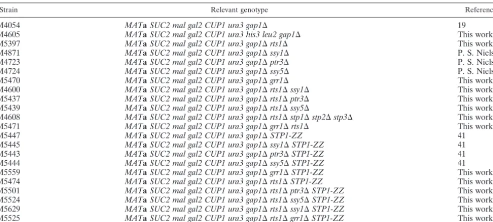
TABLE 1. Strains used in this study
Strain Relevant genotype Reference
M4054 MATaSUC2 mal gal2 CUP1 ura3 gap1⌬ 19
M4605 MATaSUC2 mal gal2 CUP1 ura3 his3 leu2 gap1⌬ This work
M5397 MATaSUC2 mal gal2 CUP1 ura3 gap1⌬rts1⌬ This work
M4871 MATaSUC2 mal gal2 CUP1 ura3 gap1⌬ssy1⌬ P. S. Nielsen
M4723 MATaSUC2 mal gal2 CUP1 ura3 gap1⌬ptr3⌬ P. S. Nielsen
M4724 MATaSUC2 mal gal2 CUP1 ura3 gap1⌬ssy5⌬ P. S. Nielsen
M5470 MATaSUC2 mal gal2 CUP1 ura3 gap1⌬grr1⌬ This work
M4600 MATaSUC2 mal gal2 CUP1 ura3 gap1⌬rts1⌬ssy1⌬ This work
M5437 MATaSUC2 mal gal2 CUP1 ura3 gap1⌬rts1⌬ptr3⌬ This work
M5439 MATaSUC2 mal gal2 CUP1 ura3 gap1⌬rts1⌬ssy5⌬ This work
M4608 MATaSUC2 mal gal2 CUP1 ura3 gap1⌬rts1⌬stp1⌬stp2⌬stp3⌬ This work
M5471 MATaSUC2 mal gal2 CUP1 ura3 gap1⌬grr1⌬rts1⌬ This work
M5447 MATaSUC2 mal gal2 CUP1 ura3 gap1⌬STP1-ZZ 41
M5445 MATaSUC2 mal gal2 CUP1 ura3 gap1⌬ssy1⌬STP1-ZZ 41
M5443 MATaSUC2 mal gal2 CUP1 ura3 gap1⌬ptr3⌬STP1-ZZ 41
M5444 MATaSUC2 mal gal2 CUP1 ura3 gap1⌬ssy5⌬STP1-ZZ 41
M5559 MATaSUC2 mal gal2 CUP1 ura3 gap1⌬grr1⌬STP1-ZZ This work
M5474 MATaSUC2 mal gal2 CUP1 ura3 gap1⌬rts1⌬STP1-ZZ This work
M5501 MATaSUC2 mal gal2 CUP1 ura3 gap1⌬rts1⌬ptr3⌬STP1-ZZ This work
M5524 MATaSUC2 mal gal2 CUP1 ura3 gap1⌬rts1⌬ssy5⌬STP1-ZZ This work
M5629 MATaSUC2 mal gal2 CUP1 ura3 gap1⌬rts1⌬ssy1⌬STP1-ZZ This work
M5525 MATaSUC2 mal gal2 CUP1 ura3 gap1⌬rts1⌬grr1⌬STP1-ZZ This work
on September 8, 2020 by guest
http://ec.asm.org/
RESULTS
Isolation of mutants constitutive in amino acid signaling.In order to select mutants with increased activity of the amino
acid-sensing pathway, a screen was designed in which ahis3
leu2derivative of the reference strain M4054 was subjected to
transposon mutagenesis. The host strain was transformed with a mixture of yeast genomic fragments representing a
mu-tagenizedLEU2-based plasmid library (46). Mutants of
inter-est were selected using three different screening criteria. First, we took advantage of the fact that dipeptide uptake (39), as well as branched-chain amino acid uptake (7), is very low in cells grown on minimal ammonium medium in the absence of leucine. Dipeptides, including Gly-His, are taken up by the peptide transporter Ptr2p (39), which is under the transcrip-tional control of the amino acid-sensing pathway (4). Mutants with constitutive signaling were thus selected on SD medium supplemented with the Gly-His dipeptide, which, upon intra-cellular cleavage, provides the histidine required for growth (39). A subsequent selection was carried out on SD supple-mented with the amino acids Ile, His, and Val, and the sulfo-nylurea herbicide metsulfuron methyl. This type of compound inhibits synthesis of branched-chain amino acids and therefore prevents growth of cells unable to take up branched-chain amino acids from the medium (53), such as mutants deficient in amino acid signaling (29, 30). Finally, colonies of interest
were tested for constitutive acitivity of theBAP2promoter.
Among the 11 mutants thus isolated, three had transposon
insertions in the PP2A subunit-encoding geneRTS1(34). The
remaining transposons were localized in VPS15, VPS34, and
VPS16, coding for proteins involved in vacuolar protein
sort-ing, andSTH1, coding for the ATPase subunit in the chromatin
remodeling complex RSC. Two others had mutations in ORFs
with unknown function,YFR044CandYFR045W. Two mutants
were not mapped. In the present work, the relationship be-tween the phenotype and the locus affected by transposon
insertion was analyzed in more detail for theRTS1gene.
Deletion ofRTS1results in constitutive activation ofBAP2
andAGP1transcription.Since transposon insertion very often completely inactivates a gene, we wished to analyze the
behav-ior of a strain deleted forRTS1. One of the selection criteria
used in the original screen was the ability to take up Gly-His on SD medium, thereby promoting growth of a histidine-requiring strain because of intracellular cleavage of the dipeptide. To
find out if an rts1⌬ strain exhibits the same phenotype, we
deleted theRTS1ORF in ahis3 derivative of the reference
strain M4054. Indeed, the lack ofRTS1enabled growth on the
dipeptide as a histidine source (not shown).
In order to be able to analyze the rts1⌬ mutation in the
absence of extracellular amino acids or dipeptides, we also
constructed aHIS3 rts1⌬strain (M5397). First we wished to
confirm the activation of target promoters for the amino acid signaling by testing for growth on YPD supplemented with metsulfuron methyl; the strain grew, as expected (not shown). Then we tested whether this activation was constitutive by yet
another criterion, using the dipeptideL-leucyl-L-ethionine. If
this dipeptide is taken up, it is cleaved to yield the toxic
me-thionine analogueL-ethionine. Also, uptake of the dipeptide
L-leucyl-L-ethionine is mediated up by the peptide transporter
Ptr2p (39) and is thus under transcriptional control of the
amino acid-sensing pathway (4). Insensitivity toL-leucyl-L
-ethi-onine therefore reflects that the pathway is not activated, while growth inhibition in the absence of an inducing amino acid indicates constitutive activation of the amino acid-sensing
pathway. Therts1⌬strain M5397 was unable to grow on
min-imal medium in the presence ofL-leucyl-L-ethionine,
suggest-ing constitutive activity of the signalsuggest-ing pathway (not shown). We also investigated activation of the promoter of the
broad-specificity AAP geneAGP1 using a fusion of the
pro-moter to theE. coli lacZgene. Cells were grown in SD medium
and incubated for 40 min with or without 100ML-leucine.
The experiment was repeated with 5 mM L-citrulline or an
equal volume of water. These concentrations have been
pre-viously found to fully or almost fully induce the AGP1
pro-moter (16, 40). The results show thatAGP1transcription in
rts1⌬cells is constitutive, i.e., unaffected by leucine or citrulline
addition. In the wild-type control experiment,AGP1promoter
activation was low in the absence of amino acids and increased strongly in response to either leucine or citrulline (Table 2).
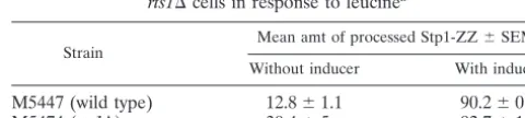
Transcriptional induction of AAP genes was previously shown to involve Stp1p endoproteolysis (3). We used the Stp1-ZZ construct previously described (40), in which the C terminus of Stp1p was fused to a doublet of the IgG-binding Z
domain of theS. aureusprotein A. This insert was integrated in
the wild-type strain and the rts1⌬ strain, resulting in strains
M5447 (40) and M5474, respectively. The fraction of processed
Stp1p inrts1⌬cells in the absence of leucine was greater than
in wild-type cells (30% versus 13%), whereas addition of leucine led to almost complete (above 90%) Stp1p processing
in both wild-type andrts1⌬cells (Table 3 and Fig. 1).
Quanti-fication of antibody binding showed that the relative amount of processed Stp1p in the absence of inducer was more than
doubled inrts1⌬cells compared to wild-type cells.
TABLE 2. AGP1promoter activity in wild-type cells andrts1⌬cells in response to leucine and citrulline
Inducer
Mean-galactosidase activitya⫾SEM
M4054 (wild type) M5397 (rts1⌬)
L-Leucine (100M)
⫺ 1.5⫾0.1 10.5⫾0.4
⫹ 8.4⫾1.0 9.9⫾0.9
L-Citrulline (5 mM)
⫺ 0.1⫾0.1 11.4⫾0.6
⫹ 8.7⫾1.4 13.5⫾0.4
a
Activity in Miller units in the absence (⫺) and 40 minutes after the addition (⫹) of inducer.
TABLE 3. Quantification of processed Stp1-ZZ in wild-type and
rts1⌬cells in response to leucinea
Strain Mean amt of processed Stp1-ZZ⫾SEM Without inducer With inducer
M5447 (wild type) 12.8⫾1.1 90.2⫾0.4
M5474 (rts1⌬) 30.4⫾5 92.7⫾1.6
aSamples were taken in the absence and 20 minutes after addition of 100M
L-leucine. Relative amounts of full-length and processed Stp1-ZZ were
deter-mined by performing the experiment twice.
on September 8, 2020 by guest
http://ec.asm.org/
Remarkably, Stp1p processing to the extent of 30% is
suffi-cient to generate an activity of theAGP1promoter comparable
to that observed in wild-type cells after amino acid addition (compare Table 2 and Table 3). In fact, this is expected from previous comparisons of dose-response relationships using
quantification of Stp1p processing versusAGP1promoter
ac-tivity as the read-out (40, 41). The sensing ofL-leucine exhibits
a 50% effective concentration of 12 M under the growth
conditions studied when Stp1p processing is monitored, but
only 1 M when AGP1 promoter-driven lacZ expression is
measured (40). The most obvious interpretation is that satu-ration of the promoter activity occurs already at modest levels of activated Stp1p.
Epistasis relationships. In order to further investigate the
role of RTS1, we constructed strains M4600 (ssy1⌬ rts1⌬),
M5437 (ptr3⌬ rts1⌬), M5439 (ssy5⌬ rts1⌬), M5470 (grr1⌬
rts1⌬), and M4608 (stp1⌬stp2⌬stp3⌬rts1⌬). STP3is
homol-ogous toSTP1and STP2, and Helge A. Andersen (personal
communication) has found that deletion ofSTP3 further
re-duces the low, remaining activity of the amino acid signaling
pathway in anstp1⌬stp2⌬mutant. All of the resulting strains
were sensitive to metsulfuron methyl on YPD, and all were
insensitive toL-leucyl-L-ethionine (not shown), suggesting that
the amino acid-sensing pathway was inactive.
-Galactosidase assays were performed on extracts from
cells in which the E. coli lacZ gene was placed under the
control of theAGP1promoter.-Galactosidase measurements
confirmed that the multiple mutants behaved similarly to the
single mutants deficient in signaling, i.e.,AGP1transcription
was very low even in the presence of extracellular amino acids (Table 4). Using the Stp1-ZZ construct, we also measured relative amounts of processed and full-length Stp1p in each of the strains, in the absence and presence of amino acids. Stp1p
was exclusively present as the full-length protein in all single
and multiple mutants (Fig. 2). In other words,ssy1,ptr3,ssy5,
andgrr1are epistatic overrts1.
DISCUSSION
In this work we have attempted to identify potential negative regulators of the amino acid-sensing pathway. For this purpose we have designed a screen allowing selection of mutants with constitutive activity of the pathway. The screen described in this report was performed in the absence of inducer in cells
lacking GAP1, and we investigated transcription levels of a
lacZ reporter gene placed under the control of the amino
acid-inducible promoters of theAGP1andBAP2genes, which
are known targets of the SPS-mediated pathway. The genes that were disrupted by transposon insertion encode proteins that could be involved at any level of the signaling.
The isolated mutants include some in which the transposon
insertion disrupted theRTS1gene. This gene encodes a
regu-latory subunit of PP2A, which is a major serine/threonine phosphatase involved in several nutrient-induced signaling pathways, in cell growth control and cell division control (9, 35, 45, 56). Protein phosphatase 2A exists in several isoforms and is mostly present in cells as a heterotrimeric complex,
consist-ing of a catalytic (C) subunit, encoded byPPH21,PPH22, or
PPH3 (45, 49), a scaffolding subunit (A), encoded byTPD3
(55), and a regulatory subunit (B or B⬘), encoded byCDC55
(22) andRTS1(47, 48), respectively. The B and B⬘subunits are
believed to regulate the activity of PP2A by determining its cellular location and modifying the substrate specificity of the
C subunit (31, 54). CDC55is required for correct cell cycle
checkpoint control (35), andcdc55⌬cells display a
cold-sensi-FIG. 1. Quantitative Western analysis of Stp1-ZZ processing in wild-type andrts1⌬cells in response to 100ML-leucine.
FIG. 2. Quantitative Western analysis of Stp1-ZZ processing in
rts1⌬,ssy5⌬, andptr3⌬single mutants and inssy5⌬rts1⌬andptr3⌬ rts1⌬double mutants, in the absence (⫺) and 20 min after the addition (⫹) of 100ML-leucine. Upper band: unprocessed Stp1-ZZ; lower band: processed Stp1-ZZ.
TABLE 4. AGP1promoter activity in cells deleted of genes encoding positive factors in amino acid signaling and/orRTS1in the absence and presence of inducing amino acids
Inducer
Mean-galactosidase activitya⫾SEM
M4054 (wild type)
M5397 (rts1⌬)
M4600 (ssy1⌬rts1⌬)
M5437 (ptr3⌬rts1⌬)
M5439 (ssy5⌬rts1⌬)
M5471 (grr1⌬rts1⌬)
M4608 (rts1⌬stp1⌬stp2⌬stp3⌬)
L-Leucine (100M)
⫺ 1.7⫾0.1 10.1⫾0.2 1.1⫾0.1 ⫺0.1⫾0.0 ⫺0.8⫾0.0 0.1⫾0.0
⫹ 7.2⫾0.6 9.6⫾0.2 1.1⫾0.0 ⫺0.1⫾0.0 ⫺0.6⫾0.0 ⫺0.1⫾0.0
L-Citrulline (5 mM)
⫺ 0.1⫾0.0 0.95⫾0.1 0.0⫾0.0
⫹ 11.3⫾1.2 0.83⫾0.1 0.0⫾0.0
aActivity in Miller units in the absence (⫺) and 40 minutes after the addition (⫹) of inducer.
on September 8, 2020 by guest
http://ec.asm.org/
tive phenotype but no phenotype at elevated temperatures
(22).RTS1was found in two independent genetic screens, as a
multicopy suppressor of hsp60(Ts) mutant alleles (48), and
later as aRoxThreeSuppressor (12);rts1⌬cells are
thermo-sensitive and exhibit a typicalcdcmutant phenotype (47).
We identified RTS1 as a negative component of the
SPS-mediated amino acid-sensing pathway. Deletion ofRTS1
re-sults in constitutive transcription ofAGP1and BAP2.
More-over,ssy1,ptr3,ssy5, andgrr1were found to be epistatic over
rts1. These results indicate that the PP2A is involved in the
SPS-mediated pathway and suggest that a dephosphorylation step is required to down-regulate signaling in the absence of extracellular amino acids. Rts1p associated with Tpd3p and the C subunit might thus dephosphorylate one of the SPS proteins. Alternatively, it could dephosphorylate the Stp transcription factors, resulting in a conformational change that perhaps lim-its accessibility of the cleavage site to the protease, thereby impairing Stp1p and Stp2p proteolysis and activation in the absence of amino acids.
While PP2A appears to down-regulate amino acid signaling, a corresponding kinase should be involved in the activation of the pathway. Casein kinase I is a candidate for this activity, since the amino acid-sensing pathway is inactive in tempera-ture-sensitive mutants affected in the casein kinase I genes
YCK1and YCK2, and these strains exhibit loss of Stp1p
pro-cessing (1). Casein kinase I and PP2A are known to act on the
same substrate inXenopusembryos (17) and we propose that
they do so in yeast as well. This hypothesis is substantiated by the finding that the yeast Cdc55p subunit forms a complex with casein kinase I (23). Moreover, casein kinase I has also been reported to be involved in the glucose-sensing pathway (50), which mediates transcriptional induction of hexose transporter
(HXT) genes in response to extracellular glucose and which
shares many similarities with the amino acid-sensing pathway. Casein kinase I is indeed required for phosphorylation of
Mth1, which is then targeted for degradation by SCFGrr1(36,
50). Thus, glucose induction of theHXT1promoter is deficient
in a yck1⌬ yck2(Ts) mutant; in addition, this mutant lacks
glucose-induced degradation of Mth1p and Std1p (36). Mth1p and Std1p interact with the transcriptional repressor Rgt1p in the absence of glucose: the resulting complex binds promoter DNA, thereby repressing transcription from the
HXT genes (33, 52). Likewise, Stp1p is phosphorylated in a
casein kinase I-dependent way prior to its endoproteolytic
ac-tivation (1). SCFGrr1 recognizes phosphorylated substrates;
this is the case for Mth1p (50) and for the G1cyclin Cln2p (25).
Although Stp1p is phosphorylated by casein kinase I (1), there
is no reason to invoke it as a target for SCFGrr1. A possible
target for SCFGrr1is Ptr3p, which in fact has been found to be
subject to posttranslational modification, exhibiting a slower-migrating band (13) that might be due to ubiquitination.
Al-ternatively, the target for SCFGrr1could be a protein not yet
known to be part of the pathway. Further work is needed to propose which protein is a target for PP2A associated with Rts1p. There may, however, be cross talk between the amino acid-sensing and the target of rapamycin (TOR) pathways, since N. Eckert-Boulet (unpublished data) has observed that
treatment ofrts1⌬ cells with rapamycin enhanced the
consti-tutive signal, also at the level of Stp1-ZZ processing. Interestingly, it has recently been found that PP2A is
in-volved in regulating the induction ofHXT1by glucose and that
Cdc55p is the regulatory subunit involved. The 14-3-3 proteins Bmh1p and Bmh2p also appear to be involved (51). These observations add to existing similarities between the glucose induction pathway and the amino acid induction pathway. It will be interesting to see whether Bmh1p and Bmh2p also are involved in the SPS-mediated pathway and whether Rts1p in-teracts with casein kinase I like Cdc55p does.
In summary, we have shown that Rts1p, one of the two known regulatory subunits of PP2A, is a down-regulator of the SPS-mediated amino acid-sensing pathway. This finding illus-trates a new role in nutrient-induced signaling for PP2A and further highlights the reuse of components in different signal-ing pathways.
ACKNOWLEDGMENTS
We acknowledge Helge A. Andersen for very fruitful discussions. Part of this work was financed by the Danish BioInstrument Centre (DABIC). We are grateful for a fellowship from the Knut and Alice Wallenberg Foundation to Katrin Larsson.
REFERENCES
1.Abdel-Sater, F., M. El Bakkoury, A. Urrestarazu, S. Vissers, and B. Andre´. 2004. Amino acid signaling in yeast: casein kinase I and the Ssy5 endopro-tease are key determinants of endoproteolytic activation of the membrane-bound Stp1 transcription factor. Mol. Cell. Biol.24:9771–9785.
2.Andre´asson, C. 2004. Ligand-activated proteolysis in nutrient signaling. Ph.D. thesis. Ludwig Institute for Cancer Research, Stockholm, Sweden. 3.Andre´asson, C., and P. O. Ljungdahl.2002. Receptor-mediated
endopro-teolytic activation of two transcription factors in yeast. Genes Dev.16:3158– 3172.
4.Barnes, D., W. Lai, M. Breslav, F. Naider, and J. M. Becker.1998.PTR3, a novel gene mediating amino acid-inducible regulation of peptide transport in Saccharomyces cerevisiae. Mol. Microbiol.29:297–310.
5.Bernard, F., and B. Andre´.2001. Ubiquitin and the SCFGrr1ubiquitin ligase
complex are involved in the signalling pathway activated by external amino acids inSaccharomyces cerevisiae. FEBS Lett.496:81–85.
6.de Boer, M., J.-P. Bebelman, P. M. Gonc¸alves, J. Maat, H. Van Heerikhui-zen, and R. J. Planta.1998. Regulation of expression of the amino acid transporter geneBAP3inSaccharomyces cerevisiae. Mol. Microbiol.30:603– 613.
7.Didion, T., M. Grauslund, M. C. Kielland-Brandt, and H. A. Andersen.1996. Amino acids induce expression ofBAP2, a branched-chain amino acid per-mease gene inSaccharomyces cerevisiae. J. Bacteriol.178:2025–2029. 8.Didion, T., B. Regenberg, M. U. Jørgensen, M. C. Kielland-Brandt, and H. A.
Andersen.1998. The permease homologue Ssy1p controls the expression of amino acid and peptide transporter genes inSaccharomyces cerevisiae. Mol. Microbiol.27:643–650.
9.Du¨vel, K., and J. R. Broach.2004. The role of phosphatases in TOR signal-ing in yeast. Curr. Top. Microbiol. Immunol.279:19–38.
10.Eckert-Boulet, N., P. S. Nielsen, C. Friis, M. M. dos Santos, J. Nielsen, M. C. Kielland-Brandt, and B. Regenberg.2004. Transcriptional profiling of ex-tracellular amino acid sensing inSaccharomyces cerevisiaeand the role of Stp1p and Stp2p. Yeast21:635–648.
11.Eckert-Boulet, N., B. Regenberg, and J. Nielsen.2005. Grr1p is required for transcriptional induction of amino acid permease genes and proper tran-scriptional regulation of genes in carbon metabolism ofSaccharomyces cer-evisiae. Curr. Genet.47:139–149.
12.Evangelista, Jr., C. C., A. M. Rodriguez Torres, M. P. Limbach, and R. S. Zitomer.1996. Rox3 and Rts1 function in the global stress response pathway in baker’s yeast. Genetics142:1083–1093.
13.Forsberg, H., and P. O. Ljungdahl.2001. Genetic and biochemical analysis of the yeast plasma membrane Ssy1p-Ptr3p-Ssy5p sensor of extracellular amino acids. Mol. Cell. Biol.21:814–826.
14.Forsberg, H., and P. O. Ljungdahl.2001. Sensors of extracellular nutrients in Saccharomyces cerevisiae. Curr. Genet.40:91–109.
15.Forsberg, H., M. Hammar, C. Andre´asson, A. Moline´r, and P. O. Ljungdahl. 2001. Suppressors ofssy1andptr3null mutations define novel amino acid sensor-independent genes in Saccharomyces cerevisiae. Genetics158:973– 988.
16.Gaber, R. F., K. Ottow, H. A. Andersen, and M. C. Kielland-Brandt.2003. Constitutive and hyperresponsive signaling by mutant forms of Saccharomy-ces cerevisiaeamino acid sensor Ssy1. Eukaryot. Cell.2:922–929. 17.Gao, Z.-H., J. M. Seeling, V. Hill, A. Yochum, and D. M. Virshup.2002.
on September 8, 2020 by guest
http://ec.asm.org/
Casein kinase I phosphorylates and destabilizes the-catenin degradation complex. Proc. Natl. Acad. USA99:1182–1187.
18.Georgakopoulos, T., G. Koutroubas, I. Vakonakis, M. Tzermia, V. Prokova, A. Voutsina, and D. Alexandraki.2001. Functional analysis of the Saccha-romyces cerevisiae YFR021w/YGR223c/YPL100wORF family suggests rela-tions to mitochondrial/peroxisomal funcrela-tions and amino acid signalling path-ways. Yeast18:1155–1171.
19.Grauslund, M., T. Didion, M. C. Kielland-Brandt, and H. A. Andersen.1995. BAP2, a gene encoding a permease for branched-chain amino acids in Sac-charomyces cerevisiae. Biochim. Biophys. Acta1269:275–280.
20.Grenson, M., C. Hou, and M. Crabeel.1970. Multiplicity of the amino acid permeases inSaccharomyces cerevisiae. IV. Evidence for a general amino acid permease. J. Bacteriol.103:770–777.
21.Gu¨ldener, U., S. Heck, T. Fielder, J. Beinhauer, J. H. Hegemann.1996. A new efficient gene disruption cassette for repeated use in budding yeast. Nucleic Acids Res.13:2519–2524.
22.Healy, A. M., S. Zolnierowicz, A. E. Stapleton, M. Goebl, A. A. DePaoli-Roach, and J. R. Pringle.1991.CDC55, aSaccharomyces cerevisiaegene involved in cellular morphogenesis: identification, characterization, and ho-mology to the B subunit of mammalian type 2A protein phosphatase. Mol. Cell. Biol.11:5767–5780.
23.Ho, Y., A. Gruhler, A. Heilbut, G. D. Bader, L. Moore, S.-L. Adams, A. Millar, P. Taylor, K. Bennett, K. Boutilier, L. Yang, C. Wolting, I. Donald-son, S. Schandorff, J. Shewnarane, M. Vo, J. Taggart, M. Goudreault, B. Muskat, C. Alfarano, D. Dewar, Z. Lin, K. Michalickova, A. R. Willems, H. Sassi, P. A. Nielsen, K. J. Rasmussen, J. R. Andersen, L. E. Johansen, L. H. Hansen, H. Jespersen, A. Podtelejnikov, E. Nielsen, J. Crawford, V. Poulsen, B. D. Sørensen, J. Matthiesen, R. C. Hendrickson, F. Gleeson, T. Pawson, M. F. Moran, D. Durocher, M. Mann, C. W. V. Hogue, D. Figeys, and M. Tyers.2002. Systematic identification of protein complexes inSaccharomyces cerevisiaeby mass spectrometry. Nature415:180–183.
24.Hora´k, J.1997. Yeast nutrient transporters. Biochim. Biophys. Acta1331: 41–79.
25.Hsiung, Y. G., H.-C. Chang, J.-L. Pellequer, R. La Valle, S. Lanker, and C. Wittenberg.2001. F-box protein Grr1 interacts with phosphorylated targets via the cationic surface of its leucine-rich repeat. Mol. Cell. Biol.21:2506– 2520.
26.Iraqui, I., S. Vissers, F. Bernard, J.-O. De Craene, E. Boles, A. Urrestarazu, and B. Andre´.1999. Amino acid signaling inSaccharomyces cerevisiae: a permease-like sensor of external amino acids and F-box protein Grr1p are required for transcriptional induction of theAGP1gene, which encodes a broad-specificity amino acid permease. Mol. Cell. Biol.19:989–1001. 27.Island, M. D., J. R. Perry, F. Naider, and J. M. Becker.1991. Isolation and
characterization ofS. cerevisiaemutants deficient in amino acid-inducible peptide transport. Curr. Genet.20:457–463.
28.Jack, D. L., I. T. Paulsen, and M. H. Saier.2000. The amino acid/polyamine/ organocation (APC) superfamily of transporters specific for amino acids, polyamines and organocations. Microbiology146:1797–1814.
29.Jørgensen, M. U., C. Gjermansen, H. A. Andersen, and M. C. Kielland-Brandt.1997.STP1, a gene involved in pre-tRNA processing in yeast, is important for amino-acid uptake and transcription of the permease gene BAP2. Curr. Genet.31:241–247.
30.Jørgensen, M. U., M. B. Bruun, T. Didion, and M. C. Kielland-Brandt.1998. Mutations in five loci affectingGAP1-independent uptake of neutral amino acids in yeast. Yeast14:103–114.
31.Kamibayashi, C., R. Estes, R. L. Lickteig, S. I. Yang, C. Craft, and M. C. Mumby.1994. Comparison of heterotrimeric protein phosphatase 2A con-taining different B subunits. J. Biol. Chem.269:20139–20148.
32.Klasson, H., G. R. Fink, and P. O. Ljungdahl.1999. Ssy1p and Ptr3p are plasma membrane components of a yeast system that senses extracellular amino acids. Mol. Cell. Biol.19:5405–5416.
33.Lakshmanan, J., A. L. Mosley, and S. O¨ zcan.2003. Repression of transcrip-tion by Rgt1 in the absence of glucose requires Std1 and Mth1. Curr. Genet. 44:19–25.
34.Larsson, K., H. A. Andersen, T. Didion, L. Du¨ring-Olsen, M. Jørgensen, P. L. Madsen, P. S. Nielsen, B. Regenberg, and M. C. Kielland-Brandt.1999. Amino acid sensing and uptake, p. 51–57. E.B.C. Symposium: Yeast physi-ology, a new era of opportunity. Monograph 28. Fachverlag Hans Carl, Nu¨rnberg, Germany.
35.Minshull, J., A. Straight, A. D. Rudner, A. F. Dernburg, A. Belmont, and A. W. Murray.1996. Protein phosphatase 2A regulates MPF activity and sister chromatid cohesion in budding yeast. Curr. Biol.6:1609–1620. 36.Moriya, H., and M. Johnston.2004. Glucose sensing and signaling in
Sac-charomyces cerevisiaethrough the Rgt2 glucose sensor and casein kinase I. Proc. Natl. Acad. USA101:1572–1577.
37.Nielsen, P. S., B. van den Hazel, T. Didion, M. de Boer, M. Jørgensen, R. J. Planta, M. C. Kielland-Brandt, and H. A. Andersen.2001. Transcriptional regulation of theSaccharomyces cerevisiaeamino acid permease geneBAP2. Mol. Gen. Genet.264:613–622.
38.Patton, E. E., A. R. Willems, and M. Tyers.1998. Combinatorial control in ubiquitin-dependent proteolysis: don’t Skp the F-box hypothesis. Trends Genet.14:236–243.
39.Perry, J. R., M. A. Basrai, H.-Y. Steiner, F. Naider, and J. M. Becker.1994. Isolation and characterization of aSaccharomyces cerevisiaepeptide trans-port gene. Mol. Cell. Biol.14:104–115.
40.Poulsen, P., B. Wu, R. F. Gaber, K. Ottow, H. A. Andersen, and M. C. Kielland-Brandt.2005. Amino acid sensing by Ssy1. Biochem. Soc. Trans. 33:261–264.
41.Poulsen, P., B. Wu, R. F. Gaber, and M. C. Kielland-Brandt.2005. Consti-tutive signal transduction by mutant Ssy5p and Ptr3p components of the SPS amino acid sensor system inSaccharomyces cerevisiae. Eukaryot. Cell4:1116– 1124.
42.Regenberg, B., L. Du¨ring-Olsen, M. C. Kielland-Brandt, and S. Holmberg. 1999. Substrate specificity and gene expression of the amino-acid permeases inSaccharomyces cerevisiae. Curr. Genet.36:317–328.
43.Regenberg, B., and J. Hansen.2000.GAP1, a novel selection and counter-selection marker for multiple gene disruptions inSaccharomyces cerevisiae. Yeast16:1111–1119.
44.Rolland, F., J. Winderickx, and J. M. Thevelein. 2001. Glucose-sensing mechanisms in eukaryotic cells. Trends Biochem. Sci.26:310–317. 45.Ronne, H., M. Carlberg, G.-Z. Hu, and J. O. Nehlin.1991. Protein
phos-phatase 2A in Saccharomyces cerevisiae: effects on cell growth and bud morphogenesis. Mol. Cell. Biol.11:4876–4884.
46.Ross-Macdonald, P., A. Sheehan, G. S. Roeder, and M. Snyder.1997. A multipurpose transposon system for analyzing protein production, localiza-tion, and function inSaccharomyces cerevisiae. Proc. Natl. Acad. Sci. USA 94:190–195.
47.Shu, Y., H. Yang, E. Hallberg, and R. Hallberg.1997. Molecular genetic analysis of Rts1p, a B’ regulatory subunit ofSaccharomyces cerevisiaeprotein phosphatase 2A. Mol. Cell. Biol.17:3242–3253.
48.Shu, Y., and R. L. Hallberg.1995.SCS1, a multicopy suppressor ofhsp60-ts mutant alleles, does not encode a mitochondrially targeted protein. Mol. Cell. Biol.15:5618–5626.
49.Sneddon, A. A., P. T. Cohen, and M. J. Stark.1990.Saccharomyces cerevisiae protein phosphatase 2A performs an essential cellular function and is en-coded by two genes. EMBO J.9:4339–4346.
50.Spielewoy, N., K. Flick, T. I. Kalashnikova, J. R. Walker, and C. Wittenberg. 2004. Regulation and recognition of SCFGrr1targets in the glucose and
amino acid signaling pathways. Mol. Cell. Biol.24:8994–9005.
51.Toma´s-Cobos, L., R. Viana, and P. Sanz.2005. TOR kinase pathway and 14-3-3 proteins regulate glucose-induced expression ofHXT1, a yeast low-affinity glucose transporter. Yeast22:471–479.
52.Toma´s-Cobos, L., and P. Sanz.2002. Active Snf1 protein kinase inhibits expression of theSaccharomyces cerevisiae HXT1glucose transporter gene. Biochem. J.368:657–663.
53.Tullin, S., C. Gjermansen, and M. C. Kielland-Brandt.1991. A high-affinity uptake system for branched-chain amino acids inSaccharomyces cerevisiae. Yeast7:933–941.
54.Turowski, P., A. Fernandez, B. Favre, N. J. C. Lamb, and B. A. Hemmings. 1995. Differential methylation and altered conformation of cytoplasmic and nuclear forms of protein phosphatase 2A during cell cycle progression. J. Cell Biol.129:397–410.
55.van Zyl, W. H., N. Wills, and J. R. Broach.1989. A general screen for mutants ofSaccharomyces cerevisiaedeficient in tRNA biosynthesis. Genet-ics123:55–68.
56.Zabrocki, P., C. Van Hoof, J. Goris, J. M. Thevelein, J. Winderickx, and S. Wera.2002. Protein phosphatase 2A on track for nutrient-induced signalling in yeast. Mol. Microbiol.43:835–842.