CASE STUDY
OSTEORADIONECROSIS
*Sayali A. Deolikar, Dr. Sangeeta J. Palaskar, Dr. Archana A. Gupta, Dr. Prakhar Kapoor
and Dr. Rasika Pawar
Department of Oral Pathology and Microbiology, Sinhgad Dental College and Hospital
S. No. 44/1, Vadgaon Budruk, off Sinhgad raod, Pune, Maharashtra
ARTICLE INFO ABSTRACT
It has long been recognized that patients who receive radiotherapy for cancer of the head and neck area are at a risk of developing osteoradionecrosis (ORN) of the jaws. Guidelines to reduce risk have been written, based upon the evidence of many studies which have looked at the incidence of ORN in different groups. Much of the research was carried out over 20 years ago and more recent analysis of data and consideration of the changes in radiotherapy raises the question as to whether modifications to the guidance is now needed. There is a wide variation in recommendations and a simpler, more unified approach to prevention of ORN could be developed as well as research on recent management techniques.
Definition: Osteoradionecrosis is defined as an area of bone tissue exposed to radiation that fails to heal over a period of 3 months without a residual or recurrent tumor (Sukh S Ryatt et al., 2007).
Copyright©2017, Sayali A. Deolikar et al. This is an open access article distributed under the Creative Commons Attribution License, which permits unrestricted
use, distribution, and reproduction in any medium, provided the original work is properly cited.
INTRODUCTION
Osteoradionecrosis of the jaw (ORN) is an infrequent yet potentially devastating complication of radiation therapy to the head and neck region (Sultan et al., 2017). In this condition, there is a non-healing wound persistent for 3 months or more characterized by the irradiated bone getting exposed and devitalized with loss of overlying skin or mucosa.
Etiology: The cause of osteoradionecrosis is radiation induced tissue damage. This damage is compounded by the fact that the mandible is essentially on end artery system supplied by the inferior alveolar artery, with minor supply from the bony attachments. In addition, both the mandible and maxilla are unique in being the only bones in the body that are exposed directly to the external environment through the gingival attachment of the teeth. In an irradiated field, minor insult such as periodontal disease, pulpal infection and procedure such as dental extractions can result delayed healing and in some cases developed into osteoradionecrosis (Reuther et al., 2003). In 1983, Marx redefined the pathophysiology of ORN by proposing that radiation therapy induces an endarteritis that results in tissue hypoxia, hypercellularity and hypovascularity
*Corresponding author: Sayali A. Deolikar,
Department of Oral Pathology and Microbiology, Sinhgad Dental College and Hospital S. No. 44/1, Vadgaon Budruk, off Sinhgad raod, Pune, Maharashtra
which in turn causes tissue breakdown and chronic non-healing wounds (Remy H Blanchaert, 2013). Meyer proposed a theory involving radiation, trauma and infection and reported that oral microbiological flora invade the underlying irradiated bone after injury. Recently, a new theory(fibro atrophic theory) has emerged proposes that fibroblast populations not only undergo total cellular depletion in response to radiation exposure, but show a reduced ability to produce and secrete collagen into surrounding tissue. This theory is based on the concept that osteoclasts suffer radiation damage earlier than the development of vascular alterations (Marx, 1983). Few studies show an increased risk of ORN development when extractions are performed before radiotherapy (Kayal et al., 2015). (Jacobson et al. Paradigm shifts in the management of osteoradionecrosis)
Various factors responsible for development of ORN
1. Radiation dose: Increasing the external beam radiation dose above 50 Gray gives a significantly increased risk of developing ORN (Fowler, 2007).
2. Time to deliver radiation dose: Radiotherapy gives a progressive injury to tissues at a cellular and humoral level. If the entire dose is given in a short period of time, this will prevent the remaining viable local non tumor cells from recovering and therefore will cause more damage. In the same fashion different schemes of
ISSN: 0975-833X
International Journal of Current Research
Vol. 9, Issue, 10, pp.58772-58776, October, 2017
INTERNATIONAL JOURNAL OF CURRENT RESEARCH
Article History:
Received 22ndJuly, 2017 Received in revised form 14thAugust, 2017
Accepted 17thSeptember, 2017
Published online 17thOctober, 2017
Citation: Sayali A. Deolikar, Dr. Sangeeta J. Palaskar, Dr. Archana A. Gupta, Dr. Prakhar Kapoor and Dr. Rasika Pawar, 2017.
“Osteoradionecrosis”, International Journal of Current Research, 9, (10), 58772-58776.
Key words:
Non-healing wounds, Tissue hypoxia, Hypocellularity and Hypovascularity, hyperbaric oxygen therapy, Irradiated bone.
CASE STUDY
OSTEORADIONECROSIS
*Sayali A. Deolikar, Dr. Sangeeta J. Palaskar, Dr. Archana A. Gupta, Dr. Prakhar Kapoor
and Dr. Rasika Pawar
Department of Oral Pathology and Microbiology, Sinhgad Dental College and Hospital
S. No. 44/1, Vadgaon Budruk, off Sinhgad raod, Pune, Maharashtra
ARTICLE INFO ABSTRACT
It has long been recognized that patients who receive radiotherapy for cancer of the head and neck area are at a risk of developing osteoradionecrosis (ORN) of the jaws. Guidelines to reduce risk have been written, based upon the evidence of many studies which have looked at the incidence of ORN in different groups. Much of the research was carried out over 20 years ago and more recent analysis of data and consideration of the changes in radiotherapy raises the question as to whether modifications to the guidance is now needed. There is a wide variation in recommendations and a simpler, more unified approach to prevention of ORN could be developed as well as research on recent management techniques.
Definition: Osteoradionecrosis is defined as an area of bone tissue exposed to radiation that fails to heal over a period of 3 months without a residual or recurrent tumor (Sukh S Ryatt et al., 2007).
Copyright©2017, Sayali A. Deolikar et al. This is an open access article distributed under the Creative Commons Attribution License, which permits unrestricted
use, distribution, and reproduction in any medium, provided the original work is properly cited.
INTRODUCTION
Osteoradionecrosis of the jaw (ORN) is an infrequent yet potentially devastating complication of radiation therapy to the head and neck region (Sultan et al., 2017). In this condition, there is a non-healing wound persistent for 3 months or more characterized by the irradiated bone getting exposed and devitalized with loss of overlying skin or mucosa.
Etiology: The cause of osteoradionecrosis is radiation induced tissue damage. This damage is compounded by the fact that the mandible is essentially on end artery system supplied by the inferior alveolar artery, with minor supply from the bony attachments. In addition, both the mandible and maxilla are unique in being the only bones in the body that are exposed directly to the external environment through the gingival attachment of the teeth. In an irradiated field, minor insult such as periodontal disease, pulpal infection and procedure such as dental extractions can result delayed healing and in some cases developed into osteoradionecrosis (Reuther et al., 2003). In 1983, Marx redefined the pathophysiology of ORN by proposing that radiation therapy induces an endarteritis that results in tissue hypoxia, hypercellularity and hypovascularity
*Corresponding author: Sayali A. Deolikar,
Department of Oral Pathology and Microbiology, Sinhgad Dental College and Hospital S. No. 44/1, Vadgaon Budruk, off Sinhgad raod, Pune, Maharashtra
which in turn causes tissue breakdown and chronic non-healing wounds (Remy H Blanchaert, 2013). Meyer proposed a theory involving radiation, trauma and infection and reported that oral microbiological flora invade the underlying irradiated bone after injury. Recently, a new theory(fibro atrophic theory) has emerged proposes that fibroblast populations not only undergo total cellular depletion in response to radiation exposure, but show a reduced ability to produce and secrete collagen into surrounding tissue. This theory is based on the concept that osteoclasts suffer radiation damage earlier than the development of vascular alterations (Marx, 1983). Few studies show an increased risk of ORN development when extractions are performed before radiotherapy (Kayal et al., 2015). (Jacobson et al. Paradigm shifts in the management of osteoradionecrosis)
Various factors responsible for development of ORN
1. Radiation dose: Increasing the external beam radiation dose above 50 Gray gives a significantly increased risk of developing ORN (Fowler, 2007).
2. Time to deliver radiation dose: Radiotherapy gives a progressive injury to tissues at a cellular and humoral level. If the entire dose is given in a short period of time, this will prevent the remaining viable local non tumor cells from recovering and therefore will cause more damage. In the same fashion different schemes of
ISSN: 0975-833X
International Journal of Current Research
Vol. 9, Issue, 10, pp.58772-58776, October, 2017
INTERNATIONAL JOURNAL OF CURRENT RESEARCH
Article History:
Received 22ndJuly, 2017 Received in revised form 14thAugust, 2017
Accepted 17thSeptember, 2017
Published online 17thOctober, 2017
Citation: Sayali A. Deolikar, Dr. Sangeeta J. Palaskar, Dr. Archana A. Gupta, Dr. Prakhar Kapoor and Dr. Rasika Pawar, 2017.
“Osteoradionecrosis”, International Journal of Current Research, 9, (10), 58772-58776.
Key words:
fractionated delivery of irradiation have been shown to reduce the complications of radiotherapy.
3. Mode of delivery, location and volume of irradiated tissue: More localized delivery by means of e.g brachytherapy limits radiation damage to surrounding tissues compared to external beam irradiation where an entire block of tissue is irradiated. Less vascularized parts of mandible such as the posterior part or surgically manipulated parts are more susceptible to osteoradionecrosis. Optimal planning and of irradiation fields will help minimize irradiation of surrounding non-involved tissues (Coskunfirat et al., 2005).
[image:2.595.310.559.219.525.2]4. Inadequate oral hygiene: Intraoral tissues are damaged by irradiation, which causes mucositis and xerostomia. These conditions, in combination with poor dental care will give rise to odontogenic and periodontal infections, this again can lead to osteoradionecrosis.
Figure 1. Legend- Prof. Oliver Hennessy (Prof. Oliver Hennessy. Mandibular osteoradionecrosis with fracture (figure 1))
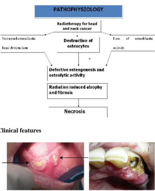
Pathophysiology
In simplistic terms, radiation produces its deleterious effects through production of free radicals, which result in mitotic cell death. The effects will be greatest on rapidly growing cells such as the mucosa. Remodelling cells such as fibroblasts, osteoblasts and osteoclasts will show changes when they try to divide, such as during healing. Damage to the microvasculature results in initial hyperemia followed by endarteritis, thrombosis and eventual obliteration. This results in the picture described
by Marx as the 3 ‘H’s or hypocellularity, hypoxia and
hypovascularity (Herskind et al., 1998; Bensadoun et al., 2001). Marx defined pathophysiology of ORN by proposing that the radiation therapy induces an endarteritis that results in tissue hypoxia, hypocellularity, hypovascularity, which in turn causes tissue breakdown and chronic non healing wounds. There are some theories proposing that ORN is not a primary infection of irradiated bone, but rather a complex metabolic and tissue homeostatic deficiency created by radiation induced cellular injury. Meyer proposed a theory involving radiation, trauma and infection and reported that oral microbiological flora invade the underlying irradiated bone after injury. Endothelium, bone and periosteum are all important tissues that have been shown to become hypoxic, hypocellular and hypovascular as a result of ORN. With this theory, the classic sequence of radiation, trauma and infection can be replaced by a sequence of metabolic and cellular changes such as cellular death and collagen lysis exceed synthesis and cellular replication resulting in chronic non healing wounds (Lyons and Ghazali, 2008).Recently, a new theory known as the ‘fibro
-atrophic theory’ has emerged and proposes that fibroblast
populations not only undergo total cellular depletion in response of radiation exposure, but also show a reduced ability
to produce and secrete collagen into the surrounding tissues. This theory is based on the concept that osteoclasts suffer radiation damage earlier than the development of vascular alterations. Accordingly, the key event in the progression of ORN is the and dysregulation of fibroblastic activity that leads to atrophic tissue within a previously irradiated area (Jacobson et al., 2010). In 1926, Ewing first recognized and reported bone changes associated with radiation therapy and described this
disease state as “radiation osteitis” (Kahenasa et al., 2012). In
1938, Watson and Scarborough described radiation osteitis as being caused by radiation, trauma and infection (Jacobson et al., 2012). Other factors include patient related facts (age, obesity), comorbidities (hypertension, diabetes), surgery in irradiated site and chemotherapy.
Clinical features
Fig. 2. Legend- Joel B Epstein et al Fig. 3. Legend- S.S Deshpande et al.
ORN is clinically observed as bony exposure for more than 3 months duration. Tell tale signs of radiotherapy may be present on the skin such as thin skin, pigmentary changes, lack of hair and telangiectasis. Intraorally the mucosa may be dry with frothy sputum but these are relatively acute changes following radiotherapy. The patient may have a non-resolving painful mucosal ulcer with evidence of exposed bone or sequestrum. This is usually posterior mandibular region. There may be trismus and this usually appears 3-6 months following radiotherapy. There may be exposed bone seen through the skin, in the form of orocutaneous fistula or the patient may present with a pathological fracture (Jacobson et al., 2012). Other symptoms include dysesthesia, halitosis, dysgeusia and food impaction.
Stages of ORN
Stage I disease represents small, superficial, localized bone resorption with cutaneous or mucosal dehiscence. Stage II disease represents larger and deeper areas of bone
[image:2.595.41.287.260.379.2]and the mucosal or cutaneous areas of breakdown are moderate in size.
Stage III ORN is defined by full thickness devitalization of bone, resorption of the inferior border of the mandible, fistula or a pathological fracture (Joel B. Epstein et al., 2012).
Prevention
Preventive measures should be put in place to help to reduce the incidence and severity of osteoradionecrosis. These measures should be applied before and after the radiotherapy. Preventive measures prior to radiotherapy:
The patient should have a full dental evaluation. This should include radiographs to show all the teeth as well as the jaws to check for unerupted teeth and any bony pathology. Periapical views of all the teeth should be taken. All the teeth should be meticulously charted for caries and periodontal pocketing. Each tooth should be given an individual prognosis and a treatment plan completed and discussed with the patient. The most important aspect is to educate the patient regarding meticulous oral hygiene and the need for life long regular follow up. All teeth should be cleaned and sealed. Patient should be encouraged to rinse with a fluoride and antibacterial mouth wash on regular basis. High risk patients should have custom trays made to assist in regular fluoride treatment. Patients with no teeth should still have a baseline radiographic evaluation to check for buried teeth. Dentures should be inspected for fitting to ensure minimal trauma to the tissues. Extractions should be done at least 2-3 weeks prior to radiotherapy and is advised to ensure timely healing, and it should be carried out in a non-traumatic manner with minimal damage to the surrounding tissues (Deshpande et al., 2015).
Hygiene during radiotherapy: During radiotherapy, the patient will experience mucositis and xerostomia. Regular review with the dentist is essential to minimize discomfort. The xerostomia will cause dryness of the mouth due to lack of saliva, which is essential to wash away debris and dilute the plaque. Regular mouthwashes and meticulous oral hygiene is
essential during this period. A dry mouth will sometimes result in patients resorting to foodstuffs that are sloppy and sticky and have more carbohydrate component which together with a low saliva flow rate may lead to increased caries (Vissink et al., 2003).
Other measures: Patient medication should be reviewed as Biphosphonates, used in various conditions such as osteoporosis, Paget's disease and metastatic breast disease have been shown to cause osteonecrosis of the jaws. The exact mechanism is not known but is related to suppression of osteoclastic activity. Paradoxically, biphosphonates have also been used in the treatment of osteoradionecrosis, highlighting our incomplete understanding of the condition (Deshpande et al., 2015; Vissink et al., 2003).
After radiotherapy: The patient should be reviewed regularly to re-enforce oral care and look for signs of dental disease or mucosal damage
Management
Medical management of osteoradionecrosis is essentially supportive with pain relief and treatment of infection. If the patient is dentate, preventative measures as outlined above should continue with advice on diet and nutrition as well as oral hygiene. Small wounds may be debrided superficially and any loose necrotic bone removed. Infection tends to be secondary and systemic antibiotics should be reserved for symptomatic cases (Ravindran Rathy et al., 2013).
Previous treatment options for osteoradionecrosis
stages of ORN especially for the effective treatment of early and advanced ORN (Delanian and Lefaix, 2002). Stage I, or early stage ORN is managed conservatively with therapies such as local wound care, HBOT and antibiotic medications. For stage II, intermediate stage ORN, it is difficult to recommend a definitive treatment procedure (Beumer et al., 1983) Stage III, advanced stage ORN is managed surgically with wide resection and immediate microvascular reconstruction. Antibiotic medications should always be instituted after bacterial identification and sensitivity testing, and any delays in surgical treatment should be avoided. Usually, penicillin with metronidazole or clindamycin is initially administered until bacterial identification is available (Delanian and Lefaix, 2002; Beumer et al., 1983).
Surgical approach for osteoradionecrosis
Surgical approaches for osteoradionecrotic jaws may encompass a series of procedures including wound debridement, which involves the removal of infected and devitalized teeth and associated soft tissues, sequestrectomy, which is the removal of devitalized bony fragments or an ivolucrum of the jaw, decortications, which is the removal of lateral and inferior cortical plates of bone to gain access to the infected medullar cavity, and resection with healthy bony margins with immediate or delayed reconstruction (Nabil and Samman, 2011; Williamson, 2007).
Hyperbaric oxygen therapy for management for osteoradionecrosis
HBOT is well known to positively affect surgical outcomes by promoting angiogenesis in irradiated tissues. HBOT not only increases the oxygen supply in hypoxic tissue, thereby inducing fibroblastic proliferation and capillary formation, but also increases tissue vascularity, viability and healing capacity (Madrid et al., 2010) HBOT most likely achieves these effects through a complex series of changes in affected tissues. Tissue swelling is probably improved through the osmotic effect of oxygen, and the steep oxygen gradient established across an irradiated tissue margin leads to the growth of new blood vessels. In addition, improving oxygen levels improves white blood cell and fibroblast function, further enhancing wound healing. However, HBOT was associated with many adverse effects including pressure induced damage to the ears, sinuses and lungs, a temporary worsening of short sightedness (myopia), claustrophobia and oxygen poisoning thus, the use of HBOT in treating established ORN has not been convincing and is still controversial (Oh et al., 2009). Recent understanding of the pathophysiology of ORN based on the concept of radiation induced fibrosis has led to the advent of new therapeutic regimens composed of pentoxyfylline and tocopherol.
New approach for the treatment of osteoradionecrosis with pentoxyfylline and tocopherol
Pentoxifylline exerts an anti-tumor necrosis factor (TNF)-α
effect, increases erythrocyte flexibility, vasodilates, inhibits inflammatory reactions in vivo, inhibits human dermal fibroblast proliferation and extracellular matrix (ECM) production, and increases collagenase activity in vitro. Pentoxifylline and its metabolites improve blood flow by decreasing its viscosity. In patients with chronic peripheral arterial disease. This effect increases blood flow to the affected
microcirculation and enhances tissue oxygenation. The usual dosage of pentoxifylline in extended-release tablet form is 400 mg, three times a day with meals. While the effect of pentoxifylline may be seen within 2 to 4 weeks, it is recommended that treatment be continued for at least 8 weeks (Wahl, 2006). Tocopherols and tocotrienols are fat-soluble antioxidants, but also seem to have many other functions in the body. The functions of endogenous tocopherol are to scavenge the reactive oxygen species generated during oxidative stress that escape the activity of in vivo antioxidant enzymes, to protect cell membranes against lipid peroxidation, and to partly inhibit TGF-ß1 and procollagen gene expression (Bennet et al., 2012).
Pentoxifylline and tocopherol combined therapy
Combined pentoxifylline-tocopherol therapy has been proven effective in reducing chronic progressive septic ORN of the mandible. Because there is currently no standard medical treatment, this approach constitutes a useful alternative to existing therapies in treating ORN. These two drugs act synergistically as potent antifibrotic agents and are available, well tolerated, inexpensive, and beneficial to the patient. Pentoxifylline is a methylxanthine derivative that exerts an negative effect on TNF-α, increases erythrocyte flexibility,
dilates blood vessels, inhibits inflammatory reactions in vivo, inhibits the proliferation of human dermal fibroblasts and the production of extracellular matrix, and increases collagenase activity in vitro. It is given with tocopherol, which reduces fibrosis by scavenging the reactive oxygen species that were generated during oxidative stress, protecting cell membranes against the peroxidation of lipids, and partially inhibiting
TGF-β1 and the expression of procollagen genes. In animal studies,
neither drug alone was capable of reversing the effects of reactive oxygen species. In addition, pentoxifylline or tocopherol alone were also unable to reverse the development of human fibrosis. However, these drugs were effective synergistically as anti-fibrotic agents (Epstein et al., 1981; McLeod et al., 2012). Recently many new innovations have been reported including ultrasound, biological molecules distraction, osteogenesis and antioxidant agents (Epstein et al., 2010).
Conclusion
ORN is more common in jaw bones as these bones have lesser bone marrow also, both maxilla and mandible are unique in being the only bones in the body that are exposed directly to the external environment through gingival attachment of teeth. It is more common in mandible than in the maxilla because of the richer blood supply to the maxilla and also it is porous in nature. In addition, mandible is more frequently irradiated. (Delanian and lefaix, 2004) Head and neck cancer patients continue to pose a challenge for surgeons and oncologists. Osteoradionecrosis can be a cruel blow to patients and their families who have already had to endure treatment for cancer. Improved radiotherapy protocols, multidisciplinary preventive care and reconstructive surgery can help to improve the quality of their lives.
REFERENCES
Bensadoun RJ, Magne N, Marcy PY, Demord F. 2001. Chemotherapy and radiotherapy induced mucositis in head and neck cancer patients: New trends in pathophysiology, prevention and treatment. Eur Arch otorhinolaryngol., 258:481-7.
Beumer J 3rd, Harrison R, Sanders B, Kurrasch M. Pre-radiation dental extractions and the incidence of bone necrosis. Head Neck Surg., 5: 514-21.
Coskunfirat OK, Wei FC, Huang WC, Cheng MH et al. 2005. Microvascular free tissue transfer for treatment of osteoradionecrosis of the maxilla. Plast Reconstr Surg., 115:54-60.
Delanian S, Lefaix JL. 2002. Complete healing of severe osteoradionecrosis by treatment combining pentoxifylline, tocopherol and clodronate Br J Radiol., 75:467-9.
Delanian S, lefaix JL. 2004. The radiation induced fibroatrophic process: Therapeutic perspective via the antioxidant pathway. Radiotherapy Oncology, 73:119-131. 10.1016/j radonc.2004.08.04.
Deshpande, S.S., M.H Thakur et al. 2015. Osteoradionecrosis of the mandible through a radiologists eyes. Clinical radiology, page 197-205.
Epstein JB, Hatcher DC, Graham M. 1981. Bone scintigraphy of fibro-osseous lesions of the jaw. Oral surgery Oral medicine Oral pathology, 51:346-350.
Epstein MS, Wicknick FW, Epstein JB, Berenson JR, Gorsky M. 2010. Management of bisphosphonate- associated osteonecrosis: Pentoxyfylline and tocopherol in addition to antimicrobial therapy. An initial case series. Oral surgery Oral medicine Oral pathology Oral radiology Endod., 110:593-596.
Fowler JF. 2007. Is there an optimum overall time for head and neck radiotherapy? : A review with new modeling. Clin Oncol(R coll Radiol), 19:8-22.
Herskind C, Bamberg M, Rodemann HP. 1998. The role of cytokines in the development of normal tissue reactions after radiotherapy. Strahlenter Onkol., 174:12-5.
Jacobson AS, Buchbinder D, Huk, Urken ML. 2010. Paradigm shifts in the management of osteoradionecrosis of the mandible. Oral Oncol., 46:795-801. 10. 1016/j. oraloncology 2010.08.007.
Jacobson AS, Buchbinder D, Huk, Urken ML. 2012. Paradigm shifts in the management of osteoradionecrosis of the mandible. Oral Radio., 113.el18-23.
Joel B. Epstein et al. 2012. Oral complications of cancer and cancer therapy from cancer treatment to survivorship. Sept CA Cancer Journals for Clinicians.
Kahenasa N, Sung EC, Nabili V, Kelly J, Garrett N et al. 2012. Resolution of pain and complete healing of mandibular osteoradionecrosis using pentoxifylline and tocopherol: A case report. Oral Surg Oral Med Oral Patho Oral Radio., 113.e18-23.
Kayal, L., S Jayachandran, Aatman Sharma, K Karthikeyan. 2015. Osteoradionecrosis of the mandible: A report of three cases. Research focused, Volume:4, Issue:1, Page 61-65. Kumuda Arvind Rao, Shishir Ram Shetty, Renita Lorina
Castelino. Osteoradionecrosis of the mandible and mastoiditis after radiotherapy for parotid mucoepidermoid carcinoma. Clinical Medicine and Research.
Lyons A, Ghazali N. 2008. Osteoradionecrosis of the jaws: Current understanding of its pathophysiology and treatment. Br J Oral Maxillofac Surg., 46: 653-660. 10. 1016/j.bjoms.2008.04.006.
Madrid C, Abarca M, Bou ferrache K. 2010. Osteoradionecrosis: An update. Oral Oncol., 46:471-474. 10.1016/j.oraloncology.2010.03.017.
Marx RE. 1983. Osteoradionecrosis: A new concept of its pathophysiology. J Oral Maxillofac Surg., 41:283-288.10. 1016/0278-2391(83).
McLeod NM, Pratt CA, Mellor TK, Brennan PA. 2012. Pentoxifylline and tocopherol in the management of patients with osteoradionecrosis, the Portsmouth experience. Br J Oral Maxillofacial Surgery, 50:41-44. Nabil S, Samman N. 2011. Incidence and prevention of
osteoradionecrosis after dental extraction in irradiated patients : a systematic review. Int J Oral Maxillofacial Surgery, 40:229-243.10.1016/j.ijom.2010.10.005.
Oh HK, Chambers MS, Martin JW, Lim HJ, Park HJ. 2009. Osteoradionecrosis of the mandible: treatment outcomes and factors influencing the progress of osteoradionecrosis. J Oral Maxillofacial Surgery, 67: 1378.10.1016/j.joms 2009.02.008
Pitak-Arnop P, Sader R, Dhanutai K, Masuratna P, Bertolus C et al. 2008. Management of osteoradionecrosis of the jaws: An analysis of evidence. Eur J Surgery Oncology, 34(10):1123-34.
Prof. Oliver Hennessy. Mandibular osteoradionecrosis with fracture. (figure 1)
Ravindran Rathy, S. Sunil, M Nivia. 2013. Osteoradionecrosis of mandible: Case report review of literature. Contemporary Clinical dentistry, 4(2):251-253.
Remy H Blanchaert. 2013. Osteoradionecrosis of the mandible: Case report with review of literature. Contempt Clin Dent., 4(2):251:253.
Reuther T, Schuster T, Mende U, Kubler A. 2003. Osteoradionecrosis of the jaw as a side effect of radiotherapy of head and neck tumor patients: A report of a thirty year retrospective review. Int. J oral Maxillofac Surg., 32:289-295
Springer IN, Nichoff P, Acil Y, Marget M, Lange A, Warnke PH, Pielenz H. et al. 2008. BMP-2 and BFGF in an irradiated bone model. J Craniomaxillofacial Surgery, 36: 210-217.
Sukh S Ryatt, Marc A.M Mureau, Stefan O.P Hofer. 2007. Osteoradionecrosis of mandible: Etiology, prevention, diagnosis and treatment. Indian Journal of Plastic Surgery, Volume 40, Issue 12, Page 65-71.
Sulaman F, Huryn JM, Zlotolow IM. 2003. Dental extractions in the irradiated head and neck patient: A retrospective analysis of Memorial Sloan- Kettering Cancer Centre protocols, criteria and end results. J Oral Maxillofac Surg., 61:1123-31.
Sultan A, Hanna GJ, Murgalit DN, Chau N et al. 2017. The use of hyperbaric oxygen for the prevention and management of osteoradionecrosis of the jaw. A
Dana-Farber/Brigham and Women’s cancer centre
Multidisciplinary Guideline. National library of medicine National institute of health, 22(3):343-350.
Vissink A, Jansma J, Spijkervet FK, Burlage FR, Coppers RP. 2003. Oral sequelae of head and neck radiotherapy. Crit Rev Oral Bio Med., 14:199-212.
Wahl MJ. 2006. Osteoradionecrosis prevention myths. Int J Radiat Oncol Biol Phys., 64:661-669.
Williamson RA. 2007. An experimental study of the use of hyperbaric oxygen to reduce the side effects of radiation treatment for malignant disease. Int. J Oral Maxillofacial Surgery, 36:533-540.