ESSENTIAL GUIDES TO METHOD
DEVELOPMENT IN TWO-DIMENSIONAL
ELECTROPHORESIS
M. J. Dunn, Imperial College School of Medicine, Harefield Hospital, Middlesex, UK
Copyright^ 2000 Academic Press
Introduction
Most one-dimensional (1-D) methods of polyacryl-amide gel electrophoresis are limited to the resolution of 100 or so protein zones. These techniques are therefore not suitable for the analysis of complex mixtures containing several thousands of proteins, such as total protein homogenates of whole cells and tissue. In addition, they are only able to separate proteins on the basis of a single physico-chemical property. For example, the observation of a particu-lar zone following SDS-PAGE does not imply protein heterogeneity, but simply indicates that any proteins present in that zone have nearly identical size proper-ties, while their charge (and other) properties could be very different.
The best approach to this problem is to combine two different 1-D methods into a 2-D procedure. Ideally, the methods used for each dimension should be selected by their ability to separate proteins ac-cording to different properties in each dimen-sion. Thus, if each method when used alone is able to resolve 100 protein zones, it would be expected that up to 10 000 proteins might be resolved when these methods are used orthogonally. This level of resolu-tion has rarely been achieved in practice, but never-theless 2-D has become the method of choice for the analysis of patterns of protein expression in whole cells, tissues and organisms; the area now known as proteomics.
History of 2-D
The Rrst protein separation by 2-D is attributed to Smithies and Poulik who in 1956 described a combina-tion of paper and starch gel electrophoresis for the separation of serum proteins. Since that time, sub-sequent advances in electrophoresis, such as the use of polyacrylamide gels, discontinuous buffer sys-tems, gradient gels, SDS-PAGE, and isoelectric focus-ing (IEF) have all resulted in the development of improved methods of 2-D. These developments
cul-minated in the 1970s with publications from several independent groups describing a combination of aR rst-dimension separation by IEF under denaturing condi-tions with a second dimension separation by SDS-PAGE. This coupling of IEF with SDS-PAGE resulted in a method of 2-D which separates proteins according to two independent parameters, charge and size.
The O’Farrell Method of 2-D
The method described by O’Farrell in 1975 has for-med the basis of almost all subsequent developments in 2-D, and several thousand papers have been pub-lished using this technique in the 25 years following its publication. This method was optimized for the separation of the proteins ofEscherichia coli(E.coli) and used a combination of IEF in cylindrical gels (cast in glass capillary tubes) containing 8 M urea and 2% w/v of the non-ionic detergent, Nonidet P-40 (NP-40), with the SDS-PAGE system of Laemmli. This method was able to resolve around 500 proteins from
E. coli. It has subsequently been applied to a wide variety of samples.
Limitations of the O’Farrell Method
The main problem with the 2-D method of O’Farrell is associated with the synthetic carrier ampholytes (SCA) which are used to generate the pH gradient in the IEF dimension. SCA are produced by a complex synthetic process which is difRcult to control re-producibly. This results in considerable batch-to-batch variability and limits the reproducibility and consistency of 2-D separations. Perhaps more impor-tantly SCA are relatively small molecules, which are notRxed within the IEF gel. As a consequence, the electroendosmotic Sow of water that occurs during IEF results in migration of the SCA molecules to-wards the cathode.
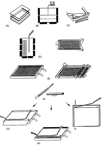
Figure 1 Schematic diagram of the procedure of 2-D using IPG IEF. (A) Assembly of the polymerization cassette for the preparation of IPG and SDS gels cast on plastic backings, (B) casting of IPG and gradient SDS gels, (C) cutting of washed and dried IPG gels into individual IPG strips, (D) rehydration of IPG strips, (E) IEF in individual IPG strips, (F) equilibration of IPG strips prior to SDS-PAGE, (G) transfer of IPG strip onto surface of laboratory-made horizontal SDS gel along cathodic wick, (H) transfer of IPG strip onto surface of commercial horizontal SDS gel along cathodic buffer strip, (I) loading of IPG strip onto the surface of a vertical SDS gel. (Courtesy of A. GoKrg, Technical University, Munich, Germany).
known as non-equilibrium pH gradient electrophor-esis (NEPHGE), for the 2-D separation of basic pro-teins. In this method, separation occurs on the basis of protein mobility in the presence of a rapidly form-ing pH gradient, but reproducibility is extremely
dif-Rcult to control. Fortunately, this problem was solved with the development of immobilized pH gradient (IPG) IEF.
2-D Using IPG IEF
IPG IEF gels are prepared using Immobilines (Amer-sham Pharmacia Biotech), a series of eight acrylamide derivatives with the structure CH2"CH}
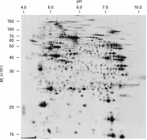
through-Figure 2 A 2-D separation of 100g heart proteins using a nonlinear pH 3.5 to 10 IPG IEF gel in the first dimension. The protein pattern was visualized by silver staining. The scale at the top indicates the nonlinear pH gradient obtained using an IPG 3-10 NL strip for the first dimension IEF separation. The scale at the left indicates the size separation in the range 15 to 150 kDa using a 15% SDS-PAGE gel in the second dimension.
out the pH range 3 to 10. The appropriate IPG reagents, calculated according to published recipes, are added to the mixture used for gel polymerization. Thus, during polymerization, the buffering groups which will form the pH gradient are covalent-ly attached via vinyl bonds to the pocovalent-lyacrylamide backbone. IPG generated in this way are, therefore, immune to the effects of electroendosmosis, so that they provide the opportunity to carry out IEF separations which are extremely stable, allowing the true equilibrium state to be attained.
Initial attempts to implement the IPG technology to 2-D separations encountered several problems. Fortu-nately, largely due to the work of GoKrg and her colleagues, these problems have been solved and IPG IEF has become the method of choice for the Rrst dimension separation of 2-D. The method is shown schematically inFigure 1. BrieSy, IPG slab gels of the desired pH range are cast (Figure 1(B)) according to the extensive library of published recipes. After poly-merization, the gels are washed, dried and stored at
!203C. The required number of gel strips (3}5 mm wide) for 2-D are cut off of the slab using a paper cutter (Figure 1(C)). Alternatively, a range of ready-made strips is available commercially from Amer-sham Pharmacia Biotech. IPG strips of any desired
length can be used, but is should be remembered that, in general, the larger the separation area of a 2-D gel, the more proteins can be resolved. Strips of 18 cm are usually employed for high-resolution separations, while shorter strips (7 or 11 cm) are used for rapid screening applications.
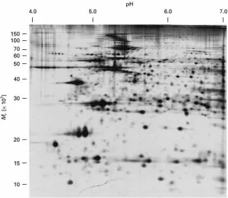
[image:3.568.141.435.382.663.2]Figure 3 A 2-D separation of 100g heart proteins using a linear pH 4 to 7 IPG IEF gel in the first dimension. The protein pattern was visualized by silver staining. The scale at the top indicates the linear pH gradient obtained using an IPG 4-7 strip for the first dimension IEF separation. The scale at the left indicates the size separation in the range 10 to 150 kDa using a 10%SDS-PAGE gel in the second dimension.
For use in 2-D, the strips are rehydrated in a re-swelling cassette (Figure 1(D)) in a solution containing 8 M urea, 0.5% non-ionic (e.g. NP-40, Triton X-100) or zwitterionic (CHAPS) detergent (3-[(cholamido-propyl)dimethylammonio]-1-propanesulfonate), 15 mM DTT and 0.2% synthetic carrier ampholyte (SCA) of the appropriate pH range. The strips are then placed directly on the surface of the cooling plate of a horizontal Sat-bed electrophoresis apparatus (Figure 1(E)). A convenient alternative is to use the special strip tray available from Pharmacia ( Fig-ure 1(E)). This tray isRtted with a corrugated plastic plate which contains grooves allowing easy alignment of the IPG strips. In addition, the tray is Rtted with bars carrying the electrodes and a bar Rtted with sample cups allowing the application of samples at any desired point on the gel surface. This tray isRlled with silicone oil which protects the gel from the effects of the atmosphere during IEF. Horizontal streaking can often be observed at the basic end of 2-D protein proRles, particularly when IPG 6}10 is used for the Rrst dimension. This problem can be resolved by applying an extra electrode strip soaked in 15 mM DTT on the surface of the IPG strip along-side the cathodic electrode strip. This has the
advant-age that the DTT within the gel, which migrates towards the anode during IEF, is replenished by the DTT released from the strip at the anode. An alterna-tive approach is to use the non-charged reducing agent, tributyl phosphine (TBP), which does not mi-grate during IEF and has been found to greatly im-prove protein solubility during IEF.
Sample Preparation
There is no universal method of sample preparation for 2-D due to the diverse nature of samples which can be analysed. Whatever method is used, it is essen-tial to minimize protein modiRcations which can re-sult in artefactual spots on 2-D protein patterns. In particular, samples containing urea should not be heated as this will lead to charge heterogeneity as a result of protein carbamylation by isocyanate ions formed from the decomposition of urea. Proteases present within samples can also readily result in arte-factual spots, so that samples should be subjected to minimal handling and kept cold at all times. Protease inhibitors can also be added.
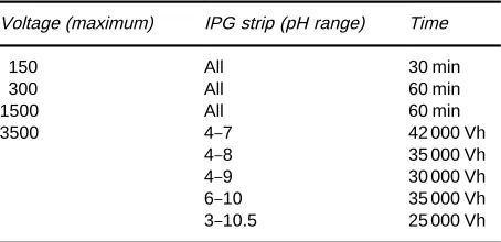
Table 1 Suggested running conditions for 18 cm IPG strips for the first, IEF dimension of 2-D. The strips should be run at 0.05 mA per strip (2 mA maximum total), 0.5 W maximum, 203C
Voltage (maximum) IPG strip (pH range) Time
150 All 30 min
300 All 60 min
1500 All 60 min
3500 4}7 42 000 Vh
4}8 35 000 Vh
4}9 30 000 Vh
6}10 35 000 Vh
3}10.5 25 000 Vh
pre-treatment prior to 2-D. However, less concen-trated solutions (e.g. urine, cerebrospinalSuid (CSF), amniotic Suid) often require concentration by methods such as lyophilization, or precipitation with trichloroacacetic acid (TCA) or acetone. Solid tissue samples must usually be disrupted in the presence of solubilization solution. For small samples this is read-ily achieved by crushing the sample in liquid nitrogen using a pestle and mortar, while larger tissue samples must be homogenized using a suitable device. Cell suspensions can be readily harvested by centrifu-gation, while cells adherent to a substrate, such as a tissue cultureSask or dish, should be collected by scraping (the use of proteases should be avoided to prevent possible sample degradation). Alternatively, the cells can be detached by lysis directly in a small volume of sample solubilization solution.
Sample Solubilization
The most popular method for protein solubilization for 2-D is that originally described by O’Farrell, using a mixture of 9.5 M urea, 4% w/v NP-40, 1% w/v DTT and 2% w/v SCA. While this method works well for the majority of samples, it is not universally applicable, with membrane proteins representing a particular challenge. The zwitterionic detergent, CHAPS has been found to be effective for the solubilization of membrane proteins, particularly when used at a concentration of 4% w/v in combina-tion with a mixture of 2 M thiourea and 8 M urea. Linear sulfobetaine detergents, such as SB 3}10 or 3}12, are also effective solubilizing agents, but these are not compatible with high concentrations of urea. This can be overcome by using these reagents at 2% w/v in combination with 5 M urea, 2 M thiourea and 2% CHAPS.
The presence of nucleic acids can be problematic during IEF. This is due to an increase in the viscosity of the sample and in some cases formation of com-plexes with the sample proteins, leading to artefactual migration and streaking. If problems of this type are suspected, it is best to degrade the nucleic acid by the addition of a suitable pure (i.e. protease free) en-donuclease to the sample solubilization solution.
Sample Reduction
Protein disulRde bonds are normally reduced with free thiol-containing reagents such as DTT or -mer-captoethanol. However, reagents such as DTT are charged so that they migrate out of the gel during IEF, leading to reoxidation of the sample proteins which can result in loss of sample solubility. It has recently been reported that replacing the thiol-containing
re-ducing agents with a non-charged rere-ducing agent such as tributyl phosphine (TBP) can greatly increase protein solubility during the IEF dimension and result in increased transfer to the second dimension gel.
Sample Application and
Running Conditions
instrument can accommodate up to 12 individual strip holders and incorporates Peltier solid-state cool-ing and a programmable 8000 V, 1.5 mA power supply.
Equilibration Between Dimensions
After the IPG IEF dimension, strips can be used im-mediately for the second dimension. Alternatively, strips can be stored between two sheets of plasticRlm at!803C for periods of several months. Prior to the second-dimension separation, it is essential that the IEF gels are equilibrated to allow the separated pro-teins to interact fully with sodium dodecyl sulfate (SDS) so that they will migrate properly during SDS-PAGE (Figure 1(F)). The recommended protocol is to incubate the IPG IEF gel strips for 15 min in 50 mM Tris buffer, pH 8.8 containing 2% w/v SDS, 1% w/v DTT, 6 M urea and 30% w/v glycerol. The urea and glycerol are used to reduce electroendosmotic effects which otherwise result in reduced protein transfer from theRrst to the second dimension. This is followed by a further 15 min equilibration in the same solution containing 5% w/v iodoacetamide in place of DTT. The latter step is used to alkylate any free DTT, as otherwise this migrates through the second-dimension SDS-PAGE gel, resulting in an ar-tefact known as ‘point-streaking’ which can be ob-served after silver staining. An alternative procedure, allowing equilibration to be achieved in a single step, is to replace the DTT in the equilibration buffer with 5 mM TBP, which is uncharged and so does not migrate during SDS-PAGE.
The Second Dimension
After equilibration, the Rrst dimension IEF gels are applied directly to the surface of the second-dimen-sion SDS-PAGE gels. The SDS-PAGE gels can be of any appropriate single or gradient polyacrylamide, and can be used either in a vertical (Figure 1(I)) or horizontal format (Figure 1(G), 1(H)). The use of vertical formats enables multiple gels to be run simul-taneously, which improves reproducibility, while the use of horizontal, 0.5 mm thin SDS gels cast on plas-tic supports improves the ease of handling the gels and gives rapid separations.
Resolution of 2-D
The resolving capacity of 2-D gels is usually con-sidered to be proportional to the total gel area avail-able for the separation. Using 18 cm long IPG IEF gels in combination with 20 cm long second-dimension SDS-PAGE gels, around 2000 proteins can be readily
resolved. Only a few hundred proteins can be separ-ated using mini-gel formats, but these are much quicker to run and can be useful for rapid screening purposes. For maximum resolution of very complex mixtures, very large format gels ('30 cm in each dimension) can be used. These are reported to be able to separate as many as 5000 to 10 000 proteins from whole cell lysates, but this is achieved at the expense of the ease of gel handling and processing.
Reproducibility of 2-D
Until recently reproducibility was a major problem limiting the more widespread application of 2-D. Using the tube gel technique of O’Farrell, it was often difRcult to obtain reproducible separations of a particular type of sample even within a single labor-atory, while comparison of 2-D separation patterns generated in different laboratories was often considered to be impossible. The use of dedicated equipment for 2-D, such as the ISO-DALT (Amer-sham Pharmacia Biotech) and the Investigator (ESA Inc) systems, helps in this regard as it allows the simultaneous electrophoresis of large numbers (between 5 and 20) of 2-D gels under reproducibly controlled conditions. More importantly, inter-laboratory studies of various types of sample (heart, barley, yeast) have unequivocally demonstrated that 2-D using IPG IEF results in 2-D protein separ-ations with very high spatial and quantitative repro-ducibility.
Proteomics
2-D separation has now matured into a technique which is capable of separating reproducibly thou-sands of proteins present in samples such as cells, tissues and even whole organisms. Recent develop-ments in methods for the microchemical characteriza-tion of proteins, particularly techniques for the analy-sis of proteins and peptides by mass spectrometry, now make it possible to identify and characterize proteins spots directly from 2-D gels. This has made 2-D separation an ideal tool to use in studies designed to determine the nature and function of the large number of structural genes being identiRed in various genome initiatives. This area has become known as ‘proteomics’ and is the subject of a separate article.
Further Reading
Corbett JM, Dunn MJ, Posch A and GoKrg A (1994) Positional r!eproducibility of protein spots in two-di-mensional polyacrylamide gel electrophoresis using im-mobilised pH gradient isoelectric focusing in the Rrst dimension: An interlaboratory study. Electrophoresis
15: 1205}1211.
Dunn MJ (1987) Two-dimensional polyacrylamide gel elec-trophoresis. In: Chrambach A, Dunn MJ and Radola BJ (eds)Advances in Electrophoresis, Vol. 1, pp. 1}109. Weinheim: VCH.
Dunn MJ (1993) Gel Electrophoresis: Proteins. Oxford: BIOS ScientiRc.
GoKrg A, Postel W and GuKnther S (1988) The current state of two-dimensional electrophoresis with immobilized pH gradients.Electrophoresis9: 531}546.
GoKrg A, Boguth G, Obermaier C, Posch A and Weiss W (1995) Two-dimensional polyacrylamide gel elec-trophoresis with immobilized pH gradients in theRrst dimension (IPG-Dalt): The state of the art and the con-troversy of vertical versus horizontal systems. Elec-trophoresis16: 1079}1086.
GoKrg A, Obermaier C, Boguth Get al. (1997) Very alkaline immobilized pH gradients for two-dimensional elec-trophoresis of ribosomal and nuclear proteins. Elec-trophoresis18: 328}337.
Herbert BR, Sanchez JC and Bini L (1997) Two-dimen-sional electrophoresis: The state of the art and future
directions. In: Wilkins MR, Williams KL, Appel RD and Hochstrasser DF (eds) Proteome Research: New Frontiers in Functional Genomics, pp. 13}33. Berlin: Springer.
Humphery-Smith I, Cordwell SJ and Blackstock WP (1997) Proteome research: Complementarity and limitations with respect to the RNA and DNA worlds. Electrophor-esis18: 1217}1242.
Klose J and Kobalz U (1995) Two-dimensional electrophor-esis of proteins: An updated protocol and implications for a functional analysis of the genome.Electrophoresis
16: 1034}1059.
O’Farrell PH (1975) High resolution two-dimensional elec-trophoresis of proteins.Journal of Biological Chemistry
250: 4007}4021.
Pennington SR, Wilkins MR, Hochstrasser DF and Dunn MJ. Proteome analysis: From protein characterization to biological function. Trends in Cell Biology 7: 168}173.
Rabilloud T, Adessi C, Giraudel A and Lunnardi J (1997) Improvement of the solubilization of proteins in two-dimensional electrophoresis with immobilized pH gradi-ents.Electrophoresis18: 307}316.
Wilkins MR, Williams KL, Appel RD and Hochstrasser DF (eds) (1997) Proteome Research: New Frontiers in Functional Genomics. Berlin: Springer.
3. ABBREVIATIONS
2,3,4,6-TeCP 2,3,4,6-tetrachlorophenol 2,3-DMP 2,3-dimethylpentane
2,4,5-T 2,4,5-trichlorophenoxyacetic acid 2,4,5-TP 2-(2,4,5-trichlorophenoxy)propionic acid 2,4,6-TCP 2,4,6-trichlorophenol
2,4-D 2,4-dichlorophenoxyacetic acid 2,4-DCP 2,4-dichlorophenol
2,4-DMP 2,4-dimethylphenol 2-CP 2-chlorophenol 2-DNP 2-dinitrophenol
2-M-4,6-DNP 2-methyl-4,6-dinitrophenol 2-NP 2-nitrophenol
4-NP 4-nitrophenol AA amino acid
AAA amino acid compositional analysis AAS atomic absorption spectrometry ACN acetonitrile
ADAM 9-anthryldiazomethane ADC analog-to-digital converter
AD-CSP amylosetris-(3,5-dimethylphenylcarbamate) CSP AE alcohol ethoxylates