A Convolution Neural Network Based Deep
Learning System for Brain Tumor Detection
towards MRI Image Segmentation
Uday Bellary, Javeriya Sadaf
Professor, Dept. of Computer Science and Engineering, P.D.A College of Engineering, Gulbarga, Karnataka, India.
PG Scholar, Dept. of Computer Science and Engineering, P.D.A College of Engineering, Gulbarga, Karnataka, India.
ABSTRACTBrain tumor segmentation towards a detection of brain tumour has been one of the major active research in biomedical image processing. A brain tumor can be detected in a brain MRI imagery. A brain MRI imagery is sequence of the images captured from different angles. Brain comprises of brain cells and in the presence of the tumor it may appear as a clotted area within the brain however the major challenge is that many parts of the brain normally looks like the tumor area therefore it is not simple to extract the tumor area from the brain image. Many of the past work focuses on extraction of brain tumor based on colour or morphological changes because the tumours can appear anywhere in the brain and have almost any kind of shape size and colour. The problem with such approach is that in an MRI imagery there are many image frame and in every frame brain tumor appear at different level.In order to avoid the lack of accuracy arising from the similarity of different brain abnormalities so in this work we use deep learning technique such as CNN and various other techniques like SOM ,FNN and fuzzy methods such as fuzzy k means and fuzzy c means ,and compare the various parameters such as specificity, sensitivity, precession, recall dice ,accuracy ,co efficient and conclude which best for detection of brain tumor. The project which has been implemented and proposed will be utilized in the fields like medical science for analysis and detection of brain tumor and give treatment .It can also be adopted in medicine development based on the type of brain tumor.
KEYWORDS: Convolution neural network, MRI image segmentation, Brain tumor detection.
I. INTRODUCTION
Tumor is an uncontrolled growth of cancer cells in any part of the body. Tumors are of different types and have different features and different treatments. Brain, which is an vital part of the body, can be divided into four parts- Grey Matter (GM), White Matter (WM), Cerebrospinal Fluid (CSF) and background. Brain tumours are abnormal proliferations of the cells. They can be either cancerous or non-cancerous.Malignant tumours consist of cancerous cells, capable of multiplying in an unpredictable fashion. Benign tumours are noncancerous tumours, usually having clear defined borders, to detect brain tumor MRI scanning is done where MRI stands for Magnetic Resonance Imaging and is a medical imaging technique used in radiology to envisage detailed internal structures. It uses magnetic field and pulses of radio wave energy to make picture of brain and other elements in brain. The proposed system is using various segmentation technique such as trilateral filtering followed by EMGM segmentation, fuzzy logic techniques such as fuzzy c means and fuzzy k means neural networks such as SOM FNN and deep learning convolution neural network(CNN). The project which has been implemented and proposed will be utilized in the fields like medical science for analysis and detection of brain tumor and give treatment .It can also be adopted in medicine development based on the type of brain tumor.
applied and an overlay based image fusion was applied to enhance the quality of the image a detection rate of 93% wit 7% of error rate was obtained using this data set the tumor was classified based on the size. In[2]. The proposed technique consisted of three modules segmentation, feature extraction and classification. the pre-proposed image is segmented using multiple kernel based probability (MKPL)clustering and the denoising was carried out using median filter. The feature is extracted on every segment based on shape, texture and intensity. After feature extraction, important feature was selected using linear discriminant analysis(LDA) for classification purpose.Finally, deep learning classifier was employed for classification into tumor or non-tumor. The proposed technique achieved an intensity with highest value 0.94.In[3].The BRATS which contained 200 patients dataset of MRI scanned images was taken and patch wise segmentation approach was done with pre-processing steps including N4ITK bias field correcting. A 93% accuracy was obtained with 2d convolution network which further suggested this approach for 3d fully convolution approach. The patch wise segmentation approach resulted in 93% accuracy. In[4].Modified K-means Clustering is used for image segmentation. In this clustering method, the MRI images are used to identify the tumor of the brain. The MRI image of the brain is given as input. The system should process the input image and detect the tumor. This method can even detect the smallest abnormality in the earlier stage itself. The different abnormality MRI scans of brain of the patients are taken for processing. In[5].The different method for segmentation technique were used and compared and the main goal was to improve the accuracy and performance and main objective was designing new algorithm to make image more simple and meaningful . Different segmentation method mentioned will help finding better and appropriate method for evaluating abilities for improving performance as well as systematically designing new segmentation method. In [6].Numerically method was introduced for finding the maximum a posterior estimation by using E-M algorithm GM distribution and reference posterior was reintroduced then E-M MAP algorithm for learning in a given image training data was introduced. In[7]proposed an image segmentation method to identify or detect tumor from the brain magnetic resonance imaging (MRI). There are many thresholding methods developed but they have different result in each image. a method by which detection of tumor can be done uniquely. Proposed a set of image segmentation algorithms which gives a satisfactory result on brain tumor images. The results depict that automatic detection of brain tumor can be done more confirm from the MRI images in comparisons to other tumor detection systems available in the market. Further improvements also can be proposed with huge amount of MRI image data if available.In[8] Many algorithms have been developed to segment and detect the tumours, taking into consideration various aspects of an image. a survey of brain tumour segmentation algorithms in image processing, and proposed approach to detect brain tumors using texture and feature vectors and one-class classification methods. In[9]Deep Neural Networks (DNNs) are often successful in problems needing to extract information from complexes, high-dimensional inputs, for which useful features are not obvious to design. presented work on applying DNNs to brain tumor segmentation for the BRATS challenge. currently experimenting with several DNN architectures, leveraging the recent advances in the field such as convolutional layers, max pooling, Max out units and Dropout regularization. , leader board dataset and challenge dataset. The results are obtained from the evaluation tool available on the Virtual Skeleton database.
III. SYSTEM DESIGN AND METHODOLOGY
The major objective of the project is to use different segmentation techniques to extract the brain tumor part from the brain and detect the tumor from which the brain is affected.
To achieve the above said functionalities we apply the following techniques.
Pre-processing is the first step to make an image more visible to human eye. De-noising is the important pre-processing step to improve the quality of an image by reducing noise component while preserving the image features.
Specificity sensitivity precession recall accuracy co efficient dice score
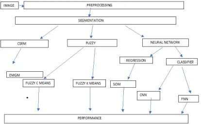
FIG 1: The proposed work flow
Following are the different techniques used as shown in figure 1:
EM-GM segmentation (CSRM) : Expectation maximization Gaussian method uses cumulative region based
segmentation method where the expected tumor region is marked out . This algorithm encloses probability density function (pdf) estimation for each tissue class (gray matter, white matter, and csf), classification, and bias field correction using the classic EM approach. Region growing is a simple region-based image segmentation method. It is also classified as a pixel-based image segmentation method since it involves the selection of preliminary seed points. This approach of segmentation examines neighboring pixels of initial seed points and determines whether the pixel neighbors should be added to the region. The process is iterated on, in the same manner as general data clustering algorithms.
SOM:A self-organizing map (SOM) or self-organizing feature map (SOFM) is a type of artificial neural network (ANN) that is trained using unsupervised learning to produce a low-dimensional (typically two-dimensional), discretized representation of the input space of the training samples, called a map, and is therefore a method to do dimensionality reduction. SOM reduces data dimensions and displays similarities among data
FUZZY K MEANS: K-means is a extensively used as a clustering algorithm to partition data into k clusters.
{cj│j=1,2,….k} by randomly selecting k feature vectors are grouped into clusters using a selected distance
measure such as Euclidean distance so that, d= ││xi-cj││.
FUZZY C MEANS: The FCM attempts to partition a finite collection of pixels into a collection of "C" fuzzy clusters with respect to some given criterion. Depending on the data and the application, different types of similarity measures may be used to identify classes. This algorithm is based on minimization of following objective function: Where ,J is the objective function N is the number of pixels in the image, C is the number
of clusters, μ is the membership table -- a table of NxC entries which contains the membership values of each data point and each cluster, m is a fuzziness factor (a value larger than1), xi is the ith pixel in N, cj is jth
cluster in C and |xi - cj| is the Euclidean distance between xi and cj .
FNN : A feedforward neural network is an artificial neural network where in connections between the units do not form a cycle. As such, it is different from recurrent neural networks. In this network, the data moves in only one direction, forward, from the input nodes, through the hidden nodes (if any) and to the output nodes. There are no cycles or loops in the network.
. CNN : A convolutional neural network (CNN, or ConvNet) is a type of feed-forwardartificial neural network in which the connectivity pattern between its neurons. The convolutional neural network is also known as shift invariant or space invariant artificial neural network (SIANN), which is named based on its shared weights architecture and translation invariance characteristics. we present an automatic brain tumor segmentation the system is deep learning convolution neural network based where by training the entire neural network vector with fixed set of feature at once neural network gets trained in several iterations where the result of 1 stage is passed onto the other stage where each of its stage determines its own feature vectors based on the probability of detection.in this way a network is build where the neural network in each stage has selected its own feature vector then the brain tumor is processed by such a network the tumor area can be segmented with extremely high accuracy in computation to other system the resulting system is very fast and the time needed to segment an entire brain with this system is very fast .
IV. RESULT ANALYSIS
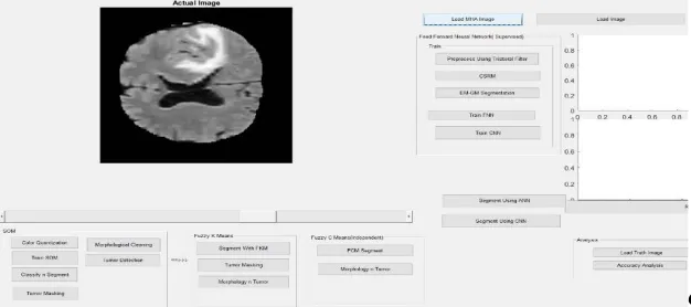
The result analysis is carried out by performing various techniques in digital image processing where the resulting image is obtained in MATLAB GUI. The dataset of brain tumor images is collected from BRATS dataset challenge where the different brain tumor images along with truth images is collected for performing various operations.The experimental result is obtained by loading a .mha image from the dataset and pre-processing it using trilateral filtering and performing the common EMGM segmentation using region based cumulative segmentation (CSRM). The result is further carried out by performing various techniques such as SOM and neural network method such as FNN ,a deep learning techniques such as CNN and fuzzy segmentation technique such as fuzzy c means and fuzzy k means hence brain tumor is detected by using various techniques and accuracy analysis of all such techniques is measured based on performance such as specificity ,sensitivity, coefficient ,dice score and accuracy and conclude which technique is best for detection of brain tumor .
In figure 2 we load the. Mha image, and in trilateral filtering method the filters are defined by a neighbourhood values like how many pixels you want to process which are defined by three sigma values as exponential and average of vertices is taken the result is nothing but the filtered image and the actual and the present filtered value is displayed via message box.
FIG 3: Image trained using SOM.
In figure 3 pre-processed image quantized and given to train via SOM. The SOM is an unsupervised training method it doesn’t require training and testing it just get trained by itself and detect the entire tumor part.
The above image is obtained by using Fuzzy k means we detect the tumor by segmenting the image the image is segmented completely and after segmentation tumor masking is done where the morphology of the image obtained will be the marked tumor area.
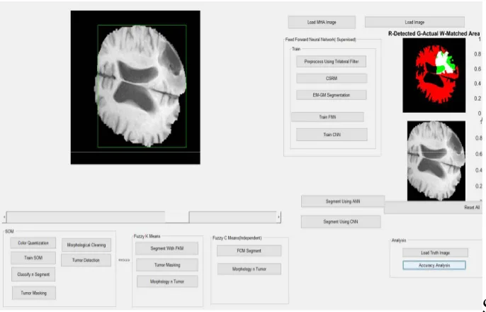
FIG5: segmented image obtained from FNN technique
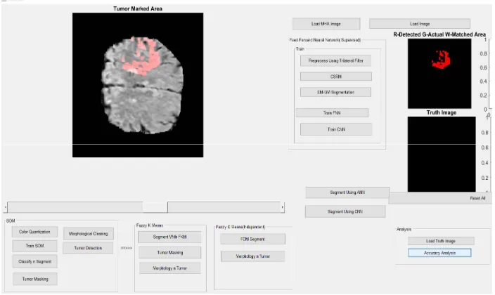
The image segmented obtained in the above figure is marked in rectangle where by loading the corresponding truth image gives the tumor detection and accuracy analysis is obtained.
The image segmented obtained in the above figure is marked in rectangle where by loading the corresponding truth image gives the tumor detection and accuracy analysis is obtained.
FIG7: CNN SEGMENTED TUMOR AREA
CNN segments in the figure 7 shows the tumor area and whereby loading the truth image in axis window 2 shows the tumordetected in the axis window 3 and the accuracy analysis is carried out respectively.
V. CONCLUSION AND FUTURE SCOPE
As brain tumor has been one of major active research in bio medical image processing the proposed work is an interactive segmentation technique obtained from various method like SOM, FNN and CNN and fuzzy segmentation technique such as fuzzy k mean and fuzzy c mean. By training with the above techniques in all aspects we detected the tumor area and segment it, the accuracy analysis is carried out based on performance like specificity,sensitivity, precession and dice score and we compare the result and conclude which technique is best for brain tumor detection. The obtained method showed CNN neural network as an accurate method from all above.
REFERENCES
[1] zhantianming,shenghuagu,canfeng. “analysis of brain mri for tumor detection and segmentation”. Proceeding of the world conference wce 2016, june 29-july1,2016, londonu.k.
[2] v.p.gladispushparathi and s.palani “brain tumor detection and classification using deep learning classifier on mriimages“.researchjurnal of applied science, enginering and technology 10(2):177-187,2015.
[3] raunaqrewari “automatic tumor segmentation from mri scans”, multimodel brain tumor segmentation brat’s proceedings.
[4] Ms. Sangeetha c., Ms. Shahin a. “brain tumor segmentation using artificial neural network”, international research journal of engineering and technology (irjet) volume: 02 issue: 04 | july-2015.
[8] evangeliai. Zacharaki, sumeiwang, sanjeevchawla, dong sooyoo, ronald wolf, elias r. Melhem,1and christosdavatzikos “classification of brain
tumor type and grade using mri texture and shape in a machine learning scheme”. Magnetic resonance in medicine 62:1609–1618 (20).
[9] bjoernh.menze, andrasjakab, stefanbauer, jayashreekalpathy-cramer, keyvanfarahani, justinkirby, yuliya burren, nicoleporz, johannesslotboom, rolandwiest,” the multimodal brain tumor image segmentationbenchmark (brats)”. Ieee transactions on medical imaging, vol.34,no.10,