JOURNAL OFVIROLOGY, May 1995, p. 2811–2818 Vol. 69, No. 5 0022-538X/95/$04.0010
Copyrightq1995, American Society for Microbiology
Cloning, Sequencing, and Functional Characterization of the
Two Subunits of the Pseudorabies Virus DNA Polymerase
Holoenzyme: Evidence for Specificity of Interaction
HERVE BERTHOMME,
1STEVEN J. MONAHAN,
2DEBORAH S. PARRIS,
2BERNARD JACQUEMONT,
3ANDALBERTO L. EPSTEIN
1*
Centre de Ge´ne´tique Mole´culaire et Cellulaire UMR 106, Centre National de la Recherche Scientifique, Universite´
Claude Bernard Lyon I, 69622 Villeurbanne Cedex,
1and Laboratoire de Neuro-virologie Mole´culaire, Faculte´
de Me´decine Alexis Carrel, Universite´ Lyon I, 69372 Lyon Cedex 08,
3France, and Department
of Medical Microbiology and Immunology, Comprehensive Cancer Center,
Ohio State University, Columbus, Ohio 43210-1292
2Received 15 July 1994/Accepted 27 January 1995
The pseudorabies virus (PRV) genes encoding the two subunits of the DNA polymerase were located on the
genome by hybridization to their herpes simplex virus type 1 (HSV-1) homologs,
pol
and UL42, and
subse-quently were sequenced. Like the HSV-1 homologs, in vitro translation products of the PRV gene encoding the
catalytic subunit (
pol
) possessed activity in the absence of the Pol accessory protein (PAP). However, the PRV
PAP stimulated the activity of Pol fourfold in the presence of 150 mM KCl, using an activated calf thymus DNA
template. The stimulation of Pol activity by PAP under high-salt conditions and the inhibition of Pol activity
by PAP when assayed in low salt (0 mM KCl) together were used to determine the specificity with which PAP
interacted with Pol. Despite functional similarity, HSV-1 UL42 and PRV PAP could neither stimulate the
noncognate Pols at high salt nor inhibit them at low salt. Furthermore, a PRV Pol mutant lacking the 30
C-terminal amino acids retained basal Pol activity but could be neither stimulated nor inhibited by the PRV
PAP. Sequence comparisons of the Pol proteins of the alphaherpesviruses reveal a conserved domain in the C
terminus which terminates immediately before the last 41 residues of both PRV and HSV-1 proteins. These
results indicate that the ability and specificity for interaction of the PRV Pol with PAP most likely resides
predominantly in the extreme Pol C terminus.
DNA replication is a complex phenomenon that has been
studied in a variety of prokaryotic and eukaryotic organisms
and viruses. Herpesviruses encode most of the enzymes
di-rectly involved in DNA replication (8, 73) and therefore
pro-vide attractive models for understanding the mechanism of this
process. In particular, they have proven to be good models for
determining the interactions among the various components of
the replication apparatus (10, 16, 23, 29, 35, 67). Moreover, the
components of the herpesvirus DNA replication complex and
the specific interactions that they share are of interest as
tar-gets for the development of antiviral chemotherapy.
Pseudorabies virus (PRV) and herpes simplex virus type 1
(HSV-1) are members of the alphaherpesvirus subfamily and
exhibit many common characteristics, including the ability to
replicate in epithelial tissues and to establish latency in the
neural tissues of swine and humans, respectively (60). These
viruses are quite similar in genomic structure and organization,
although an inversion, from approximately 0.1 to 0.4 map unit
in the UL region of the genome of PRV (4, 12), and a
con-traction of the US region, compared with HSV-1, have been
reported (66). However, the G
1
C contents of the genomes
differ considerably, and cross-hybridization experiments
indi-cate that the genomes share a maximum of 8% nucleotide
sequence homology (6, 58). While some aspects of PRV
ge-nome replication have been studied (3, 44), the essential DNA
replication functions have not been identified. Identification of
the components of the PRV DNA replication complex and
analysis of the protein-protein interactions which they form
would lend greater insight into the function, specificity, and
importance of particular interactions.
Two of the seven HSV-1 proteins which are required in trans
for the amplification of plasmids containing an HSV-1 origin of
replication (73) form a heterodimeric complex which appears
to function as the DNA polymerase holoenzyme (9, 29, 35).
The catalytic subunit of the DNA polymerase gene (pol)
en-codes a 1,235-amino-acid protein with an apparent molecular
weight of 140,000 (27, 50, 57). This polypeptide possesses
in-herent enzymatic properties including DNA chain elongation
(17, 30, 48), 3
9
-5
9
exonuclease (41, 48), and RNase H (9, 48)
activities. It has been shown to be tightly associated with a
65-kDa double-stranded DNA-binding protein encoded by the
UL42 gene of HSV-1 (9, 23, 29, 35, 55, 68), a gene that is
essential for DNA synthesis and virus replication (38, 47). The
product of the UL42 gene stimulates the basal activity and
processivity of Pol (22, 29, 35), properties which appear to be
essential for HSV-1 replication (13, 14, 59, 63).
We were interested in identifying the PRV counterparts of
this enzyme complex in an effort to examine the specificity of
their interaction. Homologs for both Pol and UL42 have been
identified for a variety of herpesviruses (1, 2, 11, 19, 40, 45, 51,
64), and for the homologs of human cytomegalovirus (HCMV)
and Epstein-Barr virus (EBV), functional interaction between
the two proteins was demonstrated (19, 40). However,
exami-nation of cross-species interaction of the components among
members of the herpesvirus family has not been published.
In this report, we describe the identification, cloning,
se-quencing, and functional characterization of the PRV Pol and
* Corresponding author. Mailing address: Centre de Ge´ne´tiqueMo-le´culaire et Cellulaire, UMR 106, CNRS, Universite´ Claude Bernard Lyon I, 43, Boulevard du 11 Novembre 1918, 69622 Villeurbanne Cedex, France.
2811
on November 9, 2019 by guest
http://jvi.asm.org/
UL42 homologs. Using in vitro transcription-translation
prod-ucts, we demonstrate that the PRV UL42 homolog stimulates
the activity of the PRV Pol and that the C-terminal 30 residues
of the PRV Pol are not essential for basal activity but are
required for stimulation by the Pol accessory protein (PAP).
Stimulation of Pol activity did not occur with heterologous
mixes between HSV and PRV proteins. Furthermore, we show
that the salt optimum for activity of the PRV Pol catalytic
subunit is substantially different from that for the PRV
holoen-zyme, and this property was used to confirm the lack of
func-tional association of heterologous components. We also have
compared Pol and PAP sequences among different
herpesvi-ruses and provide additional insight into the importance of
various domains of these proteins.
MATERIALS AND METHODS
Cells and viruses.African green monkey kidney cells (Vero) were obtained from the American Type Culture Collection (Rockville, Md.). Cells were prop-agated in Eagle’s minimum essential medium supplemented with 5% heat-inac-tivated fetal bovine serum in a 378C incubator with a 5% CO2atmosphere. HSV-1 strain F (33) and PRV strain Ka (39) were obtained from B. Roizman and T. Ben-Porat, respectively. Both virus strains were propagated in Vero cells at low input multiplicity of infection (0.01 PFU per cell). Virus yields were deter-mined by plaque assay on Vero cell monolayers at 348C in 5% CO2. For the production of PRV and HSV-1 DNA, Vero cells were infected at a multiplicity of infection of 5 PFU per cell.
Hybridization analysis.Virus DNA was purified from the cytoplasm of in-fected cells by banding in NaI equilibrium density gradients by the method of Walboomers and Ter Scheggett (69). After size fractionation of restriction en-donuclease-treated DNA by agarose gel electrophoresis, the DNA was dena-tured before being transferred to Hybond N nylon filters (Amersham). Dried and baked filters were prehybridized at 458C for 24 h in buffer containing 53SSC (13SSC is 0.15 M NaCl plus 0.015 M sodium citrate), 103Denhardt’s solution (13Denhardt’s solution is 0.02% bovine serum albumin, 0.02% polyvinylpyrro-lidone, and 0.02% Ficoll), 1% sodium dodecyl sulfate (SDS), 100mg of dena-tured herring sperm DNA per ml, and 40% deionized formamide.
Probes for hybridization were prepared by isolating, from agarose gels, frag-ments of HSV-1 DNA that had been subcloned into pSK1vectors, and the purified DNA was labelled with [a-32P]dCTP (specific activity, 3,000 Ci/mmol; Amersham) by the random priming method (20), using a kit (Amersham) as instructed by the manufacturer. Approximately 23106cpm of labelled dena-tured probe was applied to each filter. Hybridization conditions were the same as above except that the concentration of Denhardt’s solution was reduced 10-fold. Filters were washed at 458C in 63SSC–0.1% SDS, dried, and exposed to X-ray film at2708C, using an intensifying screen.
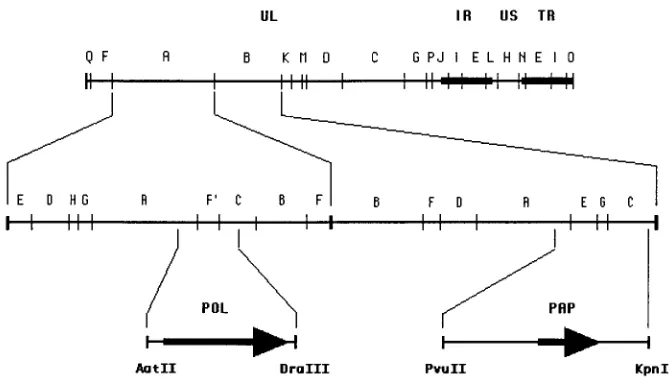
Cloning procedure.For cloning of the pol gene, a plasmid containing the 29.4-kbp BamHI A fragment of PRV DNA (kindly provided by H. J. Rziha,
Tubingen, Germany) was digested with restriction enzyme AatII and was par-tially cleaved by enzyme DraIII to yield an AatII-DraIII subfragment 3.4 kbp in size (Fig. 1). After removal of the 39overhangs with the Klenow enzyme, this fragment was cloned into the EcoRV site of the polylinker of the pBluescript SK1vector (Stratagene) to form plasmid pPRVpol. This plasmid contains the PRV pol open reading frame (ORF) as well as 133 bp upstream from the putative translation initiation codon and 142 bp downstream from the consensus polyad-enylation signal. Cloning of the PRV PAP was initiated with a plasmid containing the 18.4-kbp BamHI B fragment of PRV DNA (gift of H. J. Rziha). The 5.9-kbp PvuII-KpnI subfragment (Fig. 1) was cloned into the EcoRV and KpnI sites of pBluescript SK1to form plasmid pPRVpk5.9. This plasmid was linearized at the KpnI site, the 39overhangs were removed with the Klenow enzyme, the DNA was partially digested with NruI, and the mixture was religated, generating plasmid pPRVpn3.2. Unidirectional progressive deletion from the PvuII extremity of pPRVpn3.2 was performed by the exonuclease III method of Henikoff (34), generating plasmid pPRVpap. This plasmid contains the PRV pap ORF as well as 15 bp upstream from the putative translation initiation codon and 53 bp downstream from the consensus polyadenylation signal. Plasmids pLBN 19A, encoding full-length HSV-1 UL42 (22), and pT7-7.1, encoding HSV-1 Pol lack-ing the first 67 amino acids (17), have been described elsewhere.
DNA sequencing.Nested sets of progressive unidirectional deletions from both extremities were prepared from plasmids pPRVpol and pPRVpn3.2 (34). Se-lected clones were amplified in Escherichia coli, and DNA was isolated by an alkaline lysis procedure (62). All plasmids were sequenced by the dideoxynucle-otide chain termination method (61), using the DNA polymerase T7 sequencing kit (Pharmacia) and [a-35S]dATP (Amersham). To prevent secondary structures in DNA during sequencing, 10% dimethyl sulfoxide was added to the reaction mixtures. Deaza C-7 dGTP or dITP was used to resolve band compressions due to the high G1C nucleotide content of the PRV DNA. Both strands of DNA from each plasmid were sequenced at least two times, using progressive dele-tions. The sequences were further confirmed by subcloning and sequencing smaller restriction fragments from the parental plasmid or by direct sequencing using selected synthetic oligonucleotides as primers. DNA sequences were ana-lyzed for eukaryotic transcriptional elements by using the SIGNAL SCAN soft-ware (56).
[image:2.612.139.474.68.257.2]In vitro transcription-translation.Ten micrograms of pPRVpol, pPRVpap, pT7-7.1 (HSV-1 pol), and pLBN 19A (HSV-1 UL42) were linearized down-stream of each ORF with ClaI, PvuII, XbaI, and XbaI, respectively. The linear-ized DNA was purified by phenol-chloroform extraction and concentrated by ethanol precipitation. In vitro transcripts were generated with a kit (Promega Biotec, Madison, Wis.) as instructed by the manufacturer. Briefly, 3 mg of purified DNA was transcribed with 15 U/mg of DNA (for pPRVpap and pLBN 19A) or 10 U/mg of DNA (for pPRVpol and pT7-7.1), using T3 or T7 RNA polymerase, as appropriate, in the transcription buffer provided, with 10 mM dithiothreitol, 1.6 U of RNasin RNase inhibitor (Promega Biotec) perml, 0.5 mM each ribonucleoside triphosphate, and diethyl pyrocarbonate-treated water. After 1 h of incubation at 408C, linearized DNA was removed with RQ1 RNase-free DNase (1 U/mg of DNA; Promega Biotec) for 15 min at 378C. The RNA was purified by phenol-chloroform extraction and ethanol precipitation and analyzed on formaldehyde-agarose gels as described previously (46).
FIG. 1. Localization of the PRV pol and pap genes. The upper line shows the BamHI restriction endonuclease map of the PRV genome (52, 71). Solid boxes represent the inverted repeats that bracket the short unique region of the genome. The second line corresponds to an expanded representation of the BamHI A and B fragments. SalI subfragments of BamHI-A and NruI subfragments of BamHI-B are indicated above the line. Positions of the pol and pap genes are shown at the bottom; their orientations are indicated by arrows.
on November 9, 2019 by guest
http://jvi.asm.org/
Transcripts were translated in rabbit reticulocyte lysates as directed by the manufacturer (Promega Biotec). For each reaction, 0.5 to 1mg of RNA was incubated for 1 h at 308C with 0.8mCi ofL-[35S]methionine (specific activity, 1,000 to 1,300 Ci/mmol; Amersham) perml. Products were analyzed by SDS-polyacrylamide gel electrophoresis (PAGE) using 10 to 20% SDS-polyacrylamide gradient gels, impregnated with En3Hance (DuPont, Boston, Mass.), and ex-posed to X-ray film. For Pol enzyme assays, 5 to 10mg of RNA was translated in the presence of unlabelledL-methionine. In some cases, a coupled transcription-translation system (Promega Biotec) was used to generate proteins.
Pol assay.Pol activity was measured as incorporation of [3H]dTTP (42 Ci/ mmol; Amersham) into trichloroacetic acid-insoluble radioactivity as described previously, using activated calf thymus DNA as the template (17, 18, 22), except that KCl concentration was varied as indicated in the figure legends. In vitro translation products from the indicated pol and pap genes (25ml) were added and incubated in a final volume of 100ml for 30 min at 378C. For reactions containing both Pol and PAP, the translated products were preincubated 5 min at 378C before initiation of the reaction in order to allow complex formation. All assays were conducted in duplicate. Units of Pol activity are defined as the number of femtomoles of [3
H]dTTP incorporated into DNA at 378C during 30 min by 25ml of programmed lysate.
Nucleotide sequence accession numbers.The nucleotide sequences corre-sponding to the PRV Pol and PAP ORFs have been submitted to the GenBank database and have been assigned accession numbers L24487 and M94355, re-spectively.
RESULTS
Localization and sequencing of the PRV
pol
gene.
Heterol-ogous hybridization experiments were performed by probing a
Southern blot containing BamHI-restricted PRV genomic
DNA (strain Ka) with a subcloned 3.4-kbp BamHI R
restric-tion fragment of the HSV-1 (strain F) genome. The HSV-1
fragment was separated from plasmid DNA by gel
electro-phoresis before use as a probe for hybridization. This probe
contained 154 nucleotides upstream from the initiation codon
to nucleotide residue 3216 of the HSV-1 pol ORF. Positive
specific hybridization was observed with the 29.4-kbp BamHI
A PRV DNA fragment (Fig. 1), suggesting that PRV pol was
included within this region. Further experiments using the
same probe and PRV BamHI-A DNA cleaved with SalI
re-vealed that the HSV-1 pol gene hybridized with the PRV SalI
A, F
9
, and C subfragments (52). Preliminary DNA sequencing
revealed AatII and DraIII sites, respectively, upstream and
downstream from the PRV pol ORF. Complete sequencing of
the AatII-DraIII subfragment confirmed that it contained the
entire PRV pol gene (Fig. 1). Analysis of the PRV pol
nucle-otide sequence reveals that the gene contains a 3,144-bp
con-tinuous ORF, predicted to encode a protein of 1,048 amino
acids. At the 3
9
end of the PRV pol ORF, there is a canonical
polyadenylation signal which is 15 nucleotides downstream of
the termination codon UGA. The first AUG codon, presumed
to be the start site for translation, is found in region GAGC
GAUGG, in good agreement with the requirements of active
translation initiation site (43), with a guanine at position
1
4
and a purine at position
2
3. That this is the true initiation
codon is further suggested by the lack of another methionine
codon until codon 425 and by the fact that the N-terminal
portion of the PRV pol ORF just downstream from the
puta-tive AUG initiation codon displays conservation with that of
other alphaherpesviruses (data not shown). Using the
SIG-NAL SCAN computer program that scans DNA sequences for
eukaryotic transcription elements, we detected several
consen-sus regulatory sequences, including a consenconsen-sus TATA box at
nucleotide
2
44 and several consensus Sp1 sites between
nucle-otides
2
57 and
2
110, in the region upstream of the pol ORF.
In contrast to the genomic features in the region of the HSV-1
pol, neither a small ORF nor sequences resembling a
replica-tion origin (27, 50, 57) have been found near the PRV pol
promoter.
Localization and sequencing of the PRV
pap
gene.
To locate
the PRV sequences corresponding to the HSV-1 PAP, UL42,
a Southern blot of BamHI-digested PRV genomic DNA was
probed with a cloned 1.3-kbp MluI fragment, encompassing
sequences located entirely within the HSV-1 (strain F) UL42
ORF. Specific hybridization of the probe to the 18.4-kbp PRV
BamHI B fragment provided a rough localization of the PRV
UL42 homolog, pap (Fig. 1). To further define the location of
the gene, a plasmid containing the PRV BamHI B fragment
was digested in a series of experiments with KpnI, PvuII, and/or
NruI, Southern blots of separated fragments were prepared,
and the blots were hybridized to an HSV-1 MluI UL42 probe.
Complete sequencing of the Nru E and G subfragments
indicated that they contained the 5
9
extremity of the vhs gene
(5) and the entire putative PRV pap gene. Analysis of the pap
nucleotide sequence reveals that the gene contains a 1,152-bp
continuous ORF encoding 384 amino acids. Both the first and
second AUG codons in the ORF are found in regions which
are in relatively good agreement with Kozak’s rules for
trans-lation initiation (43). However, on the basis of conservation of
the upstream residues with those of other alphaherpesvirus
homologs, it seems more likely that the protein initiates at the
upstream AUG codon. Several putative regulatory sequences,
including a degenerate TATA box homology (TAAAATA)
located at
2
100, three overlapping Sp1 consensus elements
located between
2
86 and
2
76, and a perfect TATA box
ho-mology (ATATAA) at
2
63, were detected upstream from this
presumed initiation codon. Interestingly, at the 3
9
end of the
pap ORF is a canonical polyadenylation signal (AATAAA)
which overlaps the termination codon TAA. The relative
ori-entations of the PRV pol and pap genes are as in the HSV-1
prototype orientation (50).
Characterization of PRV
pol
and
pap
translation products.
Runoff transcripts were produced by transcription in vitro with
the T3 RNA polymerase of plasmids pPRVpol and pPRVpap
linearized downstream of the ORFs. These RNAs were
trans-lated in rabbit reticulocyte lysates in the presence of [
35S]me-thionine and analyzed by SDS-PAGE. The PRV gene products
were compared with those translated from RNA generated by
the T7 RNA polymerase runoff transcription of the linearized
plasmid pT7-7.1, coding for the HSV-1 Pol which lacks the 67
amino-terminal amino acids, and pLBN 19A, encoding the
HSV-1 UL42 gene (Fig. 2). The major translation product of
the PRV pol gene corresponded to a protein with an apparent
molecular weight of 110,000 (lane 2), whereas the major
trans-lation product produced from the HSV-1 gene migrated as a
130-kDa peptide (lane 4). The major translation product of the
HSV-1 UL42 gene has an apparent molecular mass of 62 kDa
(lane 5) as previously described (55), whereas that of the PRV
homolog is considerably smaller, migrating as a 42-kDa
polypeptide (lane 3). The sizes of both the PRV pol and pap
gene products are in good agreement with the 115.3 and 40.3
kDa predicted from the ORFs.
Biological activities of PRV Pol and PAP.
To determine
whether the PRV Pol and PAP homologs were able to function
as predicted, we performed DNA polymerase assays on in vitro
transcription-translation products of the genes. Table 1
dem-onstrates that like the HSV-1 Pol, the PRV Pol possesses
activity on activated calf thymus DNA template at 150 mM
KCl even in the absence of the accessory protein. We observed
no significant Pol activity either in unprogrammed rabbit
re-ticulocytes or in translation products of either the PRV pap or
the HSV-1 UL42 gene. However, when we mixed the PRV pap
and PRV pol translation products, we observed a 3.5-fold
stim-ulation of Pol activity compared with the activity of Pol
prod-ucts alone. In parallel assays, we observed a 3.4-fold
stimula-tion of HSV-1 Pol activity in the presence of the HSV-1 UL42.
These results demonstrate that the PRV gene that we have
VOL. 69, 1995 CHARACTERIZATION OF PRV DNA POLYMERASE SUBUNITS 2813
on November 9, 2019 by guest
http://jvi.asm.org/
designated pap encodes a Pol accessory protein and possesses
activity similar to that of its HSV-1 counterpart.
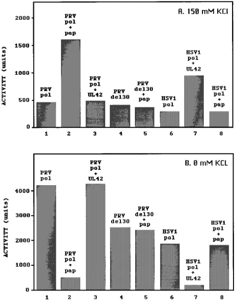
Effect of salt concentration on Pol activity.
Because the
HSV-1 Pol holoenzyme has been shown to be stimulated in the
presence of high salt, we investigated the sensitivity of the PRV
Pol or Pol-PAP complex to salt. In vitro
transcription-transla-tion products of the PRV and HSV-1 genes were assayed for
Pol activity in the presence of increasing concentrations of
KCl. As previously observed for the HSV-1 Pol catalytic
sub-unit (32), we found the activity of the PRV Pol to be extremely
sensitive to salt in the absence of its accessory protein, with
maximum activity observed when no exogenous KCl was added
(data not shown). Indeed, in the absence of exogenously added
KCl, the PRV Pol exhibited more than eight times the activity
found in the presence of 150 mM KCl (compare bar 1 in Fig.
3B with bar 1 in Fig. 3A). Although the in vitro-translated
HSV-1 Pol activity was somewhat lower than that of the PRV
Pol, we observed a similar level of decrease in activity when the
exogenous KCl concentration was raised from 0 to 150 mM
KCl (compare bar 6 in Fig. 3B with bar 6 in Fig. 3A). The
opposite effect of salt on activity was observed for both the
PRV and HSV-1 Pols in the presence of their respective
ac-cessory proteins: the activity of each Pol-PAP complex was
higher in the presence of 150 mM KCl than in the presence of
0 mM KCl (compare bars 2 and 7 in Fig. 3A with bars 2 and 7
in Fig. 3B). The net effect of these interactions is that the
accessory proteins inhibit the activities of the Pols in low salt
and stimulate their activities in the presence of high salt (Fig.
3; compare bars 2 and 1 and bars 7 and 6). Although the latter
effect has been one of several means for ascribing Pol accessory
function to HSV-1 UL42 and some of its homologs (14, 19, 22,
23, 29, 35, 40, 65), it is of particular note that the maximum
activities that we observed for the HSV-1 and PRV Pol-PAP
complexes (in high salt) never exceeded the maximum
activi-ties of the Pols without their accessory proteins (in low salt)
(Fig. 3 and results not shown).
[image:4.612.112.245.69.261.2]We took advantage of the dramatic differences in salt
sen-sitivities of the Pol-PAP complexes compared with those of the
Pol catalytic subunits alone to investigate the specificity of the
PAPs. When we mixed the heterologous HSV-1 UL42 with the
PRV Pol, we found no effect on PRV Pol activity at either 0 or
FIG. 2. In vitro translation products of the PRV and HSV-1 pol and papgenes. RNA transcribed by in vitro runoff transcription from linearized DNA templates (pPRVpol, pPRVpap, pT7-7.1 [HSV-1 pol]) and pLBN 19A [HSV-1 UL42]) were translated in rabbit reticulocyte lysate with35
[image:4.612.316.555.73.377.2]S-labelledL -methio-nine for 1 h at 308C. Products were separated through a denaturing 10 to 20% gradient polyacrylamide gel. Rabbit reticulocyte lysates were programmed with water (lane 1), PRV pol (lane 2), PRV pap (lane 3), HSV-1 pol (lane 4), and HSV-1 UL42 (lane 5) RNAs. The molecular masses (in kilodaltons) of protein standards are indicated on the left; the apparent masses of the full-length trans-lation products are shown on the right.
FIG. 3. Specificity of interaction between Pols and PAPs from PRV and HSV-1 and lack of activation of PRV Pol del30 by its cognate cofactor. Pol activity was assayed for nonlabelled translation products from PRV pol and pap, PRV pol del30, and HSV-1 pol and UL42, with an activated calf thymus DNA template, in the presence of 150 (A) or 0 (B) mM KCl. Bars indicate averages of duplicate experiments. Neither the PRV PAP nor the HSV-1 UL42 protein possessed activity in excess of that for the unprogrammed lysates, which ac-counted for less than 10% of the observed incorporation rates.
TABLE 1. Activities of PAPs on wild-type Pol proteins
Construct Pol activity (U)a
Fold stimulationb
PAP 0 0.0
PRVpol (wtc) 470649 1.0
PRVpol (wt)1PAP 1,6206115 3.5
UL42 0 0.0
HSVpol 360631 1.0
HSVpol1UL42 1,210672 3.4
a
Determined by assay of products from coupled in vitro transcription-trans-lation reactions on activated calf thymus DNA template in the presence of 150 mM KCl as described in Materials and Methods. Pol assays were performed as duplicates for four independent experiments, and the means and standard devi-ations are shown. Activities were calculated by subtracting the radioactivity incorporated by unprogrammed rabbit reticulocyte lysates from the values ob-tained with each RNA indicated. Incorporation by lysates programmed with PAP or UL42 were indistinguishable from those from unprogrammed lysates, which never exceed 10% of the level incorporated by the Pols alone.
b
Calculated as the mean activity of the indicated Pol in the presence of cofactor divided by the mean activity of that Pol in the absence of cofactor.
c
wt, wild type.
on November 9, 2019 by guest
http://jvi.asm.org/
[image:4.612.58.298.554.630.2]150 mM KCl (Fig. 3; compare bar 3 with bar 1). Likewise, a
heterologous mix of PRV PAP with HSV-1 Pol yielded neither
an increase in activity of Pol at 150 mM KCl nor a decrease in
activity at 0 mM KCl (Fig. 3; compare bar 8 with bar 6). Taken
together, these results extend those of Hart and Boehme (32)
in demonstrating the specificity of both the stimulation of Pol
activity in high salt and the inhibition of Pol activity in low salt
by PAPs. Furthermore, they suggest that cross-species
Pol-PAP complex formation does not occur among the HSV-1 and
PRV components.
Consistent with the interpretation that these effects are the
results of specific complex formation between Pol and PAP are
results obtained for one PRV Pol deletion mutant. PRV Pol
deleted of the C-terminal 30 amino acids (del30) was
gener-ated by cleavage of pPRVpol DNA with StuI prior to runoff
transcription followed by in vitro translation. PRV del30
pos-sessed activity similar to that of full-length PRV Pol at 150 mM
KCl in the absence of accessory protein (Fig. 3A; compare bar
4 with bar 1), yet the addition of PRV PAP failed to enhance
its activity (compare bars 4 and 5). Although the activity of the
PRV Pol del30 was somewhat lower than that of the full-length
Pol at 0 mM KCl, the addition of PRV PAP was not inhibitory
(Fig. 3B; compare bars 4 and 5 with bars 1 and 2). These results
demonstrate that the C-terminal 30 residues of PRV Pol are
not necessary for basal activity but are required for functional
interaction with PAP.
DISCUSSION
Comparison of PRV Pol sequences with sequences of
her-pesvirus homologs.
Using the CLUSTALV computer program
(36), we have aligned the PRV Pol and PAP sequences with
those of HSV-1 (50), varicella-zoster virus (VZV) (11), and
equine herpesvirus 1 (EHV-1) (64), all members of the
alpha-herpesvirus family. In addition, we have included the EBV,
HCMV, and human herpesvirus 6 (HHV-6) putative
ho-mologs, obtained from the SwissProt database. Employing
pairwise alignments, the PRV pol, pap, and previously reported
vhs (5) genes are most closely related to those of EHV-1, in
agreement with the placement of EHV-1 and PRV on the
same branch of an evolutionary tree (49).
In addition to the eight regions of homology (I through VII
and A) observed among a variety of prokaryotic, eukaryotic,
and viral
a
-like DNA polymerases (26, 37, 70, 72), there are
several additional regions of sequence homology among the
herpesvirus pol genes. Among these, a C-terminal domain,
extending from PRV residues 949 to 1007, is well conserved
between the alphaherpesvirus group and is somewhat more
divergent when EBV, HCMV, and HHV-6 are included in the
alignment (Fig. 4A). This domain is included in the HSV-1 Pol
region described by Digard and Coen (15) to be necessary and
sufficient for stable association with the UL42 PAP. However,
within the last 41 residues of both the PRV and HSV-1 pol
genes are only three residues identical to those of the other
alphaherpesvirus homologs, and only one if we include the
other three viruses. Interestingly, deletion of the C-terminal 30
residues of PRV Pol abolishes its ability to be stimulated by its
cognate accessory protein, while having little or no effect on
basal Pol activity (Fig. 3). The C terminus of HSV-1 Pol also
has been shown to be required for stimulation by UL42 but not
for basal polymerase activity (13, 63, 65). The HSV-1 Pol was
shown to possess catalytic activity even after deletion of
resi-dues 1073 to 1144 or of the 59 C-terminal amino acids (30).
Several other groups have confirmed these results and have
shown that the HSV-1 Pol C terminus appears to be critical for
functional interaction with UL42, although the exact
delinea-tion of the critical residues remains uncertain (13, 63, 65). Of
particular note, however, is the fact that an HSV-1 Pol mutant
lacking the C-terminal 27 residues and expressed as a
baculo-virus recombinant possessed activity which could not be
stim-ulated by UL42 in vitro and which could not function in
origin-dependent DNA replication (13, 63). Thus, it appears clear
that the residues which lie downstream of the conserved
C-terminal domain are essential for true functional interaction
FIG. 4. (A) Alignment of the predicted amino acid sequence of the C-terminal domain of the PRV pol gene product with counterparts from HSV-1, VZV, EHV-1, HCMV, HHV-6, and EBV. Sequences were aligned by using the multiple alignment CLUSTALV computer program (36). Open boxes indicate the positions of identical amino acids either in the four alphaherpesviruses or in the seven sequences. Numbers at the right indicate amino acid numbers. (B) Alignment of three conserved domains of the PAPs from four alphaherpesvi-ruses. The PRV PAP was aligned with its counterparts from HSV-1 (UL42 gene), VZV (gene 16), and EHV-1 (gene 18). Sequences were aligned by using the multiple alignment CLUSTALV computer program. The limits of the three domains were defined using arbitrary criteria (5 identical or conserved residues in 25). The positions of identical amino acids in all four sequences are indicated with darkly shaded boxes, while those of conserved amino acids are shown as lightly shaded boxes. The following residues were considered to be conserved: MILCV, DEQN, PAG, FYW, KRH, and ST. The limits of conserved domains I (solid bar), II (hatched bar), and III (open bar) are represented above the sequences. Numbers at the right indicate amino acid numbers.VOL. 69, 1995 CHARACTERIZATION OF PRV DNA POLYMERASE SUBUNITS 2815
on November 9, 2019 by guest
http://jvi.asm.org/
with their respective accessory proteins. The high level of
di-vergence of these sequences may, in fact, reflect the specificity
of interaction with the accessory proteins which are highly
divergent among the herpesviruses. It is interesting, however,
that the divergent C-terminal Pol region is much shorter in
HCMV and HHV-6 and is almost completely absent in EBV,
with the notable exception of one phenylalanine residue which
is conserved in all the seven polymerases (Fig. 4A).
Site-di-rected mutagenesis of this region of Pol will be necessary to
determine those residues which are most critical for interaction
with these proteins.
Comparison of PAP sequences.
We attempted to align PAP
sequences from the seven herpesviruses. Significant sequence
conservation was found within the alpha- and the
betaherpes-virus groups, but little conservation was found between them
(not shown). Comparison of the UL42 homologs of the four
alphaherpesviruses is consistent with the three-way alignment
previously reported for HSV-1, EHV-1, and VZV homologs
(65) and shows that the amino-terminal two-thirds to
three-fourths of the protein is best conserved (Fig. 4B). In the
con-served portion of PAP, we have distinguished three distinct
conserved domains. Domain I corresponds to PRV amino
ac-ids E-36 to Y-152, domain II corresponds to residues V-171 to
F-215, and domain III corresponds to residues A-257 to F-316.
Although these three domains correspond to only 57% of the
PRV polypeptide, they include all of the identical amino acids
and 94% of the similar residues. Although the relationship
between the structure and the function of the HSV-1 UL42
protein is not completely understood, most of the results from
mutagenesis studies support the importance of the conserved
domains in the amino-terminal portion of the HSV-1 UL42
(14, 53, 59). The strongest divergence, which occurs
down-stream of PRV residue 317, results not only from the longer
HSV-1 C-terminal protein but also from poorly conserved
ho-mology, even in pairwise alignments of this region or in
three-way alignments without the HSV-1 protein (not shown). This
C-terminal portion of the HSV-1 protein has been shown not
to be required for stimulation of Pol activity, dimerization with
Pol, binding to DNA, localization to the nucleus, or replication
of the virus in vitro (14, 25, 65), and it is easily separated from
the rest of the protein by proteolytic degradation (31). It is
especially interesting that although an HSV-1 UL42 protein
containing only the N-terminal 315 residues retains
Pol-stim-ulating activity, deletion of the next residue, which forms the
end of the N-terminal conserved motif of domain III that we
define in Fig. 4B, abolishes activity (65). Thus, it is clear that
the C-terminal portions of the PAP homologs form a distinct
domain whose function has not been determined but is not
likely to be related to Pol accessory activity.
Effect of salt on the activity of the Pol-PAP complex.
We
observed a stimulation of the HSV-1 and PRV Pol activities
with their accessory proteins in the presence of high salt as well
as an inhibition of activities by the accessory proteins under
low-salt conditions (Fig. 3). Although the assays reported
herein were performed with in vitro translation products, we
have found similar results with proteins purified from
recom-binant baculoviruses which express them alone or as a complex
(21). The stimulation of Pol activity in high salt may be
ex-plained by the lower affinity of the Pol catalytic subunit for
DNA compared with that of the Pol-PAP complex (24, 28, 32),
coupled with a further destabilization in the presence of high
ionic strength. However, it is much more difficult to reconcile
an inhibition of activity in the presence of low salt. Our results
are similar to those published by Hart and Boehme (32), who
also observed an inhibition of the purified HSV-1 Pol activity
by the purified accessory protein. However, our observation
that the heterologous accessory proteins fail to inhibit the
noncognate Pols under these conditions suggests that Pol and
PAP physically interact even at low ionic strength and it is the
interaction per se which results in inhibition. The inhibition of
Pol activity could be explained by the failure of a Pol-PAP
complex to bind to DNA at low salt. The low salt would
strengthen hydrophobic interactions, perhaps altering the
con-formation of the proteins, DNA, or both, which could be
det-rimental to the productive association of the complex with
DNA. Alternatively, the complex might bind to DNA so tightly
at low salt that the movement required for fork progression
would be inhibited. Indeed, a local ionic gradient along the
DNA may need to be established to enable the complex or
‘‘clamp’’ to slide along the DNA molecule. The ions present in
the absence of exogenously added KCl may be insufficient to
establish such a gradient.
Specificity of Pol and PAP interactions.
Our results have
demonstrated that the PRV Pol and PAP provide functions
similar to those of the HSV-1 counterparts. However, despite
these functional similarities, the PRV PAP and HSV-1 UL42
cannot substitute for each other. Although the accessory
pro-teins are less conserved than the Pol propro-teins, the reasonable
conservation of the PAPs in the amino two-thirds portions
suggests that these proteins are likely to be structurally similar.
Structural analysis of other PAPs, most notably the
b
subunit
of E. coli Pol III (42) and PCNA, the accessory protein for
mammalian cell Pol
d
(7, 54), reveals that each forms a sliding
clamp over the DNA, the former as a dimer and the latter as
a trimer. Despite the structural and functional similarities of
these proteins, they share no obvious sequence homology (54).
Nevertheless, it is clear from our studies that apparently minor
differences in overall structure can affect the ability of
acces-sory proteins to interact with Pol. The specificity by which the
accessory proteins interact with their respective Pols and the
necessity for this functional interaction in vivo (13, 14, 59, 63)
indicate the possibility for the development of antiviral
com-pounds which target this interaction and which are highly
ef-fective and specific for blocking viral DNA replication. The
assays described herein offer a rapid and facile means for
screening the specificity of such compounds.
ACKNOWLEDGMENTS
The work in France was supported by Association pour la Recher-che sur le Cancer and by Institut National de la Sante´ et de la Re-cherche Me´dicale. The work in the United States was supported in part by grant GM 34930 from the National Institutes of Health (D.S.P.). H.B. is a doctoral student receiving support from Ministe`re de la Recherche et de l’Espace (France) and from Ligue Nationale contre le Cancer. S.J.M. was supported in part by a postdoctoral fellowship (F32 AI 08566-02) from the National Institutes of Health.
REFERENCES
1. Agulnick, A. D., J. R. Thompson, S. Iyengar, G. Pearson, D. Ablashi, and R. P. Ricciardi.1993. Identification of a DNA-binding protein of human herpesvirus 6, a putative DNA polymerase stimulatory factor. J. Gen. Virol. 74:1003–1009.
2. Baer, R., A. T. Bankier, M. D. Biggin, P. L. Deininger, P. J. Farrell, T. J. Gibson, G. Hatfull, G. S. Hudson, S. C. Satchwell, C. Seguin, P. S. Tuffnell, and B. G. Barrell.1984. DNA sequence and expression of the B95-9 Epstein-Barr virus genome. Nature (London) 310:207–211.
3. Ben-Porat, T., and R. A. Veach. 1980. Origin of replication of the DNA of
a-herpesvirus (pseudorabies). Proc. Natl. Acad. Sci. USA 77:172–175. 4. Ben-Porat, T., R. A. Veach, and S. Ihara. 1983. Localization of the regions of
homology between the genomes of herpes simplex virus type 1, and pseu-dorabies virus. Virology 127:194–204.
5. Berthomme, H., B. Jacquemont, and A. Epstein. 1993. The pseudorabies virus host-shutoff gene: nucleotide sequence and comparison with alphaher-pesvirus protein counterparts. Virology 193:1028–1032.
6. Bronson, D. L., B. J. Graham, H. Ludwig, M. Benyesh-Melnick, and N. Biswal.1972. Studies on the relatedness of herpesviruses through
on November 9, 2019 by guest
http://jvi.asm.org/
RNA hybridization. Biochim. Biophys. Acta 259:29–34.
7. Burgers, P. M. J., and B. L. Yoder. 1993. ATP-independent loading of the proliferating cell nuclear antigen requires DNA ends. J. Biol. Chem. 268: 19923–19926.
8. Challberg, M. D. 1991. Herpes simplex virus DNA replication. Semin. Virol. 2:247–256.
9. Crute, J. J., and I. R. Lehman. 1989. Herpes simplex-1 DNA polymerase— identification of an intrinsic 59-39exonuclease with ribonuclease H activity. J. Biol. Chem. 264:19266–19270.
10. Crute, J. J., T. Tsurumi, L. Zhu, S. K. Weller, P. D. Olivo, M. D. Challberg, E. S. Mocarski, and I. R. Lehman.1989. Herpes simplex virus 1 helicase-primase: a complex of three herpes-encoded gene products. Proc. Natl. Acad. Sci. USA 86:1–4.
11. Davison, A. J., and J. E. Scott. 1986. The complete DNA sequence of varicella-zoster virus. J. Gen. Virol. 67:1759–1816.
12. Davison, A. J., and N. M. Wilkie. 1983. Location and orientation of homol-ogous sequences in the genomes of five herpesviruses. J. Gen. Virol. 64: 1927–1942.
13. Digard, P., W. R. Bebrin, K. Weisshart, and D. M. Coen. 1993. The extreme C terminus of herpes simplex virus DNA polymerase is crucial for functional interaction with processivity factor UL42 and for viral replication. J. Virol. 67:398–406.
14. Digard, P., C. S. Chow, L. Pirrit, and D. M. Coen. 1993. Functional analysis of the herpes simplex virus UL42 protein. J. Virol. 67:1159–1168. 15. Digard, P., and D. M. Coen. 1990. A novel functional domain of an a-like
DNA polymerase. J. Biol. Chem. 265:17393–17396.
16. Dodson, M. S., J. J. Crute, R. C. Bruckner, and I. R. Lehman. 1989. Over-expression and assembly of the herpes simplex virus type-1 helicase-primase in insect cells. J. Biol. Chem. 264:20835–20838.
17. Dorsky, D. I., and C. S. Crumpacker. 1988. Expression of herpes simplex virus type 1 DNA polymerase gene by in vitro translation and effects of gene deletions on activity. J. Virol. 62:3224–3232.
18. Dorsky, D. I., and C. S. Crumpacker. 1990. Site-specific mutagenesis of a highly conserved region of the herpes simplex virus type 1 DNA polymerase gene. J. Virol. 64:1394–1397.
19. Ertl, P. F., and K. L. Powell. 1992. Physical and functional interaction of human cytomegalovirus DNA polymerase and its accessory protein (ICP36) expressed in insect cells. J. Virol. 66:4126–4133.
20. Feinberg, A. P., and B. Vogelstein. 1983. A technique for radiolabeling DNA restriction endonuclease fragments to high specific activity. Anal. Biochem. 132:6–13.
21. Fraley, D. M., and D. S. Parris. 1994. Unpublished results.
22. Gallo, M. L., D. I. Dorsky, C. S. Crumpacker, and D. S. Parris. 1989. The essential 65-kilodalton DNA-binding protein of herpes simplex virus stimu-lates the virus-encoded DNA polymerase. J. Virol. 63:5023–5029. 23. Gallo, M. L., D. H. Jackwood, M. Murphy, H. S. Marsden, and D. S. Parris.
1988. Purification of the herpes simplex virus type 1 65-kilodalton DNA-binding protein: properties of the protein and evidence of its association with the virus-encoded DNA polymerase. J. Virol. 62:2874–2883.
24. Gallo, M. L., and D. S. Parris. 1994. Unpublished results.
25. Gao, M., S. F. DiTusa, and M. G. Cordingley. 1993. The C-terminal third of UL42, a HSV-1 DNA replication protein, is dispensable for viral growth. Virology 194:647–653.
26. Gibbs, J. S., H. C. Chiou, K. F. Bastow, Y. Cheng, and D. M. Coen. 1988. Identification of amino acids in herpes simplex virus DNA polymerase in-volved in substrate and drug recognition. Proc. Natl. Acad. Sci. USA 85: 6671–6676.
27. Gibbs, J. S., H. C. Chiou, J. D. Hall, D. W. Mount, M. Retondo, S. K. Weller, and D. M. Coen.1985. Secuence and mapping analysis of the herpes simplex virus DNA polymerase gene predict a C-terminal substrate binding domain. Proc. Natl. Acad. Sci. USA 82:7969–7973.
28. Gottlieb, J., and M. D. Challberg. 1994. Interaction of herpes simplex virus type 1 DNA polymerase and the UL42 accessory protein with a model primer template. J. Virol. 68:4937–4945.
29. Gottlieb, J., A. I. Marcy, D. M. Coen, and M. D. Challberg. 1990. The herpes simplex virus type 1 UL42 gene product: a subunit of DNA polymerase that functions to increase processivity. J. Virol. 64:5976–5987.
30. Haffey, M. L., J. T. Stevens, B. J. Terry, D. I. Dorsky, C. S. Crumpacker, S. M. Wietstock, W. T. Ruyechan, and A. K. Field.1988. Expression of herpes simplex virus type 1 DNA polymerase in Saccharomyces cerevisiae and detection of virus-specific enzyme activity in cell-free lysates. J. Virol. 62: 4493–4498.
31. Hamatake, R. K., M. Bifano, D. J. Tenney, W. W. Hurlburt, and M. G. Cordingley.1993. The herpes simplex virus type 1 DNA polymerase acces-sory protein, UL42, contains a functional protease-resistant domain. J. Gen. Virol. 74:2181–2189.
32. Hart, G. J., and R. E. Boehme. 1992. The effect of the UL42 protein on the DNA polymerase activity of the catalytic subunit of the DNA polymerase encoded by herpes simplex virus type 1. FEBS Lett. 305:97–100. 33. Heine, M. D., R. W. Honess, E. Cassai, and B. Roizman. 1974. Proteins
specified by herpes simplex virus. XII. The virion polypeptides of type 1 strains. J. Virol. 14:640–651.
34. Henikoff, S. 1984. Unidirectional digestion with exonuclease III creates tar-geted breakpoints for DNA sequencing. Gene 28:351–359.
35. Hernandez, T. R., and I. R. Lehman. 1990. Functional interaction between the herpes simplex-1 DNA polymerase and UL42 protein. J. Biol. Chem. 265:11227–11232.
36. Higgins, D. G., A. J. Bleasby, and R. Fuchs. 1992. Improved software for multiple sequence alignment. Comput. Appl. Biosci. 8:189–191.
37. Hwang, C. B. C., K. L. Ruffner, and D. M. Coen. 1992. A point mutation within a distinct conserved region of the herpes simplex virus DNA poly-merase gene confers drug resistance. J. Virol. 66:1774–1776.
38. Johnson, P. A., M. G. Best, T. Friedmann, and D. S. Parris. 1991. Isolation of a herpes simplex virus type 1 mutant deleted for the essential UL42 gene and characterization of its null phenotype. J. Virol. 65:700–710.
39. Kaplan, A. S., and A. E. Vatter. 1959. A comparison of herpes simplex and pseudorabies viruses. Virology 7:394–407.
40. Kiehl, A., and D. I. Dorsky. 1991. Cooperation of EBV DNA polymerase and EA-D(BMRF1) in vitro and colocalization in nuclei of infected cells. Virol-ogy 184:330–340.
41. Knopf, K. 1979. Properties of herpes simplex virus DNA polymerase and characterization of its associated exonuclease activity. Eur. J. Biochem. 98: 231–244.
42. Kong, X. P., R. Onrust, M. O’Donnell, and J. Kuriyan. 1992. Three-dimen-sional structure of the beta subunit of E. coli DNA polymerase III holoen-zyme: a sliding DNA clamp. Cell 69:425–437.
43. Kozak, M. 1986. Point mutations define a sequence flanking the AUG initiator codon that modulates translation by eukaryotic ribosomes. Cell 44:283–292.
44. Kupershmidt, S., J. M. DeMarchi, Z. Lu, and T. Ben-Porat. 1991. Analysis of an origin of DNA replication located at the L terminus of the genome of pseudorabies virus. J. Virol. 65:6283–6291.
45. Loh, L. C., W. J. Britt, C. Raggo, and S. Laferte´. 1994. Sequence analysis and expression of the murine cytomegalovirus phosphoprotein pp50, a homolog of the human cytomegalovirus UL44 gene product. Virology 200:413–427. 46. Maniatis, T., E. F. Fritsch, and J. Sambrook. 1982. Molecular cloning: a
laboratory manual. Cold Spring Harbor Laboratory, Cold Spring Harbor, N.Y.
47. Marchetti, M. E., C. A. Smith, and P. A. Schaffer. 1988. A temperature-sensitive mutation in a herpes simplex virus type 1 gene required for viral DNA synthesis maps to coordinates 0.609 through 0.614 in UL. J. Virol. 62:715–721.
48. Marcy, A. I., P. D. Olivo, M. D. Challberg, and D. M. Coen. 1990. Enzymatic activities of overexpressed herpes simplex virus DNA polymerase purified from recombinant baculovirus-infected insect cells. Nucleic Acids Res. 18: 1207–1215.
49. McGeoch, D. J., and S. Cook. 1994. Molecular phylogeny of the alphaher-pesvirinae subfamily and a proposed evolutionary timescale. J. Mol. Biol. 238:9–22.
50. McGeoch, D. J., M. A. Dalrymple, A. J. Davison, A. Dolan, M. C. Frame, D. McNab, L. J. Perry, J. E. Scott, and P. Taylor.1988. The complete DNA sequence of the long unique region in the genome of herpes simplex virus type 1. J. Gen. Virol. 69:1531–1574.
51. McGeoch, D. J., M. A. Dalrymple, A. Dolan, D. McNab, L. J. Perry, P. Taylor, and M. D. Challberg.1988. Structures of herpes simplex virus type 1 genes required for replication of virus DNA. J. Virol. 62:444–453. 52. Mettenleiter, T. C., N. Lukacs, H. J. Thiel, C. Schreurs, and H. J. Rziha.
1986. Location of the structural gene of pseudorabies virus glycoprotein complex gII. Virology 152:6–75.
53. Monahan, S. J., T. F. Barlam, C. S. Crumpacker, and D. S. Parris. 1993. Two regions of the herpes simplex virus type 1 UL42 protein are required for its functional interaction with the viral DNA polymerase. J. Virol. 67:5922– 5931.
54. O’Donnell, M., R. Onrust, F. B. Dean, M. Chen, and J. Hurwitz. 1993. Homology in accessory proteins of replicative polymerases-E. coli to hu-mans. Nucleic Acids Res. 11:1–3.
55. Parris, D. S., A. Cross, L. Haarr, A. Orr, M. C. Frame, M. Murphy, D. J. McGeoch, and H. S. Marsden.1988. Identification of the gene encoding the 65-kilodalton DNA-binding protein of herpes simplex virus type 1. J. Virol. 62:818–825.
56. Prestridge, D. S. 1991. SIGNAL SCAN: a computer program that scans DNA sequences for eukaryotic transcriptional elements. Comput. Appl. Biosci. 7:203–206.
57. Quinn, J. P., and D. J. McGeoch. 1985. DNA sequence of the region in the genome of herpes simplex virus type 1 containing the genes for DNA poly-merase and the major DNA binding protein. Nucleic Acids Res. 13:8143– 8162.
58. Rand, T. H., and T. Ben-Porat. 1980. Distribution of sequences homologous to the DNA of herpes simplex virus type 1 and 2, in the genome of pseu-dorabies virus. Intervirology 13:48–53.
59. Reddig, P. J., L. A. Grinstead, S. J. Monahan, P. A. Johnson, and D. S. Parris.1994. The essential in vivo function of the herpes virus UL42 protein correlates with its ability to stimulate the viral DNA polymerase in vitro. Virology 200:447–456.
VOL. 69, 1995 CHARACTERIZATION OF PRV DNA POLYMERASE SUBUNITS 2817
on November 9, 2019 by guest
http://jvi.asm.org/
60. Roizman, B. 1990. Herpesviridae: a brief introduction. Raven Press, Ltd., New York.
61. Sanger, F. S., S. Nicklen, and A. R. Coulson. 1977. DNA sequencing with chain-terminating inhibitors. Proc. Natl. Acad. Sci. USA 74:5463–5467. 62. Stephen, D., C. Jones, and J. P. Schofield. 1990. A rapid method for isolating
high quality plasmid DNA suitable for DNA sequencing. Nucleic Acids Res. 18:7463–7464.
63. Stow, N. D. 1993. Sequences at the C-terminus of the herpes simplex virus type 1 UL30 protein are dispensable for DNA polymerase activity but not for viral origin-dependent DNA replication. Nucleic Acids Res. 21:87–92. 64. Telford, E. A. R., M. S. Watson, K. McBride, and A. J. Davison. 1992. The
DNA sequence of equine herpesvirus-1. Virology 189:304–316.
65. Tenney, D. J., W. W. Hurlburt, M. Bifano, J. T. Stevens, P. A. Micheletti, R. K. Hamatake, and M. G. Cordingley.1993. Deletions of the carboxy terminus of herpes simplex virus type 1 UL42 define a conserved amino-terminal functional domain. J. Virol. 67:1959–1966.
66. Van Zilj, M., H. van der Gulden, N. de Wind, A. Gielkens, and A. Berns. 1990. Identification of two genes in the unique short region of pseudorabies virus; comparison with herpes simplex virus and varicella-zoster virus. J. Gen. Virol. 71:1747–1755.
67. Vaughan, P. J., L. M. Banks, D. J. M. Purifoy, and K. L. Powell. 1984. Interactions between herpes simplex virus DNA-binding proteins. J. Gen. Virol. 65:2033–2041.
68. Vaughan, P. J., D. J. M. Purifoy, and K. L. Powell. 1985. DNA-binding protein associated with herpes simplex virus DNA polymerases. J. Virol. 53:501–508.
69. Walboomers, J. M., and J. Ter Scheggett. 1976. A new method for the isolation of herpes simplex type 2 DNA. Virology 74:256–258.
70. Wang, T. S. F. 1991. Eukaryotic DNA polymerases. Annu. Rev. Biochem. 60:513–552.
71. Wathen, M. W., and L. M. K. Wathen. 1986. Characterization and mapping of a nonessential pseudorabies virus glycoprotein. J. Virol. 58:173–178. 72. Wong, S. W., A. F. Wahl, P. M. Yuan, N. Arai, B. E. Pearson, K. I. Arai, D.
Forn, M. W. Hunkapiller, and T. S. F. Wang.1988. Human DNA polymerase
agene expression is cell proliferation dependent and its primary structure is similar to both prokaryotic and eukaryotic replicative DNA polymerases. EMBO J. 7:37–47.
73. Wu, C. A., N. J. Nelson, D. J. McGeoch, and M. D. Challberg. 1988. Iden-tification of herpes simplex virus type 1 genes required for origin-dependent DNA synthesis. J. Virol. 62:435–443.