University of South Carolina
Scholar Commons
Theses and Dissertations
1-1-2013
The Use of Ultrasound as an Adjunct to X-Ray For
the Localization and Removal of Soft Tissue
Foreign Bodies in an Urgent Care Setting
Stacy Lane MerrittUniversity of South Carolina - Columbia
Follow this and additional works at:https://scholarcommons.sc.edu/etd
Part of theNursing Commons
This Open Access Dissertation is brought to you by Scholar Commons. It has been accepted for inclusion in Theses and Dissertations by an authorized administrator of Scholar Commons. For more information, please [email protected].
Recommended Citation
The Use of Ultrasound as an Adjunct to X-ray for the Localization and Removal of Soft Tissue Foreign Bodies in an Urgent Care Setting
By
Stacy Lane Merritt
Bachelor of Science
University of North Carolina at Pembroke, 2001
Bachelor of Science
University of South Carolina, 2006
Master of Science
University of South Carolina, 2010
Submitted in Partial Fulfillment of the Requirements
For the Degree of Doctor of Nursing Practice in
Nursing Practice
College of Nursing
University of South Carolina
2013
Accepted by:
Beverly Baliko, Major Professor
Laura Hein, Major Professor
ii
iii
Acknowledgements
First and foremost I would like to thank my co-chairs Dr. Beverly Baliko and Dr.
Laura Hein for their commitment to advising me throughout this program. I am grateful
for their constant motivation and guidance over the past two years in anticipation of the
completion of this project.
Second, I would like to thank my family for their continued support and
iv Abstract
Embedded soft tissue foreign bodies are common complaints of patients
presenting to rural urgent care centers. The removal of soft tissue foreign bodies present
challenges for the healthcare provider when objects are radiolucent and cannot be
identified on readily available diagnostic imaging modalities such as plain radiographs
(X-rays). Ultrasound has been introduced in the literature as a useful adjunct to X-rays
for the localization and removal of soft tissue foreign bodies. The purpose of this research
utilization project was to report the use of bedside ultrasound by healthcare providers as
an adjunct to X-ray for the localization and removal of foreign bodies in soft tissue
wounds among patients presenting to an urgent care setting. A total of 45 patients’
medical records were selected for this retrospective chart review. Patients’ ages ranged
from two to 88 years with a mean age of 39 years. The selected patients in the chart
review underwent soft tissue foreign body removal with the use of X-ray alone (N=24),
ultrasound and X-ray (N=8), and without the use of X-ray or ultrasound (N=13). Medical
records of the three groups of patients were compared for the following variables: time
from the onset of the foreign body removal procedure to patient discharge; the location of
the foreign body and time of removal to discharge; and types of foreign body material
and time for removal to discharge. X-ray alone detected 10 of 24 soft tissue foreign
bodies with a removal time to patient discharge of 22 minutes. X-ray and ultrasound in
parallel detected all 8 soft tissue foreign bodies with a removal time to patient discharge
v
with blind probing by the provider with a removal time to patient discharge of 16
minutes. Pertinent comparisons also yielded pain as the most common presenting
symptom associated with an embedded soft tissue foreign body while the finger was the
most commonly affected anatomical location. Wooden foreign body material required
the greatest extraction time compared to metal and glass. In this research utilization
project, the implementation of ultrasound as an adjunct to X-ray for the localization and
removal of soft tissue foreign bodies had favorable outcomes when used to remove both
vi
Table of Contents
Acknowledgements ... iii
Abstract ... iv
List of Tables ... ix
List of Figures ...x
List of Abbreviations ... xi
Chapter I. Introduction ...1
Scope of the Problem ...1
Analysis of Current Practice ...2
Discussion of Practice Innovation ...3
Ultrasound and X-ray Equipment Costs ...5
Implications for Nurse Practitioner Practice ...6
Statement of Purpose ...8
Framework Model of Research Utilization...8
Project Question ...9
Project Outcomes ...9
Summary ...11
Chapter II. Literature Review ...12
vii
Literature Inclusion and Exclusion Criteria ...14
Limitations of Literature Review ...15
Synthesis of Literature ... Guidelines ...16
Indications for the use of Ultrasound Guidance ...16
Detection of Soft Tissue Foreign Bodies ...19
Precise Localization of Soft Tissue Foreign Bodies ...24
Imaging Characteristics ...26
Imaging Modalities Cost Comparison and Risk Factors ...28
Ultrasound Guided Removal Procedure ...32
Ultrasound Limitations ...35
Literature Recommendations ...39
Future Research ...42
Summary ...44
Chapter III. Methods ...45
Framework: Stetler Model ...46
Research Design...46
Research Setting and Population ...47
Data Collection ...49
Description of Treatment Utilization Patterns ...49
Ultrasound Soft Tissue Foreign Body Removal Process ...50
Data Analysis ...51
viii
Chapter IV. Results ...53
Sample Description ...53
Summary of Patient Characteristics ...55
Analysis...59
Summary ...63
Chapter V. Discussion ...65
Outcomes ...66
Indications for Use ...67
Limitations ...73
Implications for Practice ...73
Recommendations for Future Research ...74
Conclusion ...75
References ...76
Appendix A: Literature Evidence Tables ...96
ix
List of Tables
Table 1.1 Definitions ...5
Table 2.1 SORT Grades of Recommendations ...40
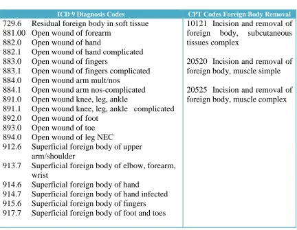
Table 3.1 ICD-9Diagnosis Codes/CPT Codes ...47
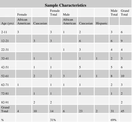
Table 4.1 Sample Characteristics ...55
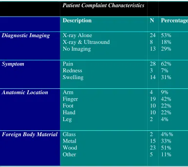
Table 4.2 Presenting Symptoms and Anatomical Location Soft Tissue Foreign Body ...56
Table 4.3 Foreign Body Removal X-ray Alone ...60
Table 4.4 Foreign Body Removal X-ray and Ultrasound ...61
Table 4.5 Foreign Body Removal No Diagnostic Imaging ...62
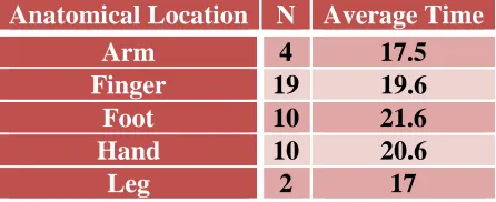
Table 4.6 Foreign Body Anatomical Location and Average Time of Foreign Body Removal to Patient Discharge ...62
Table 4.7 Foreign Body Material and Average Time of Foreign Body Removal to Patient Discharge ...63
x
List of Figures
Figure 1.1 Stetler Model ...10
Figure 2.1 Hierarchy of Evidence ...17
Figure 4.1 Inclusion and Exclusion Criteria ...54
xi
List of Abbreviations
CAT Scan ... Computed Axial Tomography
FB ... Foreign Body
MRI ... Magnetic Resonance Imaging
US ... Ultrasound
X-ray ... Plain Radiographs
1
Chapter 1 Introduction Scope of the Problem
Traumatic wounds and lacerations account for approximately 7.1 million visits to
United States emergency departments each year (Niska, Bhuiya, & Xu, 2010). A
majority of these visits (2.8 million) are from young males with complaints of upper
extremity lacerations and other wounds excluding the head and face (Niska, et al., 2010).
When the objects responsible for creating these wounds are composed of material that
shatters or splinters, such as glass or wood, the risk increases for fragments of foreign
bodies to become embedded within the wound (Capellan & Hollander, 2003; Halaas,
2007; Winland-Brown & Allen, 2010). Complications from traumatic wounds occur
when penetrating soft tissue foreign bodies are missed during the initial wound evaluation
(Levine, Gorman, Young, & Courtney, 2008).
Early detection of foreign bodies in soft tissue wounds has proven difficult as
nearly 38% are missed during initial examination by healthcare providers (Blankenship &
Baker; 2007; Boyse, Fessell, Jacobson, Lin, van Holsbeeck, & Hayes, 2001; Dean,
Groncsewski, & Constantino, 2003; Jacobson, Powell, Craig, Bouffard, & van
Holsbeeck, 1998; Manthey, Storrow, Milbourn, & Wagner, 1996; Schlager, 1997;
Tibbles & Porcaro, 2004). Puncture wounds by far are the most difficult to explore and
as many as 95% of foreign bodies isolated in these types of wounds consist of glass,
2
2010; Manson, Ryan, Ladner, & Gupta, 2011; McDevitt & Gillespie, 2008). Delayed
removal of foreign bodies can result in osteomyelitis, cellulitis, necrotizing fasciitis,
peripheral nerve damage, tendon damage, and granuloma development (Lyon, Brannam,
Johnson, Blaivas, & Duggal, 2004; Salati & Rather, 2010). As a result, undetected
foreign bodies in soft tissue wounds are the second highest cause of malpractice suits
against healthcare providers (Blankenship & Baker, 2007; Boyse et al., 2001; Dean et al.,
2003; Gibbs, 2006; Graham, 2002).
Factors contributing to the missed foreign bodies include small size and
radiolucent material composition such as wood, plastic, and other organic material.
Dried wood has only a 15% visibility on plain radiographs (X-ray) which decreases as
time progresses from the initial injury (Boyse, et al., 2001; Flarity & Hoyt, 2010).
Organic material such as splinters, thorns and other vegetative material embedded in soft
tissue for greater than 48 hours becomes saturated with body fluids rendering them
indistinguishable from surrounding tissue on X-ray (Gibbs, 2006; Peterson, Bancroft, &
Kransdorf, 2002; Shepherd, Lee, & McGahon, 2007). Glass fragments less than 2mm
prove difficult for visualization on X-ray with detection rates of 61%-83% (Orlinsky &
Bright, 2006; Steele, Tran, Watson, & Muelleman, 1998; Tuncer, Ozcelik, Mersa,
Kabakas, & Ozkan, 2011).
Analysis of Current Practices
Traditionally, healthcare providers have ordered plain radiographs (X-rays) as the
standard of care for first line screening of a suspected soft tissue foreign body without
regard to the material composition of the foreign body (Friedman, Forti, Wall, & Crain,
3
2004). Plain radiographs have proven to be an effective tool in detecting 80% of soft
tissue foreign bodies, most of which are composed of radiopaque material, such as metal,
stone, and glass (Jacobson et al., 1998; Tibbles & Porcaro, 2004). Incidentally, 85% of
soft tissue foreign bodies composed of radiolucent material, such as wood, plastic, and
thorns, are missed leading healthcare providers to search for other imaging modalities as
an adjunct to plain radiography (Jacobson et al., 1998; Tibbles & Porcaro, 2004).
Several diagnostic methods for locating foreign bodies in soft tissue exist,
including X-ray, ultrasonography, computed tomography (CT), and magnetic resonance
imaging (MRI) (Lyon et al., 2004). Although radiopaque foreign bodies such as metal,
gravel, and glass are easily detected by plain radiographs, radiolucent foreign bodies in
wounds such as wood, plastic, and vegetative material are not (Blankenship & Baker,
2007; Manthey et al., 1996). Both radiopaque and radiolucent foreign bodies can be
detected by CT and MRI but they are limited by cost, increased radiation exposure, and
availability (Lyon et al., 2004). In addition, an MRI cannot be performed if there is
suspicion for metallic foreign bodies (Lyon et al., 2004).
Discussion of Practice Innovation
Ultrasonography has been introduced as an adjunct to the conventional plain
radiographs for detecting and removing both radiopaque and radiolucent foreign bodies
in soft tissue wounds (Blankenship & Baker, 2007; Turner, Wilde, Hughes, Meilstrup, &
Manders, 1997). Ultrasound technique uses a high-frequency transducer to penetrate soft
tissue for the localization and evaluation of foreign bodies (Boyse et al., 2001; Mills &
4
bright hyperechoic foci can be visualized indicating the presence of with wooden, glass,
and metal foreign bodies (Boyse et al., 2001).
In several studies, ultrasound was found to have a sensitivity of 90% and a
specificity of 96% in the localization of foreign bodies in soft tissue without exposing the
patient to ionizing radiation (Bray, Mahoney, & Campbell, 1995; Jacobson, Powell,
Craig, Bouffard, & van Holsbeeck, 1998). Ultrasound not only gives the exact size and
depth of the foreign body but also allows for examination of nearby tendons, vessels and
muscles (Lyon et al., 2004). Other benefits include the ability of the healthcare provider
to use ultrasound at the bedside to assist with removal of the foreign body from soft tissue
wounds without painful probing and exploration (Friedman et al., 2005; Lyon et al.,
2004).
Basic ultrasound principles can be employed by healthcare providers with
relatively no ultrasound equipment experience (Hill, Conron, Greissinger, & Heller,
1997). According to a prospective study conducted by Orlinsky, Knittel, Feit, Chan and
Mandavia (2000), emergency physicians attending a two day ultrasound course had an
82% accuracy rate in identifying foreign bodies in unfrozen chicken thighs compared to
83% accuracy of radiologists and 85% accuracy of experienced sonographers (Mills &
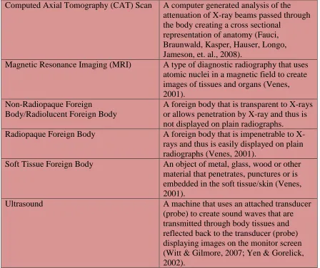
5 Table 1.1
Definitions
Computed Axial Tomography (CAT) Scan A computer generated analysis of the
attenuation of X-ray beams passed through the body creating a cross sectional
representation of anatomy (Fauci, Braunwald, Kasper, Hauser, Longo, Jameson, et. al., 2008).
Magnetic Resonance Imaging (MRI) A type of diagnostic radiography that uses
atomic nuclei in a magnetic field to create images of tissues and organs (Venes, 2001).
Non-Radiopaque Foreign
Body/Radiolucent Foreign Body
A foreign body that is transparent to X-rays or allows penetration by X-ray and thus is not displayed on plain radiographs.
Radiopaque Foreign Body A foreign body that is impenetrable to
X-rays and thus is easily displayed on plain radiographs (Venes, 2001).
Soft Tissue Foreign Body An object of metal, glass, wood or other
material that penetrates, punctures or is embedded in the soft tissue/skin (Venes, 2001).
Ultrasound A machine that uses an attached transducer
(probe) to create sound waves that are transmitted through body tissues and reflected back to the transducer (probe) displaying images on the monitor screen (Witt & Gilmore, 2007; Yen & Gorelick, 2002).
Ultrasound and X-ray Equipment Costs
The expense of conducting bedside ultrasound is comparable to X-rays when
equipment and facility requirements are taken into consideration. Ultrasound equipment
ranges in price from $50,000 to $200, 000 depending upon the functional capabilities of
6
software, various size probes and sterile probe supplies are also incurred with the
purchase of ultrasound equipment (Witt & Gilmore, 2007).
The initial cost of X-ray suite equipment starts at upwards of $100,000 or greater
based on facility operational needs and equipment features. Additional expenses include
consultation with a medical physicist regarding on site shielding requirements and
engineering plans for the dedicated office space housing the X-ray machine as specified
by South Carolina Department of Health and Environmental Control regulations. Other
costs independent of the X-ray machine include lead aprons, picture archiving and
communication system (PACS), a film reader and film cassettes.
Implications for Nurse Practitioner Practice
Since 1975 nurse practitioners have been providing emergency care in an efficient
and cost effective manner (Campo, McNulty, Sabatini, & Fitzpatrick, 2008; Cole, &
Ramirez, 2000). In 2008 the Emergency Nurses Association (ENA) developed entry
level competencies of care for nurse practitioners in emergency or urgent care settings
with ordering and interpreting radiographs, injection of local anesthetics, and removal of
foreign bodies from soft tissue listed among these competencies (ENA, 2008). A
majority of nurse practitioners employed in the emergency or urgent care settings are
board certified family nurse practitioners who have completed an accredited family nurse
practitioner program, attended continuing education workshops and received on the job
training (Cole & Ramirez, 2003). Currently no specific regulations exist delineating what
procedures are taught in family nurse practitioner programs (Cole & Ramirez, 2003). In
a survey of 71 nurse practitioners in the emergency setting, Cole and Ramirez (2000)
7
from soft tissue wounds, and 59 that the majority of their education in performing this
procedure was obtained through on the job training (Campo et al., 2008; Cole & Ramirez,
2000). In another study conducted by Cole and Ramirez (2003), 55.4% of family nurse
practitioner program directors rated bedside ultrasound as unimportant to teach family
nurse practitioner students while 71.1% rated foreign body removal from soft tissue as an
important procedure to be taught (Cole & Ramirez, 2003).
Wound management ranks among the top ten procedures performed by nurse
practitioners in an emergency care setting (Campo et al., 2008; Flarity & Hoyt, 2010).
Among the most important aspects of wound management include the history and
physical exam, wound exploration, identification of underlying structures and potential
foreign bodies harbored in the wound bed (Flarity & Hoyt, 2010). With the use of
ultrasound guided localization and removal of foreign bodies embedded in soft tissue,
underlying anatomical structures are readily visible at the bedside for identification by the
provider (Blankstein et al., 2000; Bradley, 2012; Callegari et al., 2009).
In many instances independent practice of nurse practitioners in the emergency
setting can be intimidating especially with decreased comfort levels and inadequate
educational preparation in procedures such as wound management and the use of bedside
ultrasound (Campo et al., 2008). Proper educational preparation of nurse practitioners in
the form of residency programs leads to confidence and professional development
(Campo et al., 2008). Residency programs have the capability of providing novice nurse
practitioners with the refined procedural skills, such as ultrasound and suturing, that they
8
The use of bedside ultrasound gives providers an additional assessment tool to
expedite patient care especially in the setting of wound debridement with the potential of
an embedded radiolucent foreign body (Callegari et al., 2009; McGuinness, Snaith,
Wilson, & Wolstenhulme, 2011). Other implications for practice include decreased
patient anxiety and pain along with increased provider confidence related to the foreign
body removal process as bedside ultrasound allows for direct visualization, smaller
incision sites, and the elimination of blind probing during wound exploration.
Statement of Purpose
The purpose of this research utilization project is to report the use of bedside
ultrasound by healthcare providers as an adjunct to X-ray for the localization and removal
of foreign bodies in soft tissue wounds among patients presenting to an urgent care
setting.
Framework Model of Research Utilization
Initially published in 1976, the Stetler-Marram Model for Research Utilization
centered its focus on “research-as-a-process” including the individual practitioner’s
critical thinking skills, reflective ideology, and the resulting application of research
findings into clinical practice (Melnyk & Fineout-Overholt, 2005; Stetler, 2001). The
model has undergone two revisions (1994, 2001) since the first publication but the
components of critical thinking and decision making skills among individual practitioners
have remained a mainstay for promoting evidence based practice research utilization
(Melnyk & Fineout-Overholt, 2005; Stetler, 2001). Since this model is practitioner
oriented, it was chosen to guide the implementation for the change of clinical practice
9
The refined Stetler Model addresses five phases: preparation, validation,
comparative evaluation/decision making, translation/application, and evaluation for
implementing changes in clinical practice settings (Melnyk & Fineout-Overholt, 2005).
In preparation for research utilization, the purpose of phase one of Stetler’s Model is to
define a clinical practice need and desired outcomes in conjunction with systematically
locating the best relevant evidence to support the needed change in practice (Melnyk &
Fineout-Overholt, 2005). Phase two focuses on validating and critiquing the chosen
evidence in order to determine if sufficient credible evidence exists to recommend a
change in practice (Melnyk & Fineout-Overholt, 2005). For phase three, criteria specific
to the change in practice is determined and according to these criteria the evidence is
evaluated for its applicable use in clinical practice (Melnyk & Fineout-Overholt, 2005).
Phase four translates the evidence based findings for use in persuading others regarding a
need for change in practice (Melnyk & Fineout-Overholt, 2005). During phase four the
current clinical practice is assessed for change and the formal implementation of the
planned change occurs (Burns & Grove, 2005). In the final phase of Stetler’s Model, the
planned change is evaluated according to cost-benefit analysis and the achievement of
goals set forth in phase one (Melnyk & Fineout-Overholt, 2005).
Project Question
Is ultrasound useful as a clinical tool in addition to wound exploration and X-ray to
ensure complete removal of soft tissue foreign bodies?
Project Outcomes
1
0
Figure 1.1 Stetler Model
11
2. Ultrasound has the potential to become an alternative imaging approach to plain radiographs for locating radiolucent foreign bodies, such as wood, plastic, and
other vegetative materials, in soft tissue.
Summary
Common complaints for patients presenting to urgent care include an embedded
soft tissue foreign body after a work injury; stepping on glass, a splinter or toothpick; and
various other encounters of soft tissue puncture wounds, penetrating wounds and
traumatic lacerations. Even though plain radiographs detect a majority of these foreign
bodies, some materials go undetected placing patients at high risk for complications. By
using bedside ultrasound as an adjunct to plain radiographs for the identification and
removal of foreign bodies, patients become more involved in their plan of care and
experience less pain and anxiety during the removal process (Shiels, 2007). In the
following chapter, an analysis of the literature and evidence supporting the practice
innovation surrounding the use of bedside ultrasound as an adjunct to plain radiographs
12
Chapter 2 Literature Review
A comprehensive search of the literature was performed using the research
question as a framework for search terms. The purpose of this search was to identify
high quality evidence in the form of meta-analysis, randomized control trials, systematic
reviews, quantitative or qualitative research studies, practice guidelines, reports from
expert consensus and peer reviewed clinical articles to demonstrate the use of ultrasound
as an additional assessment tool for localization and removal of soft tissue foreign bodies
in wounds.
Databases searched include: CINAHL, Cochrane Library, PubMed/MEDLINE,
Ovid, and Web of Science. Other online searches performed include the following:
American College of Emergency Physicians (ACEP), American College of Radiology
(ACR), American Institute of Ultrasound in Medicine (AIUM), Google Scholar, the Joint
Commission on Accreditation of Healthcare Organizations (JCAHO), National
Guidelines Clearing House (NGC), United States Preventative Task Force (USPTF), and
Agency for Healthcare Research and Quality (AHRQ).
Literature Search Strategies
Initially for the literature search broad key terms were used followed by title
searches and author searches. Using the broad search terms of foreign body removal,
13
articles were retrieved pertaining to foreign bodies located in other anatomical spaces
besides soft tissue.
Several search term combinations were used and included the following:
ultrasound, foreign body, extraction, laceration, puncture, soft tissue, X-ray, radiolucent,
and radiopaque. In addition, other synonym key words searched were ultrasound guided
procedures, examination, removal, skin, plain radiographs, radiography, sonography,
ultrasonography, and wounds.
Narrowing the literature search to the search terms of ‘ultrasound, foreign body,
and soft tissue’ yielded more articles pertinent to the research question. Limiting the
search terms to ‘penetrating, puncture, laceration, and soft tissue foreign bodies’ further
narrowed the results. As supporting articles were retrieved, the reference citations were
examined for additional evidence.
After review of bibliographic references from articles retrieved in the search
process, several authors were noted to have performed extensive research on the use of
ultrasound guidance for removal of foreign bodies from soft tissue. A subsequent search
was performed using the authors’ last name yielding numerous other research studies.
Several common themes were identified throughout the literature review and
include the following: indications for the use of ultrasound guided soft tissue foreign
body removal; detection of soft tissue foreign bodies with ultrasound; the use of
ultrasound versus plain radiographs (X-rays), computed tomography (CT), and magnetic
resonance imaging (MRI) for the detection of radiolucent soft tissue foreign bodies; soft
tissue foreign body imaging characteristics emitted by ultrasound frequency; precise
14
cost effectiveness and risk assessment associated with the use of ultrasound versus other
diagnostic modalities to detect and remove soft tissue foreign bodies. A majority of the
literature did not specifically address the procedure for bedside ultrasound guided
removal of soft tissue foreign bodies.
Literature Inclusion and Exclusion Criteria
Inclusion criteria for the literature search addressed those items less than 20 years
old and written in the English language. Since many of the in vitro research studies
addressing ultrasound and the localization of radiolucent foreign bodies were performed
in the 1990s, a 10 year limitation of the literature was not feasible. Articles referencing
the use ultrasound guided removal of soft tissue foreign bodies were first available in the
late 1990s.
The population of interest included adults and children, both males and females in
all ethnic groups, ages 5 and above. Children were included since the results of
ultrasound localization and removal of soft tissue foreign bodies in this population can be
extrapolated to the adult population. Children in particular benefit from the ultrasound
guided procedure because they can participate in the real time procedural guidance hence
causing decreased anxiety (Cohen, 2008).
Articles over 20 years old were excluded. Furthermore, studies pertaining to
animal bites or foreign bodies in the breast, ear canal, esophagus, eye, genitals, nose,
peritoneum, trachea, and rectum were excluded. Many of these anatomical locations
inhibit examination by ultrasound, create the potential for significant scarring, or demand
15
Limitations of Literature Review
Limitations of the literature review include a paucity of evidence in the form of
meta-analysis, systematic reviews and randomized controlled trials to support the
ultrasound guided removal of foreign bodies in soft tissue; many studies were greater
than 10 years old; 34 of the articles retrieved to support the research question were case
studies and six were literature reviews from peer reviewed sources. In addition, a
majority of the randomized controlled studies selected for review were in vitro and not
specifically conducted using live, human skin tissue.
However, extending the literature search to include acute wound management
strategies and ultrasound policy guidelines resulted in relevant evidence based resources.
Several sources cited the association of wound location and retained foreign bodies in
traumatic lacerations with increased risk of infection (Hollander, Singer, Valentine, &
Shofer, 2001; Nicks, Ayello, Woo, Nitski-George, & Sibbald, 2010; Zehtabchi, Tan,
Yadav, Badawy, & Lucchesi, 2012). Other articles addressed the significance of
thorough wound exploration in the setting of small penetrating wounds and the benefit of
bedside ultrasound as an additional assessment tool to detect potential tendon injuries
obscured by foreign bodies (Tuncali et al., 2005; Wu, Roque, Green, Drachman, Khor,
Rosenberg, & Simpson, 2012).
Synthesis of Literature
Articles chosen for analysis were ranked according to the Melnyk and
Fineout-Overholt (2005) hierarchy of evidence (Figure 2.1). The rating system for the hierarchy
of evidence includes levels of evidence I through VII with level I representing the highest
16
controlled trials and level VII indicating the lowest quality of evidence in the form of
expert opinion (Melnyk & Fineout-Overholt, 2005).
Guidelines
The National Guideline Clearinghouse (NGC) guideline summary for the
American College of Radiology (ACR) appropriateness criteria for acute trauma to the
foot (2010) recommends X-ray for the initial diagnostic study of acute penetrating trauma
to the foot and ultrasound for radiolucent foreign bodies. In addition if initial X-rays are
negative, ultrasound is recommended as the next best study for penetrating trauma with a
foreign body (NGC, 2010).
Indications for the use of Ultrasound Guidance
Mechanism of injury, wound characteristics, location and patient perception of retained
foreign bodies have been an indication for meticulous wound exploration during the
initial physical exam performed by the healthcare provider (Bray, Mahoney, & Campbell,
1995; Capellan & Hollander, 2003; Friedman, Forti, Wall, & Crain, 2005; Nicks, et al.,
2010; Orlinsky & Bright, 2006; Ozsarac, Demircan, & Sener, 2011; Steele, Tran,
Watson, & Muelleman, 1998; Zehtabchi et al., 2012). In their prospective patient series,
Avner and Baker (1992) questioned the accuracy of visual wound exploration by
providers for detecting all glass fragments embedded deep in a laceration. Furthermore, a
cross sectional study conducted by Hollander et al. (2001) determined that the increased
risk for infection in traumatic lacerations was associated with patient age, medical history
of diabetes mellitus, laceration width, and foreign body contamination. However, the
best form of imaging modality to assist providers in identifying underlying foreign bodies
1
7
Adapted from: Fineout-Overholt, E., Melnyk, B, & Schultz, A. (2005). Transforming Evidence-Based Practice in the 21
Figure 2.1 Hierarchy of Evidence
Overholt, E., Melnyk, B, & Schultz, A. (2005). Transforming Healthcare from the Inside Out: Advancing Based Practice in the 21st Century. Journal of Professional Nursing, 21, 335
18
Friedman et al. (2005) conducted a prospective cohort study in a pediatric
emergency department to investigate bedside ultrasound or a combination of ultrasound
and patient perception of a foreign body as a screening tool for the detection of foreign
bodies in wounds. A total of 105 patients with 131 wounds were evaluated with foreign
bodies removed from 12 wounds (Friedman et al., 2005). The wounds containing foreign
bodies were isolated to the hands or feet with ultrasound detecting six out of nine
radiopaque foreign bodies while plain radiographs detected eight (Friedman et al., 2005).
Ultrasound detected two of three radiolucent foreign bodies while plain radiographs were
unsuccessful at detecting any of the radiolucent material (Friedman et al., 2005).
Subsequently, a repeat ultrasound was performed revealing the third radiolucent foreign
body. Significant results were found with the specificity of bedside ultrasound alone
compared to the specificity of ultrasound and plain radiographs in parallel or the use of
plain radiographs alone for the detection of foreign bodies in wounds (Friedman et al.,
2005). However, the highest sensitivity resulted with the use of bedside ultrasound and
plain radiographs in parallel (Friedman et al., 2005). Beneficial evidence gathered from
this study was the potential application of bedside ultrasound as an adjunct screening tool
prior to plain radiographs for the detection of foreign bodies in wounds.
In a prospective study using six cadavers, Crystal, Masneri, Hellums, Kaylor,
Young, Miller, and Levsky (2007) explored use of beside ultrasound in order to detect
various small foreign bodies in traumatic lacerations that might have been missed during
the initial wound exploration. In a total of 150 extremity wounds, researchers randomly
inserted various foreign materials consisting of metal, plastic, glass, wood and in some
19
were blinded to location, type, and number of foreign bodies (Crystal et al., 2007).
Sonographic detection of foreign bodies by the physicians yielded an overall sensitivity
of 52.6% and specificity of 47.2% (Crystal et al., 2007). The authors concluded that
ultrasound for the detection of foreign bodies is rarely used alone but instead is often
used in conjunction with the provider’s physical exam and plain radiographs (Crystal et
al., 2007).
Historically, research has shown that small penetrating and puncture lacerations to
the hand, wrist, and forearm can disguise deeper structural injuries or harbor fragments of
foreign bodies (Tuncali, Yavuz, Terzioglu, & Aslan, 2005; Tuncer, Ozcelik, Mersa,
Kabakas, & Ozkan, 2011). In a prospective study, Soubeyrand, Biau, Jomaah, Pradel,
Dumontier, and Nourissat (2008) explored the efficacy of ultrasound for the detection of
deep structural or neurovascular injury as a result of penetrating lacerations to the volar
surface of the hand. The ultrasound examinations were performed prior to surgical
exploration of the 30 wounds (Soubeyrand et al., 2008). Ultrasound located 17 tendon
tears (Se 100%; Sp 100%; PPV 100%; NPV 100%), 14 arterial injuries (Se 87.5%; Sp
100%; PPV 100%; NPV 96.7%), and 12 nerve injuries (Se 75%; Sp 90.8%; PPV 66.7%;
NPV 93.7%) (Soubeyrand et al., 2008). However, ultrasound missed two arterial injuries
and four nerve injuries detected during surgical exploration (Soubeyrand et al., 2008).
During surgical exploration ultrasound was employed for post procedure imaging and
detected three foreign bodies prior to wound closure (Soubeyrand, et al., 2008).
Detection of Soft Tissue Foreign Bodies
Bray et al. (1995) randomly inserted various foreign bodies composed of wood,
20
ultrasound detection of foreign bodies in the hand. X-rays performed prior to ultrasound
examination revealed all metal foreign bodies, 50 out of 54 glass foreign bodies, and
none of the wooden foreign bodies (Bray et al., 1995). Ultrasound detected 156 of 166
foreign bodies in the cadaver hands resulting in a diagnostic sensitivity of 94% and a
specificity of 99% (Bray et al., 1995). In this study the researchers noted the diagnostic
sensitivity for both X-ray and ultrasound detection of glass was 93% with ultrasound
capable of detecting those glass foreign bodies not visualized on X-ray (Bray et al.,
1995). This prospective study advocated for the use of ultrasound and X-ray in
combination for detecting foreign bodies in the hand instead of blind probing during
wound exploration (Bray et al., 1995).
A retrospective study of 23 patients in an outpatient orthopedic clinic was
conducted by Shrestha, Sharma, Mohammad, and Dhoju (2009) to detect radiolucent soft
tissue foreign bodies in extremities with the use of ultrasound. Nineteen patients were
found to have the characteristic ultrasound hypoechoic appearance of a foreign body
while all plain radiographs were negative for foreign body (Shrestha et al., 2009). The
material of the foreign body identified consisted of wood (12), plant thorn (4), bamboo
twig (2), and granuloma (1) (Shrestha et al., 2009). The authors concluded that plain
radiography lacks sensitivity to detect radiolucent foreign bodies and ultrasound is
superior with greater sensitivity and specificity for the identification of radiolucent
foreign bodies in soft tissue of the extremities (Shrestha et al., 2009).
Levine, Gorman, Young and Courtney (2008) performed a retrospective case
series of patients diagnosed with foreign body in a wound over a period of four years in
21
of 490 patients selected according to certain inclusion criteria (Levine et al., 2008). Most
complaints were lacerations with foreign bodies or stepping on an object with wood,
metal, glass and ceramic being among the top three materials responsible for the injury
(Levine et al., 2008). Plain radiographs were determined to have a sensitivity of 75.5%
for glass, 98.6% for metal, and 7.4% for wood (Levine et al., 2008). Approximately 90%
of these foreign bodies were removed in the emergency department with limited
specialist consultation (Levine et al., 2008). Post removal imaging by X-rays were
completed (Levine et al., 2008). Recommendations from this study included the use of
ultrasound to achieve greater sensitivity in the detection of wood foreign bodies (Levine
et al., 2008). No comparison of ultrasound and plain radiography for the detection of
wooden foreign bodies in wounds was addressed in this study.
A retrospective cohort study by Rubin, Chezar, Raz, and Rozen (2010)
investigated the management of 96 adult patients that received a nail puncture wound to
the plantar surface of the foot while wearing rubber soled shoes. All patients underwent
X-ray and 22 patients had ultrasound examinations of their injured foot (Rubin et al.,
2010). The X-rays detected a metal foreign body in one patient while ultrasound
indicated 9 foreign bodies (Rubin et al., 2010). The authors recommend ultrasound
examination of all patients presenting with nail puncture wounds through shoes (Rubin et
al., 2010).
In another retrospective study conducted by Salati and Rather (2010), 61 cases of
missed foreign bodies in the hand were treated from 2003 to 2009. Patients related
various complaints from non-healing, draining wound, pain, foreign body sensation,
22
previous medical treatment, 18 had X-rays and wound care, and 9 had wound care
without X-rays (Salati & Rather, 2010). Most significant was the fact that 37 patients
(61%) had wooden splinters retained in their hands and X-ray only detected two (3%)
wooden foreign bodies while ultrasound detected 35 (97%) (Salati & Rather, 2010).
However, ultrasound did not detect any of the four stone fragments and only detected one
of 13 metallic fragments and one of 7 glass pieces in the hands of patients (Salati &
Rather, 2010).
Several in vitro studies have been conducted regarding the use of ultrasound for
the identification of radiolucent and semi-radiopaque foreign bodies embedded in turkey,
cow tongue, cadaver extremities, pork shoulder and chicken models in which the
sensitivity for ultrasound detection ranged from 85-100% (Harcke & Levy, 2003; Harcke,
Levy, & Lonergan, 2002; Hill, Conron, Greissinger, & Heller, 1997; Jacobson, Powell,
Craig, Bouffard, van Holsbeeck, 1998; Manthey, Storrow, Milbourn, & Wagner, 1996;
Mizel, Steinmetz, & Trepman, 1994; Oikarinen, Nieminen, Makarainen, & Pyhtinen,
1993; Turkcuer, Atilla, Topacoglu, Yanturali, Kiyan, Kabakci, et al., 2006; Turner,
Wilde, Hughes, Meilstrup, & Manders, 1997). Turkcuer et al. (2006) conducted a
randomized, blinded descriptive in vitro study in which rubber and wooden foreign
bodies were inserted into chicken thighs for the comparison of plain and soft tissue
radiographs with ultrasound for accurate detection of non-radiopaque foreign bodies.
Their hypothesis stated that soft tissue and plain radiographs could be eliminated for the
examination of non-radiopaque soft tissue foreign bodies and replaced with
high-frequency ultrasound (Turkcuer et al., 2006). Forty foreign bodies of rubber shoe sole
23
chicken thighs used as the control group with similar tissue damage (Turkcuer et al.,
2006). Two veteran radiologists were blinded to the chicken thigh preparation and each
other’s interpretation of the diagnostic imaging studies (Turkcuer et al., 2006). Plain
radiography detected no wooden foreign bodies in 20 model preparations and two false
positive wood foreign bodies from the control group while two out of 20 rubber foreign
bodies were detected in the model preparations with two false positive rubber foreign
bodies detected from the control group (Turkcuer et al., 2006). The same results were
obtained with soft tissue plain radiographs. Ultrasound detected 17 of 20 wooden foreign
bodies (85%) in 20 chicken thighs with four false negative wooden foreign bodies from
the control group while 19 out of 20 rubber foreign bodies were detected in the 20
chicken thighs with four false positive detected in the control group (Turkcuer et al.,
2006). Ultrasound was found to have 90% sensitivity and 80% specificity for rubber and
wood foreign bodies in the model preparations of chicken thighs. The authors suggest
that plain radiographs should not be used to detect non-radiopaque foreign bodies and
ultrasound should be considered as an option (Turkcuer et al., 2006).
Hill et al. (1997) and Jacobson et al. (1998) conducted randomized, controlled
cadaver studies to explore the efficacy, sensitivity and specificity of ultrasound for
detecting radiolucent foreign bodies in the legs and feet. Ultrasound results revealed a
93% sensitivity for wood and 73% sensitivity for plastic with an overall sensitivity of
83% and specificity of 59% in the Hill et al. (1997) study. Jacobson et al. (1998) found a
sensitivity of 86.7% and a specificity of 96.7% for ultrasound detection of 2.5mm pieces
of wood which increased to a sensitivity of 93.3% and a specificity of 96.7% for the
24
Manthey et al. (1996) challenged the ability of ultrasound to detect soft tissue
foreign bodies in the distal extremities. In their randomized, blinded descriptive study,
the researchers randomly inserted various foreign bodies consisting of metal, wood,
plastic, cactus, needles, glass and gravel into 60 chicken thighs in order to mimic
puncture wounds in the hand (Manthey et al., 1996). Another 60 chicken thighs were
used as a control group. All chicken thighs received X-rays and ultrasound imaging with
radiologists blinded to type and number of foreign bodies along with their preliminary
ultrasound read (Manthey et al., 1996). The X-rays were interpreted after completion of
the ultrasound analysis and detected 98% of the radiopaque foreign bodies (Manthey et
al., 1996). Results of this study yielded a sensitivity of 43% and specificity of 70% with
a 50% false negative rate and 30% false positive rate for ultrasound detection of foreign
bodies (Manthey et al., 1996).
Precise Localization of Soft Tissue Foreign Bodies
Gibbs (2006) conducted a retrospective study to determine the efficacy of
ultrasound in locating soft tissue foreign bodies. A total of 20 patients were selected
based on chart review from April 2001 to February 2005 (Gibbs, 2006). Plain
radiographs (X-ray) were used for the initial imaging screening in 17 out of the 20
patients detecting eight foreign bodies composed of metal and inorganic material (Gibbs,
2006). X-ray did not detect wood or glass in nine patients. All 20 patients underwent
ultrasound examination revealing eight organic, eight inorganic and four metallic foreign
bodies (Gibbs, 2006). In addition, ultrasound was used to assist with the removal of
foreign bodies in 11 patients allowing for precise localization and faster removal times
25
removal attempt by a physician probing for glass foreign bodies in the hand as
demonstrated on X-ray. Other applications of ultrasound guided foreign body removal
include post procedure imaging, smaller incision sites, and the elimination of blind
probing resulting in fewer complications and faster recovery for the patient (Gibbs,
2006).
A retrospective review of 20 patients conducted by Rockett, Gentile, Gudas,
Brage, and Zygmunt (1995) demonstrated the efficacy of ultrasound in the localization of
wooden soft tissue foreign bodies in the foot prior to surgical removal. Plain radiographs
(X-rays) performed prior to ultrasound failed to reveal any of the wooden foreign bodies
in 20 patients (Rockett et al., 1995). At the time of ultrasound examination, the
anatomical location of suspected point of foreign body entry was marked and then
scanned in transverse and longitudinal planes (Rockett et al., 1995). Positive ultrasound
findings of wooden foreign bodies in 10 patients were then marked by the
ultrasonographer in anticipation of surgical excision (Rockett et al., 1995). Surgical
pathology results correlated with the positive ultrasound findings of wooden foreign
bodies (Rockett et al., 1995).
Several case studies demonstrate the effectiveness of bedside ultrasound for the
localization of radiolucent wooden foreign bodies retained in soft tissue wounds
(Borgohain, B., Borgohain, N., Handique, & Gogoi, 2012; Bu, Overgaard, Viegas, 2008;
Dean, Gronczewski, & Constantino, 2003; Firth, Roy, & Moroz, 2011; Graham, 2002;
Harris, 2010; Hung Y.T., Hung, L.K, Griffith, Wong, & Ho, 2004; Sidharthan & Mbako,
2010; Teng & Doniger, 2012). Wooden foreign bodies have the potential to cause
26
(Sidharthan & Mbako, 2010). Wood also has the ability to splinter creating draining
sinus tracts and migrating into tendons and joint capsules (Borgohain, et al., 2012; Bu, et
al., 2008; Graham, 2002; Harris, 2010; Sidharthan, & Mbako, 2010).
A majority of the patients detailed in the case reports had plain radiographs
completed at the time of presentation to a healthcare provider which were all interpreted
as negative for foreign body (Borgohain et al., 2012; Bu et al., 2008; Firth et al., 2011;
Graham, 2002; Harris, 2010; Hung et al., 2004; Sidharthan & Mbako, 2010). In addition,
five patients had wooden foreign bodies that were missed on initial examination by a
healthcare provider and they were discharged home with oral antibiotics only to return
with complaints of foul smelling wound, drainage, non-healing wounds, pain with weight
bearing activities, and difficulty walking (Borgohain et al., 2012; Graham, 2002; Harris,
2010; Sidharthan & Mbako, 2010). Based on the patients’ complaints along with a high
clinical index of suspicion and wound characteristics, providers in these case studies
employed the use of ultrasound which successfully localized wooden radiolucent foreign
bodies for removal.
Imaging Characteristics
Davae, Sofka, DiCarlo, and Adler (2003) retrospectively reviewed sonographic
examinations from 1998 to 2001 with possible soft tissue foreign bodies. A total of 25
patients underwent ultrasound for possible foreign body but only 12 were included in this
study (Davae et al., 2003). Ultrasound was performed by experienced radiologists and
detected all foreign bodies in patients that had subsequent surgical exploration (Davae et
al., 2003). Material composition of the foreign bodies identified included glass (2), wood
27
two false positive results (Davae et al., 2003). Ultrasound imaging revealed a hypoechoic
halo in eight of the 10 patients and hyperemia with power doppler in all of the patients
with proven foreign bodies as correlated with histopathology (Davae et al., 2003). Under
power doppler the hyperemia was a consistent ultrasound finding for foreign body and
can represent inflammation (Davae et al., 2003). The authors relate that their facility
conducts ultrasounds routinely if plain radiographs are negative for foreign body but the
provider remains with a high index of clinical suspicion for the existence of a soft tissue
foreign body (Davae et al., 2003).
There are characteristic sonographic images for type and age of foreign body
which can assist the provider in accurately identifying a soft tissue foreign body for
removal (Gibbs, 2006). Several authors suggest using a high-frequency linear array
transducer (7.5MHz or higher) and scanning in two planes creating longitudinal and
transverse images for increased localization of the foreign body (Blankenship & Baker,
2007; Gibbs, 2006; Teng & Doniger, 2012). Orientation of the ultrasound probe parallel
to the foreign body has proven to display the greatest signal for visualization of the
foreign body (Bradley, 2012; Turner et al., 1997).
Superficial foreign bodies composed of organic materials can present with many
different images on ultrasound based on the time progressed from initial injury (Gibbs,
2006). In the beginning of the acute phase of injury, the organic material, such as wood,
displays as a bright hyperechoic structure with clean shadowing but over time as the
material decomposes and absorbs body fluids, the foreign body is not as bright and a
hypoechoic ring develops which can benefit from color doppler imaging for better
28
artifact while glass appears as more scattered “comet tail” artifact (Blankenship & Baker,
2007; Schlager, 1997; Teng & Doniger, 2012).
Imaging Modalities Cost Comparison and Risk Factors
Several imaging modalities exist for the identification and localization of soft
tissue foreign bodies. Diagnostic imaging available for selection consists of computed
tomography (CT), fluoroscopy, magnetic resonance imaging (MRI), plain radiography
(X-ray), and ultrasound. Ultrasound and fluoroscopy are the only two bedside modalities
that exist to visualize and guide the removal of soft tissue foreign bodies under real time
conditions.
Any form of diagnostic imaging is subject to vary in cost due to insurance
regulations, patient co-payments and claim reimbursements. However, CT and MRI
account for the most expensive forms of diagnostic imaging. CT costs average $1500 to
$2000 while MRI averages $2000 to $4000 (Sistrom & McKay, 2005; Williams,
Rousseau, & Glaudemans, 2005). Extremity x-rays average around $65 to $75 to several
hundreds of dollars depending on the number of image views associated with the
procedure and the facility location (outpatient diagnostic centers versus not-for –profit
and for-profit hospital systems) (Sistrom & McKay, 2005). In addition, ultrasounds
average approximately $200 to $400 (Sistrom & McKay, 2005). Other charges related to
fees for radiologist interpretations and supplies associated with the diagnostic imaging
procedures also may be incurred.
Computed tomography (CT) is best utilized during the initial presentation of the
injury when the foreign body is composed of radiolucent material or if the foreign body is
29
Srisuwan, Sivasomboon, Nasuto, Suwannahoy, Settakorn, et al., 2012; Shepherd, Lee, &
McGahon, 2007; Sidharthan & Mbako, 2010). Metallic foreign bodies create artifact
making them difficult to detect with CT (Aras et al., 2010). In a case report Dumarey, De
Maeseneer, & Ernst (2004) demonstrated CT to be ineffective at locating fragmented
splinters of wood adjacent to larger wooden foreign body structures. In this case
ultrasound was completed prior to the CT which identified the splinter fragments
avoiding a second surgical procedure for the patient (Dumarey et al., 2004).
Furthermore, another study by Al-Zahrani, Kremli, Saadeddin, Ikram, Takroni, & Zeidan
(1995), demonstrated CT to be only 70% effective in diagnosing foreign bodies. Other
potential risks associated with CT include the high amount of ionizing radiation
especially for young patients; increased expense of imaging and insurance requirement of
pre-authorization prior to conducting the study; and potential complications from contrast
dye (Bernardy, Ullrich, Rawson, Allen, Jr., Thrall, Keysor, et al., 2009; Bierig & Jones,
2009; Soudack, Nachtigal, & Gaitini, 2003).
Magnetic Resonance Imaging (MRI) has limited use for foreign body
identification. MRI is best used to identify retained wood in fat or to diagnose
complications such as cellulitis or necrotizing fasciitis resulting from foreign bodies
(Shepherd et al., 2007). MRI is contraindicated when the material composition of the
foreign body is unknown or if the foreign body is metallic (Aras et al., 2010; Sidharthan
& Mbako, 2010). In addition, certain patients are prohibited from undergoing an MRI
and include those with implanted pacemakers, aneurysm clips and other medical devices
or embedded metallic fragments of any kind (American College of Radiology, 2011).
30
from most insurance companies prior to ordering the exam (Bernardy et al., 2009;
Soudack et al., 2003). Furthermore, an MRI often uses intravenous gadolinium and
requires cooperation from the patient lying still on the exam table for long periods of time
which usually results in the need for procedural sedation of pediatric patients and some
adult patients with intense claustrophobia (Firth et al., 2011; Graham, 2002; Harris, 2010;
Read, Conolly, Lanzetta, Spielman, Snodgrass, & Korber, 1996; Sidharthan & Mbako,
2010).
Plain radiography (X-ray) is often the preferred initial radiographic imaging for
suspected foreign body due to its availability and cost (Aras et al., 2010; Blankenship &
Baker, 2007; Peterson, Bancroft, & Kransdorf, 2002; Shepherd et al., 2007; Teng &
Doniger, 2012). X-ray commonly allows for visualization of radiopaque foreign bodies
such as metal, gravel, and glass (Teng & Doniger, 2012; Turkcuer et al., 2006).
However, X-ray has been reported to detect only 15% of radiolucent wooden foreign
bodies (Ando, Hatori, Hagiwara, Isefuku, & Itoi, 2009; Graham, 2002; Lee, Chung, &
Kam, 2008; Shepherd et al., 2007). X-ray has also been shown to have limitations in the
detection of foreign bodies less than 5mm in size (Peterson et al., 2002; Teng & Doniger,
2012). Risks pertaining to using X-ray include the exposure to ionizing radiation which
is unnecessary if the foreign body composition is known to be radiolucent (Friedman et
al., 2005).
Similar to ultrasound, fluoroscopy can be used in real time to visualize and
remove retained foreign bodies (Shepherd et al., 2007). Results from a prospective,
randomized masked investigation by Wyn, Jones, McNinch, and Heacox (1995) indicate
31
embedded in soft tissue but only limited detection of wood and plastic radiolucent foreign
bodies. Considerable risks are encountered with the use of fluoroscopy. Not only can
patients experience high doses of ionizing radiation based on the length of the procedure
but providers and medical staff are also exposed (American College of Radiology, 2008;
Shiels, 2007).
Bedside ultrasound has the unique ability to detect and locate foreign bodies in
superficial soft tissue wounds and lacerations regardless of material composition,
presence of infection, size of foreign material, or age of injury (Ozsarac, Demircan, &
Sener, 2011). Other advantages of ultrasound include the lack of ionizing radiation; the
ability to localize and remove soft tissue foreign bodies with real time guidance; allows
visualization of nearby important anatomical structures during the removal procedure;
capable of identifying size, shape and depth of the foreign body; decreased incision size
and time for removal; safer for patients; and it is relatively inexpensive (Aras et al., 2010;
Blankstein, Cohen, Heiman, Salai, Diamant, Heim, & Chechick, 2001; Dean et al., 2003;
Konez et al., 1999; Orlinsky, Knittel, Feit, Chan, & Mandavia, 2000; Teng & Doniger,
2012). Ultrasound also can produce pre and post removal images to ensure complete
foreign body removal while providing reassurance to the patient and provider in those
circumstances where the foreign body has the potential to splinter or fragment during the
removal process (Sidharthan & Mbako, 2010; Young, Shiels, Murakami, Coley, &
Hogan, 2010). Furthermore, ultrasound is the only form of imaging modality that is safe
32
Ultrasound Guided Removal Procedure
Several studies were located in which researchers performed ultrasound guided
removal of soft tissue foreign bodies (Bradley, 2012; Callegari, Leonardi, Bini, Sabato,
Nicotera, Spano, et al., 2009; Lee, Chung, & Kam, 2008; Levsky, McArthur, & Abell,
2007; Manson, Ryan, Ladner, & Gupta, 2011; Paziana, Fields, Rotte, Au, & Ku, 2012;
Young, Shiels, Murakami, Coley, & Hogan, 2010). These studies defined various
techniques for ultrasound guided removal of superficial and deep soft tissue foreign
bodies. Two studies cited the use of ultrasound in combination with fluoroscopy for
ultrasound guided removal of soft tissue foreign bodies (Bradley, 2012; Young et al.,
2010).
In a single-blinded, randomized, crossover study, Manson et al. (2011) randomly
assigned 14 emergency medicine residents to use either ultrasound or plain radiographs in
order to remove metal pins from pig’s feet. Ultrasound guided removal was dynamic in
nature with the resident physician directly viewing the foreign body with ultrasound
while inserting hemostats into the soft tissue for retrieval of the metallic pin (Manson et
al., 2011). Three veteran emergency physicians, who were blinded to imaging methods
and resident identity, were asked to evaluate the cosmetic outcome post foreign body
removal (Manson et al., 2011). Findings revealed all 28 foreign bodies successfully
located and removed from the pig’s feet with no significant difference between imaging
modalities, removal time or cosmetic outcomes (Manson et al., 2011).
In a prospective study, Bradley (2012) described his evaluation of 350 patients for
suspected foreign bodies located in penetrating wounds using ultrasound. The author,
33
ultrasounds as negative, thus 287 ultrasounds were positive for various foreign bodies
(Bradley, 2012). A total of 27 patients were referred to surgeons due to location of
foreign body or unsuccessful extractions and eight additional foreign bodies were left in
wounds since no symptoms were exhibited (Bradley, 2012). The author removed a total
of 252 (88%) foreign bodies of which 45 superficial foreign bodies were localized by
ultrasound and the skin marked for incision site guidance (Bradley, 2012). Dynamic or
continual ultrasound guidance was used to successfully remove 207 foreign bodies that
were deeply embedded within the wounds with fluoroscopy used in 19 cases after
ultrasound (Bradley, 2012). After conducting his study, the author realized that by
localizing and mapping superficial foreign bodies with ultrasound instead of continuous
guidance saved procedural time (Bradley, 2012).
Another descriptive study by Callegari et al. (2009) presented the technique of
ultrasound guided removal of soft tissue foreign bodies and its superiority to standard
surgical intervention. A total of 62 patients with 95 foreign bodies received both X-ray
and ultrasound evaluation (Callegari et al., 2009). X-ray successfully detected 76 of the
foreign bodies composed of metal, glass, and stone while ultrasound detected foreign
bodies in 94 cases regardless of foreign body material composition (Callegari et al.,
2009). In one case the foreign body was indistinguishable from surrounding tissues on
ultrasound requiring radioscopy (Callegari et al., 2009). Under continuous sterile
ultrasound guidance, performed by a radiologist, 94 foreign bodies including glass, metal,
vegetable, plastic, and stone were removed under real time guidance using surgical
forceps from 62 patients within a total procedure time of 15-30 minutes (Callegari et al.,
34
employed in practice by reducing incision sizes, allowing for adequate visualization of
surrounding anatomical structures, minimizing bleeding and complications (Callegari et
al., 2009). However, certain foreign body characteristics and locations may still require
surgical consultation.
In a retrospective study of 11 adolescent patients with 76 self-embedded foreign
bodies, Young et al. (2010) reported the use of ultrasound or a combination of ultrasound
and fluoroscopy in order to identify and guide the removal of 68 foreign bodies in the
interventional radiology suite. In 43 cases ultrasound was used exclusively for the
dynamic guidance of soft tissue foreign body removal (Young et al., 2010). The authors
reported the use of ultrasound guided removal of foreign bodies enhances the patient’s
self-esteem due to minimal incision size resulting in reduced scarring (Young et al.,
2010). The time associated with ultrasound guided foreign body removal was not
addressed in this study. However, the authors did mention intravenous sedation was
required in seven cases but no details were provided regarding the removal procedure or
patient monitoring time (Young et al., 2010).
Three case studies demonstrated the detection, localization and ultrasound guided
removal of soft tissue foreign bodies (Lee et al., 2008; Levsky et al., 2007; Paziana et al.,
2012). Paziana et al. (2012) presented two case studies of ultrasound guided removal of
thorns and wooden splinters using a portable ultrasound. Both patients had previously
undergone plain film X-ray exams with negative results (Paziana et al., 2012). Prior to
foreign body removal, emphasis was placed on the importance of foreign body
identification in relation to nearby important anatomical structures by scanning in both
35
ultrasound guided removal technique of the wooden foreign bodies under direct real-time,
visualization using careful blunt dissection (Paziana et al., 2012).
The remaining two case studies demonstrate ultrasound guided foreign body
removal with the use of a finder needle in order to mark the orientation and path of the
retained foreign body (Lee et al., 2008; Levsky et al., 2007). In the Levsky et al. (2007)
case study, the patient experienced a puncture wound to the plantar surface of her toe
after stepping on a sewing needle, leaving a broken piece of the needle embedded in her
toe. The first attempt by the authors using ultrasound guidance and a finder needle to
locate the foreign body at the entry point of the puncture wound was unsuccessful
(Levsky et al., 2007). During the second attempt the authors moved the finder needle
approximately 5mm from the initial injury site and only then were they able to locate the
orientation of the foreign body (Levsky et al., 2007). Interestingly, this case study
demonstrated that foreign bodies have the potential to migrate from the initial site of
injury and if the authors had blind probed the original puncture site without the use of
ultrasound and a finder needle, serious complications could have occurred (Levsky et al.,
2007).
Ultrasound Limitations
The literature cited several limitations regarding the use of ultrasound for the
detection, localization and removal of soft tissue foreign bodies. One common limitation
cited throughout the literature is the operator skill required to accurately diagnose soft
tissue foreign bodies by meticulously scanning the area parallel to the foreign body and in
both longitudinal and transverse orientations (Blankenship & Baker, 2007; Bonatz,
36
Gibbs, 2006; Graham, 2002; Hill et al., 1997; Lee et al., 2008; Levine & Leslie, 1993;
Levsky et al., 2007; Paziana et al., 2012; Read et al., 1996; Teng & Doniger, 2012).
Many studies exploring the use of ultrasound for the detection and localization of soft
tissue foreign bodies used animal and cadaver models which do not provide actual skin or
live anatomical features; lack tissue interfaces; and show no inflammation or edema
(Bray et al., 1995; Crystal et al., 2009; Dean et al., 2003; Mizel et al., 1994; Teng &
Doniger, 2012). False positives captured on ultrasound are often attributed to air
surrounding the injury, scar tissue from previous removal attempts, calcifications,
sesmoid bones in the hand, fresh hematomas, and pus (Aras et al., 2010; Blankenship &
Baker, 2007; Bonatz et al., 1998; Boyse et al., 2001; Bray et al., 1995; Davae et al., 2003;
Dean et al., 2003; Gibbs, 2006; Graham, 2002; Hung et al., 2004; Jacobson et al., 1998;
Manthey et al., 1996; Orlinsky et al., 2000; Saboo et al., 2009; Teng & Doniger, 2012)
False negatives result from a small foreign body located near bone or tendon or beneath
subcutaneous gas (Boyse et al., 2001; Bray et al., 1995; Davae et al., 2003; Gibbs, 2006;
Hung et al., 2004; Manthey et al., 1996; Rockett, Gentile, Gudas, Brage, & Zygmunt,
1995; Saboo et al., 2009).
Ultrasound is most effective at identifying nonradiopaque superficial foreign
bodies and its accuracy decreases below two centimeters deep when using
high-frequency probes (Aras et al., 2010; Blankenship & Baker, 2007; Boyse et al., 2001;
Callegari et al., 2009; Dean et al., 2003; Gibbs, 2006; Lee et al., 2008; Teng & Doniger,
2012; Turkcuer et al., 2006). In addition the size of the ultrasound transducer can make