30
A SURVEY ON ARTIFICIAL INTELLIGENCE BASED BRAIN
PATHOLOGY IDENTIFICATION TECHNIQUES IN
MAGNETIC RESONANCE IMAGES
(Survey Paper)
D. JUDE HEMANTH, C. KEZI SELVA VIJILA, J. ANITHA Department of ECE, Karunya University, Coimbatore, India
Phone no: +91-9443001874 E-mail: [email protected]
ABSTRACT
Brain tumor pathologies are the most common fatality in the current scenario of health care society. Hence, accurate detection of the type of the brain abnormality is highly essential for treatment planning which can minimize the fatal results. Accurate results can be obtained only through computer aided automated systems. Besides being accurate, these techniques must converge quickly in order to apply them for real time applications. Even though several automated methods are available with these desirable performance measures, there is no clear differentiation between these techniques about the suitability for various applications. Many reports claim its work to be superior but a complete comparative analysis is lacking in these works. In this survey paper, an extensive comparative analysis is performed to illustrate the merits and demerits of various available techniques. This work also explores the applicability of the techniques in brain disorder diagnosis in MR images. The main objective of this work is to highlight the position of various automated techniques which can indirectly aid in developing novel techniques for solving the health care problem of the current society.
Key words:Accuracy, Brain tumor images, Convergence, Diagnosis techniques, Magnetic Resonance and
Survey
1. INTRODUCTION
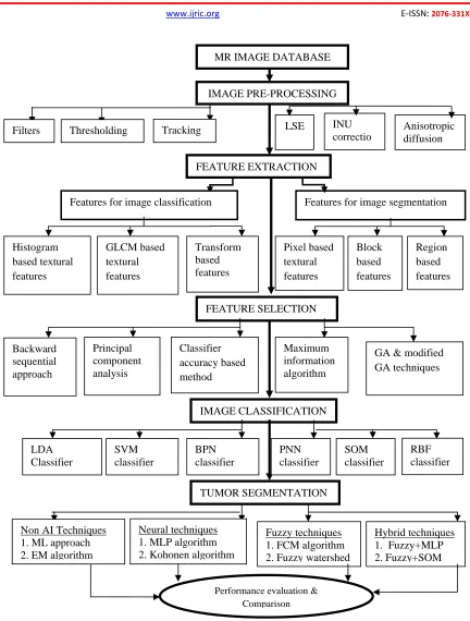
Computational applications are gaining significant importance in the day-to-day life. Specifically, the usage of the computer aided systems for computational biomedical applications has been explored to a higher extent. Medical image analysis is an important biomedical application which is highly computational in nature and requires the aid of the automated systems. These image analysis techniques are often used to detect the abnormalities in the human bodies through scan images. Automated brain disorder diagnosis with MR images is one of the specific medical image analysis methodologies. The automated diagnosis involves two major steps: (a) Image classification & (b) Image segmentation. Image classification is the technique of categorizing the abnormal input images into different tumor groups (brain tumors
31
Figure 1. Framework of the automated identification system
2. LITERATURE SURVEY ON IMAGE PRE-PROCESSING
Image pre-processing is one of the preliminary steps which are highly required to ensure the high accuracy of the subsequent steps.
The raw MR images normally consist of many artefacts such as intensity in-homogeneities, extra cranial tissues, etc. which reduces the overall accuracy. Several researches are reported in the literature to minimize the effects of artefacts in the
Anisotropic diffusion INU
correctio LSE
FEATURE EXTRACTION
Features for image classification Features for image segmentation Filters Thresholding Tracking
Block based features Pixel based
textural features
Region based features
FEATURE SELECTION
TUMOR SEGMENTATION IMAGE CLASSIFICATION Histogram
based textural features
GLCM based textural features
Transform based features
Performance evaluation & Comparison
IMAGE PRE-PROCESSING
Maximum information algorithm
GA & modified GA techniques Backward
sequential approach
Principal component analysis
Classifier accuracy based method
LDA Classifier
SVM classifier
BPN classifier
PNN classifier
SOM classifier
RBF classifier
Non AI Techniques 1. ML approach 2. EM algorithm
Neural techniques 1. MLP algorithm 2. Kohonen algorithm
Fuzzy techniques 1. FCM algorithm 2. Fuzzy watershed
32
MR images. An analysis on filtering techniques such as Gabor & QMF filters for noise reduction is performed by [1]. These primitive methods along with reducing the noise blur the important and detailed structures necessary for subsequent steps. The colour ray casting method to differentiate the region of interest from the background is implemented by [2]. But this technique is image dependent and not applicable for gray level images. Expectation maximization segmentation (EMS) software package is also used for image pre-processing [3]. The main advantage of this technique is that it is a fully automatic technique. Diffusion filtering combined with simple non-adaptive intensity thresholding is used to enhance the region of interest [4]. The main drawback of this technique is the non-adaptive nature of the threshold value. Fuzzy connectedness based intensity non uniformity correction has been implemented by [5]. A sequential approach with fuzzy connectedness, atlas registration and bias field correction is used in this approach. The conclusions revealed that the proposed technique can be used only if the intensity variations between the images are of a limited range.
The effect of inter-slice intensity variation is minimized with the weighted least square estimation method [6]. The selection of weights for the least square method is the major disadvantage of this approach. The noise removal technique using wavelets and curve lets is implemented in [7]. Hybrid approaches involving Variance Stabilizing Transform (VST) are also used in this work. But this technique is applicable for images with Poisson noise. Tracking algorithm based de-noising technique is performed by [8]. Since the seed point for tracking is random in nature, this technique is not much efficient. A contrast agent accumulation model based contrast enhancement is implemented by [9]. This improves only the contrast of the image and the unwanted tissues are not eliminated. The wiener filtering technique for noise removal in MR brain images is used in [10]. Apart from noise removal, several other pre-processing steps are also reported in the literature. This includes image format conversion, image type conversion etc. The combination of three modalities of MR images for further processing is proposed in [11]. All the above mentioned techniques remove only specific artefacts which is not sufficient for high classification accuracy and segmentation efficiency. Apart from eliminating the noises, techniques for the removal of unwanted tissues
such as the skull tissues in MR images are highly essential for accurate identification of the diseases.
3. LITERATURE SURVEY ON FEATURE EXTRACTION
The next step in the automated diagnosis process is feature extraction. Feature extraction is the technique of extracting specific features from the pre-processed images of different abnormal categories in such a way that the within - class similarity is maximized and between - class similarity is minimized. Earlier research works report many feature extraction techniques employed for medical image processing. 2D wavelet transform based textural features for classification is used by [12]. In this report, initially basic statistical features are used and then co-occurrence based textural features are used to improve the accuracy. But the effects of usage of different wavelets are not dealt in the report. A comparison of 2D wavelet transform based textural features and 3D wavelet transform based textural features is performed by [13]. This work concluded that the combination of 2D and 3D wavelet based textural features yield better results than the 2D wavelet features. Feature extraction technique using the complimentary wavelet transformed image is implemented by [14]. The report claimed that the features extracted from all the four sub-bands are more efficient than the features from the only the approximation sub-band. All these techniques used the basic Discrete Wavelet Transform (DWT) which does not yield superior results.
33
A comparative analysis with the conventional algorithms is presented in this report. First and second order statistical features are also extracted from each training points and used for segmentation applications. These types of features are used by [19]. This report suggested that the combination of histogram based statistical features is more effective than the intensity based features. The effects of usage of moment based features are analyzed by [20]. Though this approach yields considerable improvement in the accuracy, the lack of generalization capability of these features is a drawback of this methodology. Feature extraction based on block processing technique is reported by [21]. Several other researchers analyzed the efficiency of each texture features individually. The entropy is the most dependable feature among the textural features. This information is revealed by [22]. These earlier works on feature extraction techniques clearly lay an emphasis on the necessity for high quality for successful image classification and segmentation techniques.
4. LITERATURE SURVEY ON FEATURE SELECTION
All the extracted features do not guarantee high accuracy. The presence of insignificant features reduces the output accuracy besides increasing the computational time period. Since both these parameters are highly essential, a methodology must be framed to eliminate the insignificant features. This technique of selecting an optimal feature set is called as feature selection. Though it is an optional step, many earlier works reported the usage of feature selection techniques to enhance the quality of the output. The applicability of Genetic Algorithm (GA) for pattern classification is explored by [23]. The samples belonging to the same class are accepted by this GA approach and the other samples are rejected based on the strength of association measure. A comparative analysis is also performed with the maximum likelihood classifier. The selection of optimal fuzzy rules for a complex model using Genetic Algorithm (GA) is illustrated by [24]. A comparative analysis with the un-optimized model suggested that the learning time has been reduced significantly because of genetic based optimization. Optimal fuzzy rule selection for classification is also implemented by [25]. A hybrid neuro fuzzy approach is used in this work with the architecture performing the selection
procedure. But this technique is highly sensitive to change in the parameters of the membership functions. A hybrid approach involving MLP and GA for pattern recognition is proposed by [26]. GA is used for optimizing several parameters of the neural network to improve the performance in terms of accuracy and convergence time period.
34
Another evolutionary approach namely, Particle Swarm Optimization (PSO) is used by [35]. This method is used for optimizing the weights of Artifical Neural Networks for image classification applications. Since PSO involves many random parameter initializations, the weights obtained using this approach may not be a stabilized set of weights. An enhanced version of PSO is implemented by [36]. The premature convergence of the conventional PSO is eliminated in this approach by performing modifications in the velocity update equations. The results concluded that the proposed approach is simple and easy for implementation. A survey on the applications of GA for image classification and segmentation is conducted by [37]. The basic theory behind GA is covered in this report. Even though applications are mentioned in the report, no practical implementation is carried out to justify the hypothesis. An extensive analysis on comparison of GA and PSO algorithms for feature selection is performed by [38]. The results concluded that PSO is better than GA in terms of accurate results. Feature selection using PSO for SVM based classification is performed by [39]. The conclusion of this work is that PSO converges to the optimal solution quickly than the other optimization algorithms. Weight based feature quality assessment is reported by [40]. RELIEF algorithm has been used in this work for feature selection. Experiments are also conducted on various datasets to show the applicability of this approach for feature selection. A comparative study between the various optimal search based techniques for cancer classification is done by [41]. The various techniques are analyzed in terms of convergence rate and accuracy. These techniques are also implemented using the real time datasets to highlight the best technique.
The combined rough set theory and the PSO technique for knowledge extraction from fMRI data is used by [42]. They concluded that the PSO requires less time to obtain better results than GA. Unstable nature of the optimal feature set is the major drawback of this approach. PSO technique is also used to obtain an optimal rule set for fuzzy image segmentation. One such work is reported by [43]. Different membership functions are used for experimental analysis in this work. An improved PSO algorithm is reported by [44]. This methodology claims that the modified PSO reduces the convergence time period to a higher extent. A comparison with Ant Colony
Optimzation (ACO) technique is also analyzed in this work. A combination of fuzzy approach and GA is used by [45]. Though this approach yields high accuracy because of fuzzy theory, the time taken by the GA is significantly high which makes the system practically non-feasible. PSO technique for ultrasound applications are dealt by [46]. The hybrid techniques of wavelets and Principal Component Analysis (PCA) are used by [47]. But this technique requires fresh training whenever a new image is encountered. Thus, this survey reveals the merits and demerits of the various optimization techniques used for feature selection. Though these techniques are rarely used for abnormality detection in MR brain images, the results are highly promising in terms of classification accuracy and convergence time period. Hence, the applicability of evolutionary algorithms for feature selection will be explored in this work for accurate classification and segmentation.
5. LITERATURE SURVEY ON BRAIN
IMAGE CLASSIFICATION TECHNIQUES
35
for image classification applications. The four different types of tumor is classified using LDA technique by [51]. But the classification accuracy reported in the paper is in the order of 80% which is relatively low. This work also suggested the various reasons for misclassifications.
Support Vector Machine based classification of various levels of MR glioma images is performed by [52]. This method claimed to be better than rule based systems but the accuracy reported in the paper is low. This work dealt with only glioma images and thus the lack of generalizing capability of this work is another drawback of this sytem. The application of Kohonen neural networks for image classification is explored by [53]. Some modifications of the conventional Kohonen neural network are also implemented in this work which proved to be much superior to the conventional neural networks. A hybrid approach such as combination of wavelets and Support Vector Machine (SVM) for classifying the abnormal and the normal images is used by [54]. This report revealed that the hybrid SVM is better than the Kohonen neural networks in terms of performance measures. But the major drawback of this system is the small size of the dataset used for implementation. The classification accuracy results may reduce when the size of the dataset is increased. A modification of conventional SVM such as Least Square SVM (LS-SVM) for brain tumor recognition is proposed by [55]. Both bi-level classification and multiclass classification are performed in this work to show the superior nature of the proposed method over the conventional classifiers. This report also specified an important note that the differences between various algorithms increase when the number of classes increase. Thus, this work suggested the necessity for multiclass classification techniques than bi-level classification techniques. Another version of LS-SVM is proposed and successfully implemented by [56]. An extensive comparative analysis is performed between the SVM, neural classifier and the statistical classifiers. Results suggested the advantages of SVM in terms of classification accuracy. Only bi-level classification is performed in this work which is inadequate for judging the nature of the automated system. The modified Probabilistic Neural Network for tumor image classification is used by [57]. Abnormal images such as metastase, glioma and meningioma are differentiated using the least square feature transformation based PNN. A comparative analysis is also performed with SVM. This work
inferred that the transform based PNN is superior to the SVM in terms of classification accuracy.
36
classifiers. A hybrid approach for pattern classification is reported by [66]. The combination of SVM and fuzzy rules is experimented in this work. The results revealed that the proposed hybrid approach is accurate, fast and robust.
6. LITERATURE SURVEY ON BRAIN
IMAGE SEGMENTATION TECHNIQUES
Pathology identification is performed by the image classification technique and then the treatment is planned based on the nature of abnormality. After treatment, it is highly essential to estimate the response of the patient to the treatment. In case of brain tumor abnormalities, the size of the tumor may decrease which indicates a positive effect and sometimes it may increase which shows a negative effect. In any case, it is important to perform a volumetric analysis on MR brain tumor images. Image segmentation covers this objective by extracting the abnormal portion from the image which is useful for analyzing the size and shape of the abnormal region. This method is also called as “pixel based classification” since the individual pixels are clustered unlike the classification techniques which categorizes the whole image. Several research works are reported in the area of medical image segmentation. All the research works performed on image segmentation can be classified into two broad categories: (a) Non-AI techniques and (b) AI techniques. Initially, a survey is performed on Non-AI techniques followed by the report on AI techniques.
6.1. Image Segmentation Based on Non- AI Techniques
The maximum likelihood approach to segment the pathological tissues from the normal tissues is used by [67]. The drawback of this approach is that the proposed system is dependent on class probabilities and threshold values. A model based tumor segmentation technique was implemented by [68]. This approach uses a modified Expectation Maximization (EM) algorithm to differentiate the healthy and the timorous tissues. A set of tumor characteristics are presented in this paper which is highly essential for accurate segmentation. But the drawback of this work is the lack of quantitative analysis on the extracted tumor region. Level set method based tumor segmentation technique is developed by
[69]. This method involves the method of boundary detection with the seed point. Watershed algorithm is also used to capture the weak edges. The main problem of this approach is the selection of seed point. Random selection of seed point may lead to inappropriate results and also consumes large convergence time period. A complete analysis of various types of brain tumors and the effect of MR image segmentation techniques on the treatment is studied by [70]. The report concluded that the enhanced MR image segmentation techniques play a major role for brain tumor treatment. This study shows the requirement for an accurate and quick image segmentation technique. The merits and demerits of various statistical segmentation techniques is elaborated by [71]. This work analyzed the performance measures of histogram based method, EM technique and the statistical parameter mapping (SPM2) package in detail. This report aimed at differentiating four different types of brain tissues. Experiments are carried out on simulated brain images. But the statistical techniques fail in the case of large deformations.
37
based brain tumor extraction is performed by [77]. The ability of this approach is limited since it can detect only the densely packed tumor tissues. The application of pseudo-conditional random fields for brain tumor segmentation is demonstrated by [78]. This technique is implemented on images of different tumor size. This system also claimed to be highly accurate and much faster than other conventional techniques. The mode of training used in this approach is patient - specific training which is one of the limitations of this technique. Bayesian model based tissue segmentation technique for tumor detection is implemented by [79]. This method proved to be computationally efficient besides yielding improved results over the conventional techniques. K-Nearest Neighbour technique based MR brain image classification is performed by [80]. An extensive comparative analysis is performed with other techniques. The dependency on threshold values for accurate output is the drawback of this approach. A volumetric image analysis based on mesh and level set method is illustrated by [81]. This work concentrated on 3D image processing and employed on different tumor types. But the results yielded by this approach are inferior and not comparable to the other pixel based classifiers. A survey on various medical image segmentation algorithms is performed by [82]. The drawbacks of several techniques are clearly illustrated in this report and also suggested suitable techniques for tumor segmentation.
6.2. Image Segmentation Based on Artificial Neural Networks
Another group of researchers depend on AI techniques for brain image segmentation. Among the AI techniques, ANN and Fuzzy theory are the predominantly used methodologies for segmentation. ANN is preferred by the researchers because of its adaptive nature, accuracy, etc. The application of Linear Vector Quantization (LVQ) for brain image segmentation is deomonstrated by [83]. A comparative analysis is performed with the Back Propagation neural network (BPN) and the experimental results proved the superior nature of LVQ. The report also concluded that the ANN is faster than the conventional classifiers. Self Organizing Map based segmentation technique on MR brain images is implemented by [84]. Though the proposed system is faster, the segmentation efficiency is comparatively low since it employs unsupervised mode of training. Automate brain image segmentation using conventional LVQ is
proposed by [85]. The convergence rate of this approach is high but this system failed to distinguish the outer layers of brain which is normally seen in MR brain images. Also the results are not quantitatively analyzed in this report.
38
the SOM architecture is done by [93]. Hierrarchial clustering technique has been incorporated in this approach to yield better results. An enhanced version of LVQ neural network is implemented by [94]. The concept of GA is incorporated in this technique to improve the performance of conventional LVQ. An analysis in terms of segmentation efficiency and convergence time period is provided in the report. A hybrid expert system based SOM for interpretation of MR brain images is developed by [95]. This system proved to be much superior than the individual ANN since the accuracy is increased to a higher extent. This work also highlighted the application of neuro-fuzzy approach for high quality results. But the lack of availability of an expert knowledge database for all the applications is the major drawback of this system. A novel neural network such as Incremental Supervised Neural Network (ISNN) is proposed by [96]. This method depends on Continuous Wavelet Transform (CWT) and Zernike moments for the analysis of six different types of brain tissues. The architecture of the proposed neural network is adaptive in nature and the number of neurons is added incrementally. Experimental results suggested the applicability of this network for noisy environment.
6.3. Image Segmentation Based on Fuzzy Techniques
Several earlier works based on fuzzy logic theory are also reported in the literature. A modified Fuzzy C-Means (FCM) algorithm for brain image segmentation is implemented by [97]. This method is shown to provide significant time saving when compared with the conventional FCM algorithm. Lack of quantitative analysis on segmentation efficiency is the drawback of this approach. A more accurate FCM algorithm is proposed by [98]. The outlined approach in this paper is applicable for MR images with intensity inhomogeneties. But the execution time of this approach is directly proportional to the amount of inhomogeneties present in the images. This leads to slower convergence rate which makes the system practically non-feasible. A complete survey of various segmentation algorithms is presented by [99]. The merits and the demerits of various techniques are analyzed in detail. Appropriate techniques for different applications are also suggested in this review paper. The applicability of conventional FCM algorithm for 3-D image processing is reported by [100]. This work highlighted the methodology for the
selection of number of clusters in FCM algorithm. Expert knowledge is also required for this automated approach. Lack of availability of accurate expert knowledge is the drawback of this approach. The significance of weights for fuzzy rules in image processing applications is revealed in [101]. The necessity of weights for fuzzy rules is proven through computer simulations on real time data sets. The report also has provided an efficient method for framing fuzzy IF-THEN rules. A robust fuzzy clustering algorithms proposed by [102]. This approach has eliminated the dependency of FCM algorithm on similarity measures such as distance measures. The proposed approach is highly generalized in terms of the parameters used in the algorithm. Time efficient FCM for real time applications is proposed by [103]. The requirement of two updates equations have reduced to a single update equation in this approach. This approach has solved the high memory requirement problem of conventional FCM.
39
paper. Iterative watersheds based fuzzy tumor segmentation is reported by [109]. This approach claimed to be fast and accurate but the segmentation efficiency is comparatively low. The report also suggested the usage of prior knowledge to improve the efficiency. An image segmentation technique using fuzzy connectedness is proposed by [110]. The requirement for initialization of several initial parameters including the seed pixel is one of the disadvantages of this system. A FCM algorithm to achieve high accuracy for MR brain images is developed by [111]. Several methods for initializing the cluster centres such as Silhoutte method are highlighted in this work.
Only qualitative analysis is reported in the paper which is not sufficient for judging the effectiveness of the system. A possiblistic fuzzy clustering approach is reported by [112]. This approach is experimented on brain image segmentation and promising results are obtained by this technique.
An improved FCM algorithm is also proposed by [113]. This technique is based on block processing where each block is processed by a parallel processor. Though this approach is faster, the requirement for additional hardware is the major drawback of this system. An extensive survey on tissue segmentation algorithms for MR brain images is conducted by [114]. A technique to minimize the intensity nonuniformity artifact is also proposed in this paper. Several suggestions on pixel based approaches, region based approaches, model based approaches are provided in this work. The merits of incorporating the fuzzy information in MR brain image segmentation techniques are illustrated by [115]. Several fuzzy models are created and the fuzzy features are extracted from these models. Experimental results have suggested the usage of fused fuzzy features for improving the accuracy of the techniques. Even though performance measures are specified, an extensive analysis in terms of the measures is not done in this work. Diffusion model based segmentation technique is reported by [116]. K-means clustering is used in this work for tumor extraction. Lack of the fuzzy knowledge is clearly visible in the segmented results which are of low quality. No quantitative analysis is performed in this work which is another drawback of this method. Sriparna etal. have mixed the GA concept with the fuzzy theory to achieve more efficient results [117]. The experimental results are compared with the EM algorithm. Results have revealed the applicability of GA for cluster centre selection in
the FCM algorithm. This methodology has significantly increased the accuracy. The report also suggested that the proposed work is more appropriate for noise free MR images.
A modified FCM algorithm for MR brain image segmentation is implemented in [118]. The method of initial selection of membership values are highlighted in this report. This work has not conducted significant experiments which is evident from the lack of quantitative analysis. The usage of fuzzy localization map for structure identification in MR brain images is discovered by [119]. The experiments conducted on MR images revealed the efficient and the robust nature of the proposed system. An adaptive Mean-Shift clustering based tissue segmentation is performed by [120]. Spatial information is also incorporated in this approach to minimize the effect of artefacts. In this work, experiments are conducted to cluster the normal brain images. This technique can be extended to extract abnormal tissues. A new kernelised weighted C-means algorithm is developed by [121]. High quality results are reported in this work but the experiments are conducted on a small size dataset which is highly insufficient to estimate the quality of the proposed system. Lack of generalization is another drawback of the approach since the analysis is performed only on glioma tumour images. Gaussian smoothing based FCM algorithm to achieve improved performance measures is reported by [122]. This approach has incorporated a feature selection algorithm for improved accuracy. Experimental analysis has revealed the suitability of this approach for noisy MR images. But the computational complexity of this approach is significantly high due to the bootstrap based feature selection techniques.
6.4. Image Segmentation Based on Hybrid (Neural + Fuzzy) Techniques
40
the major drawback of this approach. An optimal neuro-fuzzy possiblistic C-means algorithm is proposed by [125]. A comparative analysis between the conventional techniques is also performed in this work. This work mainly aimed at improving the segmentation efficiency of noisy MR images. The drawback is the slow convergence rate due to the fact that many complex steps are involved in this algorithm.
A combinational approach of SOM, SVM and fuzzy theory is implemented by [126]. An extensive analysis is performed with the other segmentation techniques to show the superior nature of the proposed approach. A reflex fuzzy min-max neural network for clustering techniques is developed by [127]. The reflex mechanism of brain together with fuzzy rules has been used to reduce the misclassification rate. The results are also compared with the conventional techniques and found to be useful for improving the segmentation efficiency. No specific application is reported in this work. Wavelet based neuro fuzzy segmentation is proposed by [129]. A neural architecture with fuzzy rules is used for implementation in this work. This work is not experimented on brain images. The combination of SOM and FCM algorithm for brain image segmentation is experimented by [129]. But the drawback of this technique is that it is not suitable for tumors of varying size. The convergence rate reported in this work is also very low which another drawback of this technique. A fuzzy kohonen neural network is implemented by [130]. This technique is completely dependent on the input features which are the drawback of this system. The qualitative and quantitative analysis results are inadequate when compared with the other techniques.
7. CONCLUSION
In this work, the merits and demerits of various automated techniques for brain tumor identification is analyzed in detail. The suitability of the techniques for various applications is also illustrated in this survey. Several novel hybrid approaches may be developed through the ideas conveyed in this report. This report also aid in highlighting the significant contributions of engineering theory to the medical field.
ACKNOWLEDGMENT
The authors wish to thank Dr. S. Alagappan, M/s. Devaki Scan Centre, India for his valuable support in image acquisition. The authors also wish to thank Dr. D. Selvathi for her valuable suggestions and guidance.
REFERENCES
[1]Sebe N, Michael S.L. Wavelet based texture classification. 15th Int. Conf. on Pattern Recognition 2000; 3: 3959-62.
[2]Jiang C, Zhang X, Huang W, Meinel C. Segmentation and quantification of brain tumor. IEEE Int. Conf. on Virtual Environments, Human Computer Interfaces and Measurement Systems 2004; 61-6.
[3]Greenspan H, Ruf A, Goldberger J. Constrained Gaussian mixture model framework for automatic segmentation of MR brain images. IEEE Trans. Med. Imag. 2006; 25: 1233-45.
[4]Yang Y, Huang S. Novel statistical approach for segmentation of brain magnetic resonance imaging using an improved expectation maximization algorithm. Optica Applicata 2006; 36: 125-36.
[5]Zhou Y, Bai J. Atlas based fuzzy connectedness segmentation and intensity non-uniformity correction applied to brain MRI. IEEE Trans. Biomed. Eng. 2006;54:122-9. [6]Morris M, Greiner R, Sander J, Murtha A,
Schmidt M. Learning a classification based glioma growth model using MRI data. Journal of Computers 2006; 1:21-31.
[7]Zhang B, Jalal M, Starck J. Wavelets, ridgelets and curvelets for Poisson noise removal. IEEE Trans. Imag. Proc. 2008; 17: 1093-1108. [8]Jaya J, Thanushkodi K., Karnan M. Tracking
algorithm for de-noising of MR brain images. International Journal of Computer Science and Network Security 2009; 9:262-7.
[9]Prastawa M, Bullitt E, Gerig G. Simulation of brain tumors in MR images for evaluation of segmentation efficacy. Medical Image Analysis 2009; 13: 297-311.
[10]Ratan R, Sharma S, Sharma S. Multiparamter segmentation and quantization of brain tumor from MR images. International Journal of Science and Technology 2009; 2:11-5.
41
[12]Arivazhagan S, Ganesan L. Texture classification using wavelet transform. Pattern Recognition Letters 2003; 24:1513-21.
[13]Jafari-Khouzani K, Soltanian-Zadeh H, Elisevich K, Patel S. Comparison of 2D and 3D wavelet features for TLE lateralization. Proc. of SPIE 2004;5369:593-601.
[14]Hiremath P, Shivashankar S. Wavelet based features for texture classification. Graphics, Vision and Image Processing Journal 2006;6:55-8.
[15]Hiremath P, Shivashankar S. Texture classification using wavelet packet decomposition. Graphics, Vision and Image Processing Journal 2006; 6:77-80.
[16]Ryszard S. Image feature extraction techniques and their applications for CBIR and biometrics system. International Journal of Biology and Biomedical Engineering 2007;1:6-16.
[17]Georgiadis P, Cavouras D, Kalatzis J, Daskalakis A, Kagadis G, Sifaki K, Solomou E. Non-linear least square feature transformations for improving the performance of probabilistic neural networks in classifying human brain tumors on MRI. Lecture Notes on Computer Science 2007;4707:239-47. [18]Huang K, Aviyente S. Wavelet feature
selection for image classification. IEEE Trans. Imag. Proc. 2008; 17:1709-20.
[19]Jesus C, Noah L, Ebadollahi S, Andrew L, John K. Concept detection in longitudinal brain MR images using multi modal cues. IEEE International Symposium on Biomedical Imaging 2009; 418-21.
[20]Kowaliw T, Banzhaf W, Kharma N, Harding S. Evolving novel image features using Genetic programming based image transforms. IEEE Congress on Evolutionary Computation 2009;2502-7.
[21]Pradhan N, Sinha K. Development of a composite feature vector for the detection of pathological and healthy tissues in FLAIR MR images of brain. Bioinformatics and Medical Engineering Journal 2009;10:1-11.
[22]Kekre H, Tanuja S, Saylee G. Detection and demarcation of tumor using vector quantization in MR images. International Journal of Engineering Science and Technology 2009; 1:59-66.
[23]Kishore K, Patnaik M, Mani V, Agrawal K. Application of Genetic programming for multi-category pattern classification. IEEE Trans. Evolutionary Computation 2009;4:242-58.
[24]Marco R. Genetic fuzzy learning. IEEE Trans. Evolutionary Computation 2009;4:259-73. [25]Chakraborty D, Nikhil P. A neuro fuzzy
scheme for simultaneous feature selection and fuzzy rule based classification. IEEE Trans. Neural Networks 2004;15:110-23.
[26]Alsutanny Y, Aqel M. Pattern recognition using multilayer neural genetic algorithm. Neurocomputing 2003;51:237-47.
[27]Sindhwani V, Rakshit S, Deodhare D, Erdogmus D, Jose C. IEEE Trans. Neural Networks 2004;15:937-48.
[28]Arjan S, Willem M, Edelenyi F, Jack A, Lutgarde B. Combination of feature reduced MR spectroscopic and MR imaging data for improved brain tumor classification. Nucl. Mag. Res. Biomed. 2004;18:34-43.
[29]Fan Y, Shen D, Davatzikos C. Classification of structural images via high dimensional image warping, robust feature extraction and SVM. Proc. of MICCAI, Lecture Notes on Computer Science 2005;3749:1-8.
[30]Auralia I, Andries P. Comparing different optimization strategies for non-linear mapping and mapping large datasets using neural networks. World Congress on Evolutionary Computation 2005;306-13.
[31]Hong J, Cho S. Efficient huge scale feature selection with speciated genetic algorithm. Pattern Recognition Letters 2006;27:143-50. [32]Vlasis K, Christos P. A sawtooth genetic
algorithm combining the effects of variable population size and re-initialization to enhance performance. IEEE Trans. Evolutionary Computation 2006;10:19-28.
[33]Hoai N, Mckay I, Essam D. Representation and structural difficulty in genetic programming. IEEE Trans. Evolutionary Computation 2006;10:157-66.
[34]Julian M, Stephen S. Redundancy and Computational Efficiency in Cartesian genetic programming. IEEE Trans. Evolutionary Computation 2006;10:167-74.
[35]Chandramouli K, Izquierdo E. Image classification using chaotic particle swarm optimization. IEEE Int. Conf. on Image Processing 2006;3001-04.
[36]Liang J, Qin K, Suganthan P, Baskar S. Comprehensive learning particle swarm optimizer for global optimization of multimodal functions. IEEE Trans. Evolutionary Computation 2006;10:281-95. [37]Paulinas M, Usinskas A. A survey of genetic
42
enhancement and segmentation. Information Technology and Control 2007;36:278-84. [38]Garcia-Nieto J, Jourdan L. A comparison of
PSO and GA approaches for gene selection and classification of microarray data. Proc. of the 9th Annual conference on Genetic and Evolutionary Computation 2007;427.
[39]Tu C, Chuang L, Chang J, Yang C. Feature selection using PSO-SVM. IAENG International Journal of Computer Science 2007;33:138-43.
[40]Sun Y. Iterative RELIEF for feature weighting: Algorithms, Theories and Applications. IEEE Trans. Pattern Anal. Machine Intell. 2007;29:1036-51.
[41]Lin J, Sa H, Chen H, Bernard F. Optimal search based gene subset selection for gene array cancer classification. IEEE Trans. Information Technology in Biomedicine 2007;11:398-405.
[42]Lin H, Abraham A, Ye H. Extracting multi knowledge from fMRI data through swarm based rough set reduction. Proc. of the 3rd Int. Workshop on Hybrid Artificial Intelligence Systems 2008;281-88.
[43]Masooleh M, Moosavi S. An improved fuzzy algorithm for image segmentation. World Academy of Science, Engineering and Technology 2008;38:400-4.
[44]Gomez Y, Bello R, Puris A, Maria G. Two step swarm intelligence to solve the feature selection problem. Journal of Universal Computer Science 2008;14:2582-96.
[45]Ganesan R, Radhakrishnan S. Segmentation of Computed Tomography Brain Images using Genetic Algorithm. International Journal of Soft Computing 2009;4:157-61.
[46]Ijaz U, Richard P, Andrew H, Graham M. Particle swarm optimization for invivo 3D ultrasound volume registration. Proc. of the 30th Int. Symposium on Acoustical Imaging 2009;1-10.
[47]El-Sayed A, Abdel-Badeeh M, Tamer H. A hybrid technique for automatic MRI brain images classification. Informatica 2009;54:55-67.
[48]Ronald G, Stephen A, Kromhout-Schiro S, Suresh K. The role of neural networks in improving the accuracy of MR spectroscopy for the diagnosis of head and neck squamous cell carcinoma. AJNR 2000; 21:1133-38. [49]Michael R, Simon K, Nabavi A, Peter M,
Ferenc A, Kikinis R. Automated segmentation of MR images of brain tumors. Radiology 2000;218:586-91.
[50]Egmont P, De D, Handels H. Image processing with neural networks-a review. Pattern Recognition 2002;35:2279-301.
[51]Majos C, Julia-Sape M, Alonso J, Serrallonga M, Aguilera C, Juan J, Gilli J. Brain tumor classification by proton MR spectroscopy: Comparison of diagnostic accuracy at short and long TE. AJNR 2004;25:1696-704.
[52]Li G, Yang J, Ye C, Geng D. Degree prediction of malignancy in brain glioma using support vector machines. Computers in Biology and Medicine 2006;36:313-25.
[53]Messen W, Wehrens R, Buydens L. Supervised Kohonen networks for classification problems. Chemometrics and Intelligent Laboratory Systems 2006;83:99-113.
[54]Chaplot S, Patnaik M, Jagannathan N. Classification of magnetic resonance brain images using wavelets as input to support vector machine and neural network. Biomedical Signal Processing and Control 2006;1:86-92.
[55]Luts J, Heerschap A, Johan A, Huffel S. A combined MRI and MRSI based multiclass system for brain tumour recognition using LS-SVMs with class probabilities and feature selection. Artificial Intelligence in Medicine 2007;40:87-102.
[56]Selvaraj S, Thamaraiselvi S, Selvathi D, Gewali L. Brain MRI slices classification using least squares support vector machine. International Journal of Intelligent Computing and Medical Sciences in Image Processing 2007;1:21-33.
[57]Georgiadis P, Cavouras D, Kalatzis J, Daskalakis A, George C, Sifaki K, Ekaterini Solomou. Improving brain tumor characterization on MRI by probabilistic neural networks and non-linear transformation of textural features. Computer Methods and Programs in Biomedicine 2008;89:24-32. [58]Yamashita K, Yoshiura T, Arimura H, Mihara
F, Noguchi T, Hiwatashi A, Honda H. Performance evaluation of radiologists with artificial neural network for differential diagnosis of intra-axial cerebral tumors on MR images. AJNR 2008;29:1153-58.
[59]Ibrahiem M, Ramakrishnan S. On the application of various probabilistic neural networks in solving different pattern classification problems. World Applied Sciences Journal 2008;4:772-780.
43
discrimination between primary brain tumors on MRI: From 2D to 3D texture analysis. e-Journal of Science and Technology 2008;9-18.
[61]Felix N, Lluis M. Feature selection in proton magnetic resonance specrtroscopy for brain tumor classification. European Symposium on Artificial Neural Networks –Advances in Computational Intelligence and Learning 2008;77-82.
[62]Palaniappan R, Eswaran C. Using genetic algorithm to select the presentation order of training patterns that improves simplified fuzzy ARTMAP classification performance. Applied Soft Computing 2009;9:100-06. [63]Merinsky Z, Hostalkova E, Prochazka A.
Brain tumour diagnostic support based on medical image segmentation. Proc. of 17th Annual Conference Technical Computing 2009;1-7.
[64]Chandra S, Bhat R, Singh H, Chauhan S. Detection of brain tumors from MRI using Gaussian RBF kernel based support vector machine. International Journal of Digital Content Technology and its Applications 2009;1:46-51.
[65]Hamilton-Wright A, Daniel W, Hamid R. Fuzzy classification using pattern discovery. IEEE Trans. on Fuzzy Systems 2007;15:772-83.
[66]Lin C, Yeh C, Liang S, Chung J, Kumar N. Support-vector based fuzzy neural network for pattern classification. IEEE Trans. on Fuzzy Systems 2006;14:31-41.
[67]Zavaljevski A, Dhawan A, Gaskil M, Ball W, Johnson D. Multi-level adaptive segmentation of multi-parameter MR brain images. Computerized Medical Imaging and Graphics 2000;24:87-98.
[68]Moon N, Bullitt E, Leemput K, Gerig G. Model based brain and tumor segmentation. Int. Conf. on Pattern Recognition 2002;528-531.
[69]Zhu F, Tian J. Modified fast marching and level set method for medical image segmentation. Journal of X-ray Science and Technology 2003;11:193-204.
[70]Deorah S, Charles L, Zita A, Timothy R. Trends in brain cancer incidence and survival in the United States: Surveillance, Epidemiology and End results program. Neurosurgery Focus 2006;20:1-7.
[71]Zaidi H, Ruest T, Schoenahl F, Montandon M. Comparative assessment of statistical brain MR image segmentation algorithms and their
impact on partial volume correction in PET. Neuroimage 2006;32:1591-607.
[72]Khotanlou H, Colliot O, Bloch I. Automatic brain tumor segmentation using symmetry analysis and deformable models. Int. Conf. on Advances in Pattern Recognition 2007;198-202.
[73]Moussaoui A, Ferahta N, Chen V. A new hybrid RMN image segmentation algorithm. International Journal of Applied Sciences, Engineering and Technology 2007;1:171-8. [74]Withey D, Koles Z. Three generations of
medical image segmentation methods and available software. International Journal of Bioelectromagnetism 2007; 9:67-68.
[75]Unnikrishnan R, Pantofaru C, Hebert M. Toward objective evaluation of image segmentation algorithms. IEEE Trans. Pattern. Anal. Machine Intell. 2007;29:929-44.
[76]Bazin P, Dzung P. Topology preserving tissue classification of magnetic resonance brain images. IEEE Trans. Med. Imag. 2007;26:487-496.
[77]Ray N, Greiner R, Murtha A. Using symmetry to detect abnormalities in brain MRI. Computer Society of India Communications 2008;31:7-10.
[78]Lee C, Wang S, Murtha A, Mathew B, Greiner R. Segmenting brain tumors using pseudo conditional random fields. Proc. of the 11th Int. Conf. on Medical Image Computing and Computer Assisted Intervention (MICCAI) 2008;359-66.
[79]Jason J, Sharon E, Dube S, El-Saden S, Sinha U, Yuille A. Efficient multilevel brain tumor segmentation with integrated Bayesian model classification. IEEE Trans. Med. Imag. 2008;27:1-7.
[80]Anbeek P, Koen L, Max V. Automated MS-Lesion segmentation by K-Nearest Neighbour classification. The MIDAS online Journal 2008. (http://hdl.handle.net/10380/1448). [81]Aloui K, Naceur S. 3D brain tumor
segmentation using level set method and meshes simplification from volumetric MR images. World Academy of Science, Engineering and Technology 2009;57:127-31. [82]Ma Z, Joao T, Natal M. A review on the
current segmentation algorithms for medical images. Int. Conf. on Imaging Theory and Applications 2009;135-40.
44
[84]Reyes-Aldasoro C, Aldeco A. Image segmentation and compression using neural networks. Proceedings of Advances in Artificial Perception and Robotics 2000;23-5. [85]Carlos A, Iftekharuddin K, Kozma R.
Automated brain data segmentation and pattern recognition using ANN. Int. Conf. on Computational Intelligence, Robotics and Autonomous systems 2003;pp. 27-31.
[86]Valdes-Cristerna R, Medina-Banuelos V, Yanez-Suarez O. Coupling od radial basis network and active contour model for multispectral brain MRI segmentation. IEEE Trans. Biomed. Eng. 2004;51:459-70.
[87]Li Y, Chi Z. MR brain image segmentation based on self-organizing map network. International Journal of Information Technology 2005;11:45-53.
[88]Martin-Landrove M, Villalta R. Brain tumor image segmentation using neural networks. Proc. of International Society of Magnetic Resonance in Medicine 2006;14:1610. [89]Chartier S, Boukadoum M. A bidirectional
hetroassociative memory for binary and grey-level patterns. IEEE Trans. on Neural Networks 2006;17:385-96.
[90]Huang G, Zhu Q, Siew C. Real-time learning capability of neural networks. IEEE Trans. on Neural Networks 2006;17:863-78.
[91]Lange O, Meyer-Baese A, Hurdal M, Foo S. A comparison between neural and fuzzy cluster analysis techniques for functional MRI. Biomedical Signal Processing and Control 2006;1:243-52.
[92]Bhattacharya S, Manlik U, Dutta P. A pruning algorithm for efficient image segmentation with neighbourhood neural networks. IAENG International Journal of Computer Science 2008;35.
[93]Liao W, Chen H, Yang Q, Lei X. Analysis of fMRI data using improved self-organizing mapping and spatio-temporal metric hierarchial clustering. IEEE Trans. Med. Imag. 2008;27:1472-83.
[94]Yeh J, Fu C. A hierarchial genetic algorithm for segmentation of multi-spectral human brain MRI. Expert Systems with Applications 2008;34:1285-95.
[95]Guler I, Demirhan A, Karakis R. Interpretation of MR images using self-organizing maps and knowledge based expert systems. Digital Signal Processing 2009;19:668-77.
[96]Iscan Z, Dokur Z, Olmez T. Tumor detection by using Zernike moments on segmented
magnetic resonance brain images. Expert Systems with Applications 2010;37:2540-49. [97]Cheng T, Dmitry G, Lawrence H. Fast fuzzy
clustering. Fuzzy Sets and Systems 1998;93:49-56.
[98]Dzung P, Jerry P. An adaptive fuzzy C-means algorithm for image segmentation in the presence of intensity inhomogeneties. Pattern Recognition Letters 1999;20:57-68.
[99]Dzung P, Xu C Jerry P. Current methods in medical image segmentation. Annual Review of Biomedical Engineering 2000;2:315-37. [100]Lynn F, Lawrence H, Dmitry G, Reed M.
Automatic segmentation of non-enhancing brain tumors in magnetic resonance images. Artificial Intelligence in Medicine 2001;21:43-63.
[101]Ishibuchi H, Nakashima T. Effect of rule weights in fuzzy rule based classification systems. IEEE Trans. Fuzzy Systems 2001;9:506-15.
[102]Rajesh N, Sen S. Robust fuzzy clustering of relational data. IEEE Trans. Fuzzy Systems 2002;10:713-27.
[103]John F, Hutcheson T. Reducing the time complexity of the fuzzy C-means algorithm. IEEE Trans. on Fuzzy Systems 2002;10:263-267.
[104]Javier G, Shen Q. From approximate to descriptive fuzzy classifiers. IEEE Trans. on Fuzzy Systems 2002;10:484-97.
[105]Khalighi M, Soltanian-Zadeh H, Lucas C. Unsupervised MRI segmentation with spatial connectivity. International Symposium on Medical Imaging 2002;1-9.
[106]Auephanwiriyakul S, James K. Analysis and efficient implementation of a linguistic fuzzy C-means. IEEE Trans. on Fuzzy Systems 2002;10:563-82.
[107]Eschrich S, Ke J, Lawrence H, Dmitry G. Fast accurate fuzzy clustering through data reduction. IEEE Trans. on Fuzzy Systems 2003;11:262-70.
[108]Liew A, Yan H. An adaptive spatial clustering algorithm for 3D MR image segmentation. IEEE Trans. on Med. Imag. 2003;22:1063-75.
[109]Mancas M, Gosselin B. Fuzzy tumor segmentation based on iterative watersheds. Proc. of the Medical Imaging Conference of the International Society for Optical Imaging 2003;pp. 1598-608.
45
International Journal of Applied Mathematics and Computer Sciences 2004;2:25-30. [111]Kannan S. Segmentation of MRI using new
unsupervised fuzzy C-means algorithm. Graphics, Vision and Image Processing Journal 2005;5:17-24.
[112]Masulli F, Rovetta S. Soft transition from probabilistic to possiblistic fuzzy clustering. IEEE Trans. on Fuzzy Systems 2005;14:516-27.
[113]Murugavalli S, Rajamani V. A high speed parallel fuzzy C-mean algorithm for brain tumor segmentation. Bioinformatics and Medical Engineering Journal 2006;6:29-34. [114]Liew A, Yan H. Current methods in the
automatic tissue segmentation of 3D magnetic resonance brain images. Current Medical Imaging Reviews 2006;2:1-13. [115]Dou W, Ruan S, Chen Y, Bloyet D, Constans
J. A framework of fuzzy information fusion for the segmentation of brain tumor tissues on MR images. Image and Vision Computing 2007;25:164-71.
[116]Masroor M, Mohamad D. Segmentation of brain MR images for tumor extraction by combining k-means clustering and Perona-Malik anisotropic diffusion model. International Journal of Image Processing 2008;2:27-34.
[117]Saha S, Bandyopadhyay S. MRI brain image segementation by fuzzy symmetry based genetic clustering technique. IEEE World Congress on Evolutionary Computation 2007;4417-24.
[118]Kannan S. A new segmentation system for brain MR images based on fuzzy techniques. Applied Soft Computing 2008;8:1599-606. [119]Scherrer B, Forbes F, Garbay C, Dojat M.
Distributed local MRF models for tissue and structure brain segmentation. IEEE Trans. Med. Imag. 2009;28:1278-95.
[120]Mayer A, Greenspan H. An adaptive mean shift framework for MRI brain segmentation. IEEE Trans. Med. Imag. 2009;28:1238-50. [121]Szwarc P. Segmentation of brain tumors in
MR images. 11th International PhD Workshop 2009;206-11.
[122]Xiao K, Ho S, Bargiela A. Automatic brain MRI segmentation scheme based on feature weighting factors selection on fuzzy C-means clustering algorithms with Gaussian smoothing. International Journal of Computational Intelligence in Bioinformatics and Systems Biology 2010;1:316-31.
[123]Jang J. ANFIS: Adaptive network based fuzzy inference system. IEEE Trans. on Systems, Man and Cybernatics 1993;23:665-84.
[124]Boskovitz V, Guterman H. An adaptive neuro fuzzy system for automatic image segmentation and edge detection. IEEE Trans. on Fuzzy Systems 2002;10:247-62. [125]Moussaoui A. A neuro fuzzy image
segmentation algorithm. International Journal of Soft Computing 2006;1:232-38.
[126]Juang C, Chiu S, Chang S. A self organizing TS-type fuzzy network with support vector learning and its application to classification problems. IEEE Trans. on Fuzzy Systems 2007;15:998-1008.
[127]Nandedkar A, Biswas K. A general reflex fuzzy min-max neural network. Engineering Letters 2007;14:1-11.
[128]Acharyya M, Malay K. Image segmentation using wavelet packet frames and neuro fuzzy tools. International Journal of Computational Cognition 2007;5:27-43.
[129]Murugavalli S, Rajamani V. An improved implementation of brain tumor detection using segmentation based on neuro fuzzy technique. Journal of Computer Science 2007;3:841-46.