Plant Disease Detection in Image Processing
Using MATLAB
Sandesh Raut 1, Amit Fulsunge 2
P.G. Student, Department of Electronics & Communication Engineering, Tulsiramji Gaikwad College Engineering &
Technology, Nagpur, Maharashtra, India1
Associate Professor, Department of Electronics Engineering, Tulsiramji Gaikwad College Engineering & Technology,
Nagpur, Maharashtra, India2
ABSTRACT: For increasing growth and productivity of crop field, farmers need automatic monitoring of disease of
plants instead of manual. Manual monitoring of disease do not give satisfactory result as naked eye observation is old method requires more time for disease recognition also need expert hence it is non effective. So in this paper, we introduced a modern technique to find out disease related to both leaf and fruit. To overcome disadvantages of traditional eye observing technique, we used digital image processing technique for fast and accurate disease detection of plant. In our proposed work, we developed k-means clustering algorithm with multi SVM algorithm in MATLAB software for disease identification and classification.
KEYWORDS:Plant disease, K-means clustering, GLCM, Multi SVM algorithm.
I. INTRODUCTION
The old and classical approach for detection and recognition of plant diseases is based on naked eye observation, which is very slow method also gives less accuracy. In some countries, consulting experts to find out plant disease is expensive and time consuming due to availability of expert. Irregular check up of plant results in growing of various diseases on plant which requires more chemicals to cure it also these chemicals are toxic to other animals, insects and birds which are helpful for agriculture. Automatic detection of plant diseases is essential to detect the symptoms of diseases in early stages when they appear on the growing leaf and fruit of plant. This paper introduces a MATLAB based system in which we focused on both leaf & fruit diseased area and used image processing technique for accurate detection and identification of plant diseases.
II. LITERATUREREVIEW
In paper [1] authors focused on Rice disease identification and considered the two diseases, namely Leaf Blast & Brown Spot. Boundary detection & spot detection methods are used for feature extraction of the infected parts of plant’s leaves. Authors introduced SOM (Self Organising Map) neural network in zooming algorithm for classification of rice diseased images. Method of making of input vector in SOM is padding of zeros & interpolation of missing points, zooming algorithm gives satisfactory result.
In paper [2] authors considered five plant diseases namely Late scorch, Cottony mold, Early scorch, Ashen mold and Tiny whiteness from Jordan’s Al-Ghor area for testing. K-Means clustering method is used for segmentation of leaf images and the CCM (Colour Co-occurrence Method) method is used for infected leaf texture analysis. For classification of plant diseases, back propagation algorithm in neural network is used.
In paper [3] authors used LABVIEW vision & MATLAB for detection of chili plant disease. Leaf inspection in early stage is possible due to combined technique of two softwares. The LABVIEW is used for capturing images of leaf and MATLAB is used as image processing software. Edge detection, fourier filtering, morphological operations are done with help of image pre-processing and color clustering method is used for separating chili and non-chili leaves in feature extractions. Image recognition and the classification shows chili plant healthiness.
In paper [4] authors introduced technique for detection of Malus Domestica leaves disease. Grayscale images are obtained by histogram equalization and the texture analysis in image segmentation is done with help of co-occurrence matrix method algorithm also color analysis is obtained using K-means clustering algorithm. In threshold matching process, there is comparison between individual pixels value and threshold value. For detection of plant diseases, texture & color images are compared with previously obtained images of leaf.
In paper [5] authors described technique for detection of Bacterial leaf scorch infection in plant. In image segmentation, K-means clustering algorithm is applied for separating foreground and background images. Clustering in segmentation is based on subtracting the clustered leaf images and intensity mapping for highlighting leaf area. K-means is very effective and simple for detection of infected area.
In paper [6] authors introduced technique of Citrus leaf disease detection and diseases are: Anthracnose, Citrus canker, Overwatering and Citrus greening. Image pre-processing involved color space conversion by applying YCbCr color system & L*a*b* color space also color image enhancement by applying discrete cosine transform. Gray-Level Co-Occurrence Matrix is used for feature extraction to see various statistics such as energy, contrast, homogeneity and entropy. Lastly SVMRBF and SVMPOLY are used for citrus leaf diseases detection.
In paper [7] authors presented technique for detection of Sun scorch Orchid Black leaf & spot leaf disease. Pre-processing is obtained by histogram equalization, intensity adjustment and filtering for image enhancement. Segmentation involved thresholding process and three morphological processes which are applied for removing & preserving the small & large object respectively. Finally classification is done by calculation of white pixels in leaf image and diseases are recognised.
In paper [9] authors presented technique in which pre-processing involved conversion RGB images to grey using the equation f(x)=0.2989*R+0.5870*G+0.114*B and removing objects and noise in image. Boundary & spot detection algorithms are configured in segmentation to find leaf infected part. After that H&B components and color co-occurrence methods are used to extract various features. Binary images are created from grey images by Otsu threshold algorithm and diseases are classified and identified using both artificial neural network and back propagation network along with K-means method.
In paper [10] authors described technique to detect Spot & Scorch disease in which by creating color transformation structure, color values are converted to space value in image pre-processing. Masked cells inside the boundaries are removed by masking of green-pixels after applying K-means method. Color co-occurrence method extracts the features such as color, texture & edge and lastly neural network is used for recognition and disease classification.
In paper [11] authors introduced technique of Groundnut plant disease detection and diseases are: Late leaf spot and Early leaf spot disease. In pre processing involved the conversion from RGB leaf image to HSV color image also used co-occurrence matrices to extract color features and statistical approach in texture feature extraction to analyze texture images. Back propagation algorithm is applied for disease recognition and classification.
In paper [12] authors introduced technique in which after image acquisition, by creating color transformation structure, color values are converted to space value in image pre-processing also applied K-means method for segmentation. Leaf unnecessary area is removed by masking of green pixels and texture features are calculated for segmented object also masked cells are removed. Infected clusters are converted from RGB to HSI and after that SGDM matrix is generated for H & S. GLCM calculations are made for extraction of features which are then passed through the neural network for disease recognition and classifications.
In paper [13] authors described technique of Sugarcane leaf disease detection and diseases are: Brown Spot, Downy mildew, Sugarcane Mosaic, Downy Fungal, Red stripe and Red rot. Pre-processing involved conversion of RGB image to grayscale and unwanted parts are removed. Healthy area and potentially infected area are located by segmentation. Linear, Non linear and Multiclass SVM are applied for disease detection.
In paper [14] authors described technique of Apple fruit disease detection and diseases are: Apple Scab, Apple Rot and Apple Blotch. Image pre-processing involved RGB to L*a*b* color space image transformation. In feature extraction Global Color Histogram, Local Binary Pattern, Completed Local Binary Pattern and Color Coherence Vector features are used for disease detection. K-means clustering is applied for 'a*b*' space in segmentation to label each pixel and segment image by color. CLBP features and multiclass SVM are used for classification of fruit disease.
In paper [15] authors presented technique of Grapes and Apple fruit disease detection and diseases are: Black Rot, Powdery Mildew, Rot, and Apple Scab disease. With the help of morphology, image components are extracted for boundaries and various visual patterns are described by texture feature. RGB color space is converted to HSI color space and ANN neural network & back propagation algorithms are used for disease classification and fruit grading.
In paper [16] authors described technique of Pomegranate fruit diseases detection and diseases are: Bacterial Blight, Leaf Spot, Fruit Rot and Fruit Spot. In image pre-processing noise is removed using (3*3), (5*5), (7*7) filtering masks. In segmentation, k-means clustering method is applied for dividing image into object and region also GLCM formula is configured for statistical texture features extraction. Lastly authors used Multilayer Perceptrons for training the neural networks using back-propagation algorithm.
relevant image with carrying out boundary detection for getting required area of image. Centroid value is calculated using improved K-Means method and compared with database to obtain result.
In paper [18] authors described technique of Apple, Grape & Pomegranate fruits disease detection. Grape fruit diseases are Downy Mildew, Powdery Mildew and Black Rot; Apple fruit diseases are Apple Blotch, Apple Scab and Apple Rot; Pomegranate fruit diseases are Bacterial Blight, Gray Mold and Aspergillus Fruit Rot. Image segmentation is configured for locating objects & bounding line of image and used K-means clustering for labelling each pixel. In SURF (Speed up Robust Feature) algorithm, with help of blob detector and local descriptor four features are extracted also artificial neural network (ANN) is applied for pattern matching & classification of disease.
In paper [19] authors introduced technique of Mango fruit disease detection. Authors made video of mango fruit disease in image acquisition and original image is converted into binary also histogram is computed. Watershed algorithm in image segmentation is applied to identify defected regions and features are extracted using blob extraction using template matching algorithm. Diseases are classified by normalized correlation method and shows defected region in the fruit image.
In paper [20] authors described technique of Apple fruit disease detection and diseases are: apple blotch and apple rot. In K-Means method, euclidean distance is used for finding infected region and fruit image is converted to L*a*b* color space from RGB. Color, shape & texture features are extracted and feature level fusion is carried out for fusing more than two features. Color features are Global color histogram and Color coherence vector. Texture features are Gabor Features, Local binary pattern, complete local binary pattern and Local ternary pattern. Lastly Random forest classifier is applied for classification result.
In paper [21] authors introduced technique of Apple fruit disease detection and diseases are: Rot Infections, Apple Scab and Blotch Fungal disease. After image acquisition-means method is used to detect region of interest and selection of only infected part. Then features are extracted and stored in database also support vector machine is configured for disease classification and recognition.
In paper [22] authors described technique of Pomegranate fruit diseases detection and diseases are: Alterneria, Bacterial Blight and Anthracnose. Pre-processing involved image resizing, filtering and morphological operations. RGB, La*b, HSV and YCbCr are used to create clusters in segmentation. In feature extraction color, morphology and texture features are extracted and gabor filter is used in texture and morphology for obtaining boundary of image. Shape vectors are extracted from healthy fruit image and minimum distance classifier (MDC) is applied for training and classification of diseased or non-diseased images.
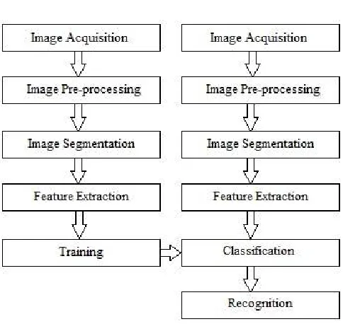
III.PROPOSEDMETHODOLOGY
Fig. 1 Framework of the proposed system A. Image Acquisition
Image acquisition is the first method of digital image processing and it is described as capturing the image through digital camera and stores it in digital media for further MATLAB operations. It is also an action of retrieving an image from hardware, so it can be passed through further process. In our work, using digital camera we captured healthy and diseased images of leaf & fruit as shown in fig. 2 for MATLAB image processing system.
Fig. 2 Original image of diseased leaf and fruit
B. Image Pre-processing
Fig. 3 Contrast enhanced and RGB to gray converted image
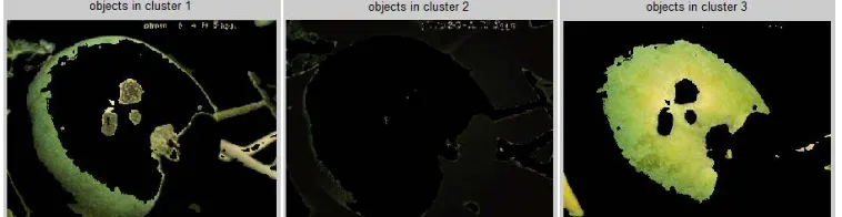
C. Image Segmentation
Image segmentation is the method for conversion of digital image into several segments and rendering of an image into something for easier analysis. Using image segmentation is used for locating the objects and bounding line of that image. In segmentation, we used K-means clustering method for partitioning of images into clusters in which at least one part of cluster contain image with major area of diseased part. The k-means clustering algorithm is applied to classify the objects into K number of classes according to set of features. The classification is done by minimizing sum of square of distances between data objects and the corresponding cluster. Image is converted from RGB Color Space to L*a*b* Color Space in which the L*a*b* space consists of a luminosity layer 'L*', chromaticity-layer 'a*' and 'b*'. All of the color information is in the 'a*' and 'b*' layers and colors are classified using K-Means clustering in 'a*b*' space. From the results of K-means, labelling of each pixel in the image is done also segmented images are generated which contain diseases. In this experiment we used segmentation technique so input image is partitioned into three clusters for good segmentation result. The following fig. 4 shows leaf image segmentation with three clusters formed by K-means clustering method.
Fig. 4 Diseased leaf image clusters
The following fig. 5 shows fruit image segmentation with three clusters formed by K-means clustering method.
D. Feature Extraction
In feature extraction desired feature vectors such as color, texture, morphology and structure are extracted. Feature extraction is method for involving number of resources required to describe a large set of data accurately. Statistical texture features are obtained by Gray level co-occurrence matrix (GLCM) formula for texture analysis and texture features are calculated from statistical distribution of observed intensity combinations at the specified position relative to others. Numbers of gray levels are important in GLCM also statistics are categorized into order of first, second & higher for number of intensity points in each combination. Different statistical texture features of GLCM are energy, sum entropy, covariance, information measure of correlation, entropy, contrast and inverse difference and difference entropy.
E. Training & Classification
Support vector machine is based on maximizing the minimum distance from the separating hyper plane to the nearest example. Only binary classification is supported in basic SVM, but in extension multiclass classification case can be possible. In these extensions, additional constraints and parameters are added to optimization problems for handling the separation of the different classes. SVM is a binary classifier that means the class labels can only take two values ±1. To get M-class classifiers, set of binary classifiers are
constructed in this way f
1
, f
2
, . . . , f M
and each are trained for separating one class from the rest.
The g j
(x) function returns the signed real value that can be interpreted as distance from separation of hyper plane to point x. Value can also be interpreted as a confidence value. The larger the value the more confident one is that the point x belong to the positive class. Hence, assign point x to the class whose confidence value is largest for this point. We used both K-means clustering & Multi SVM technique for classification and recognition of leaf and fruit disease. For creating database, image is acquired and passed through pre-processing, segmentation, features extraction then disease name is selected for given leaf or fruit and lastly data is stored in database.
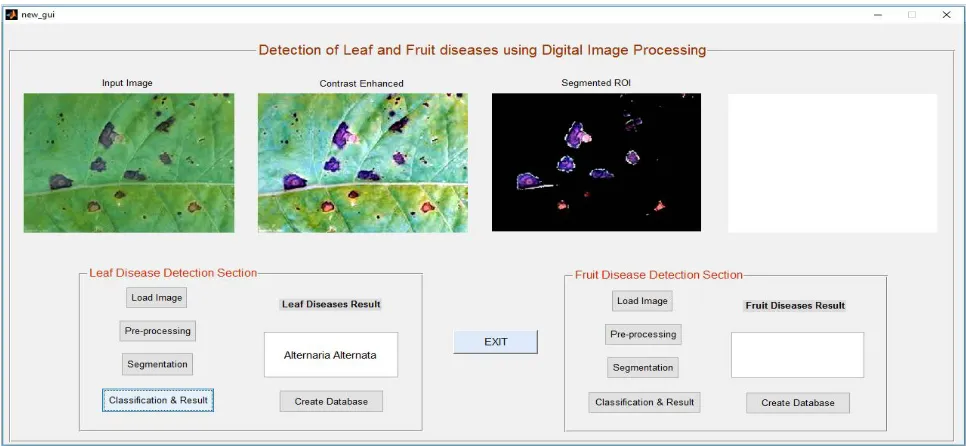
IV.EPERIMENTALRESULTS
Twenty one images of fruit are used for learning and fruit disease detection result is shown in right side section of fig. 7.
Fig. 7 System shows fruit disease detection result
V. CONCLUSION
This paper provides efficient and accurate plant disease detection and classification technique by using MATLAB image processing as shown in fig. 6 and fig. 7. The proposed methodology in this paper depends on K-means and Multi SVM techniques which are configured for both leaf & fruit disease detection. The MATLAB software is ideal for digital image processing. K-means clustering and SVM algorithm provides high accuracy and consumes very less time for entire processing. In future work, we will extend our database for more plant disease identification.
REFERENCES
[1] Santanu Phadikar and Jaya Sil “Rice Disease identification using Pattern Recognition Techniques” IEEE Proceedings of 11th International Conference on Computer and Information Technology (ICCIT 2008), Khulna, Bangladesh, pp. 1-4244-2136-7/08, 25-27 December, 2008. [2] Dheeb Al Bashish, Malik Braik and Sulieman Bani-Ahmad “A Framework for Detection and Classification of Plant Leaf and Stem Diseases”
IEEE International Conference on Signal and Image Processing, pp. 978-1-4244-8594-9/10, 2010.
[3] Zulkifli Bin Husin, Abdul Hallis Bin Abdul Aziz, Ali Yeon Bin Md Shakaff and Rohani Binti S Mohamed Farook “Feasibility Study on Plant Chili Disease Detection Using Image Processing Techniques” IEEE Third International Conference on Intelligent Systems Modelling and Simulation, pp. 978-0-7695-4668-1/12, 2012.
[4] Sabah Bashir and Navdeep Sharma “Remote Area Plant Disease Detection Using Image Processing” IOSR Journal of Electronics and Communication Engineering (IOSRJECE) ISSN : 2278-2834 Volume 2, Issue 6, PP 31-34,Sep-Oct 2012.
[5] Murali Krishnan and Dr.M.G.Sumithra “A Novel Algorithm for Detecting Bacterial Leaf Scorch (BLS) of Shade Trees Using Image Processing” IEEE 11th Malaysia International Conference on Communications, Kuala Lumpur, Malaysia pp. 978-1-4799-1532-3/13, 26th - 28th November 2013.
[6] Ms. Kiran R. Gavhale, Prof. Ujwalla Gawande and Mr. Kamal O. Hajari “Unhealthy Region of Citrus Leaf Detection Using Image Processing Techniques” IEEE International Conference for Convergence of Technology, pp. 978-1-4799-3759-2/14, 2014.
[8] Usama Mokhtar, Mona A. S. Alit, Aboul Ella Hassenian, Hesham Hefny “Tomato leaves diseases detection approach based on support vector machines” IEEE pp. 978-1-5090-0275-7/15, 2015.
[9] Sachin D. Khirade, A. B. Patil, “Plant Disease Detection Using Image Processing” IEEE International Conference on Computing Communication Control and Automation, pp. 978-1-4799-6892-3/15, 2015.
[10] Ghulam Mustafa Choudhary and Vikrant Gulati “Advance in Image Processing for Detection of Plant Diseases” International Journal of Advanced Research in Computer Science and Software Engineering 5(7), [ISSN: 2277 128X], pp. 1090-1093, July- 2015.
[11] Ramakrishnan.M and Sahaya Anselin Nisha.A “Groundnut Leaf Disease Detection and Classification by using Back Probagation Algorithm” IEEE ICCSP conference, pp. 978-1-4 799-8081-9/15, 2015.
[12] Prakash M. Mainkar, Shreekant Ghorpade and Mayur Adawadkar “Plant Leaf Disease Detection and Classification Using Image Processing Techniques” International Journal of Innovative and Emerging Research in Engineering Volume 2, Issue 4, e-ISSN: 2394 – 3343, p-ISSN: 2394 – 5494, 2015.
[13] Prajakta Mitkal, Priyanka Pawar, Mira Nagane, Priyanka Bhosale, Mira Padwal and Priti Nagane “Leaf Disease Detection and Prevention Using Image processing using Matlab” International Journal of Recent Trends in Engineering & Research (IJRTER) Volume 02, Issue 02, [ISSN:2455-1457], February– 2016.
[14] Anand Singh Jalal, Shiv Ram Dubey “Detection and Classification of Apple Fruit Diseases Using Complete Local Binary Patterns” IEEE Third International Conference on Computer and Communication Technology, pp. 978-0-7695-4872, 2012.
[15] Monika Jhuria, Rushikesh borse, Ashwani Kumar “Image Processing for Smart Farming: Detection of Disease and Fruit Grading” Proceeding of the IEEE Second International Conference on Image Information Processing, pp. 978-1-4673-6101, 2013.
[16] Mrunmayee Dhakate, Ingole A.B. “Diagnosis of Pomegranate Plant Diseases using Neural Network” IEEE pp. 978-1-4673-8564, 2015. [17] Ridhuna Rajan Nair, Swapnal Subhash Adsul, Namrata Vitthal Khabale,Vrushali Sanjay Kawade “Analysis and Detection of Infected Fruit
Part Using Improved k-means Clustering and Segmentation Techniques” IOSR Journal of Computer Engineering (IOSR-JCE), pp. 37-41, 2015.
[18] Ashwini Awate, Damini Deshmankar, Prof. Samadhan Sonavane “Fruit Disease Detection using Color, Texture Analysis and ANN” IEEE International Conference on Green Computing and Internet of Things (ICGCIoT), pp. 978-1-4673-7910, 2015.
[19] Pujitha N, Swathi C, Kanchana V “Detection Of External Defects On Mango” International Journal of Applied Engineering Research ISSN 0973-4562 Volume 11, Number 7, pp. 4763-4769, 2016.
[20] Bhavini J. Samajpati, Sheshang D. Degadwala “Hybrid Approach for Apple Fruit Diseases Detection and Classification Using Random Forest Classifier” IEEE International Conference on Communication and Signal Processing, pp. 978-5090-0396, 2016.
[21] Sherlin Varughese, Nayana Shinde, Swapnali Yadav, Jignesh Sisodia “Learning-Based Fruit Disease Detection Using Image Processing” International Journal of Innovative and Emerging Research in Engineering Volume 3, Issue 2, p-ISSN: 2394-5494, 2016.