University of South Carolina
Scholar Commons
Theses and Dissertations
1-1-2013
Assessment of the Effectiveness of an Innovative
Screening Colonoscopy Protocol in Producing
High Quality Performance and Outcomes by
Trained Primary Care Physicians
Yi Jhen Li
University of South Carolina
Follow this and additional works at:https://scholarcommons.sc.edu/etd Part of theHealth Services Administration Commons
This Open Access Dissertation is brought to you by Scholar Commons. It has been accepted for inclusion in Theses and Dissertations by an authorized administrator of Scholar Commons. For more information, please [email protected].
Recommended Citation
ASSESSMENT OF THE EFFECTIVENESS OF AN INNOVATIVE SCREENING COLONOSCOPY PROTOCOL IN PRODUCING HIGH QUALITY PERFORMANCE AND OUTCOMES BY TRAINED
PRIMARY CARE PHYSICIANS
by Yi Jhen Li
Master of Business Administration Taipei Medical University, 2008 Bachelor of Health Administration
Taipei Medical University, 2006
Submitted in Partial Fulfillment of the Requirements For the Degree of Doctor of Philosophy in
Health Services Policy & Management Arnold School of Public Health
University of South Carolina
2013 Accepted by:
Sudha Xirasagar, Committee Chair James Hardin, Committee Member Jiajia Zhang, Committee Member Zaina Qureshi, Committee Member
iii
iv
Abstract
This study assessed the effectiveness of specific elements of an innovative colonoscopy clinical protocol, namely use of a hands-on 2-person technique (vs. solo performance) and use of propofol sedation in enhancing the performance quality of screening colonoscopies by trained primary care physicians (PCP) and specialists. The study used data from a state-licensed ambulatory surgery center for endoscopy in South Carolina from September 4, 2001 to February 4, 2011. This center has trained 54 PCPs in colonoscopy performance since 2001. Post training, PCPs are credentialed to perform colonoscopy only with the 2-person technique with a specialist available onsite to provide rescue assistance. A total of 59 physicians performed colonoscopies, and 57 physicians (54 PCPs and 3 specialists) consistently complied with the 2-person technique, while 2 non PCPs (one colorectal surgeon and one general surgeon) used the conventional solo performance technique. Propofol sedation in lieu of the conventionally used midazolam-meperidine (MM) sedation was implemented since April 1, 2006.
v
vi
Table of Contents
Abstract ... iv
Table of Contents ... vi
List of Tables ... viii
List of Figures ... x
Chapter 1 RESEARCH BACKGROUND AND SIGNIFICANCE ... 1
1.1 Study Background ... 1
1.2 Objectives ... 4
1.3 Significance of the research and methodology ... 4
1.4 Limitations ... 5
1.5 Conclusions ... 6
Chapter 2 LITERATURE REVIEW ... 8
2.1 Overview of colorectal cancer in the United States ... 8
2.2 Colorectal cancer prevention and the role of polyps ... 10
2.3 Identifying at-risk population ... 15
vii
2.5 History of CRC screening in the US ... 24
2.6 CRC screening methods ... 28
2.7 Quality of colonoscopy / effectiveness ... 40
2.8 Factors associated with CRC screening rates and adenoma rates ... 46
2.9 Significance of the research ... 61
Chapter 3 METHODS ... 63
3.1 Research questions and Hypothesis ... 63
3.2 Methodology ... 66
3.3 Preparing and cleaning the data ... 73
3.4 Inclusion and exclusion criteria... 74
3.5 Sample selection ... 75
3.6 Defining the key variables of interest ... 76
3.7 Data analysis ... 80
3.8 Statistical analysis ... 83
3.9 Preliminary review of sample distribution by key dependent variables ... 87
Chapter 4 CONCLUSIONS AND DISCUSSION ... 95
References ... 135
viii
List of Tables
Table 2-1 American Cancer Society Recommendations for the Early Detection of Colon
Cancer in Asymptomatic Persons (1992) ... 26
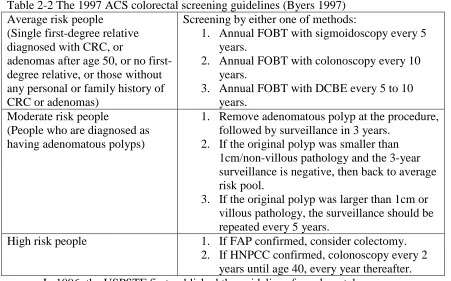
Table 2-2 The 1997 ACS colorectal screening guidelines (Byers 1997) ... 27
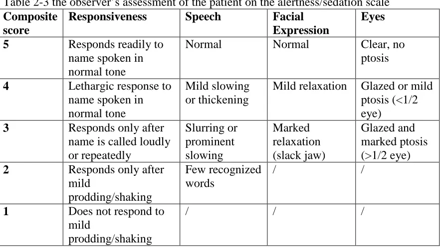
Table 2-3 the observer’s assessment of the patient on the alertness/sedation scale ... 58
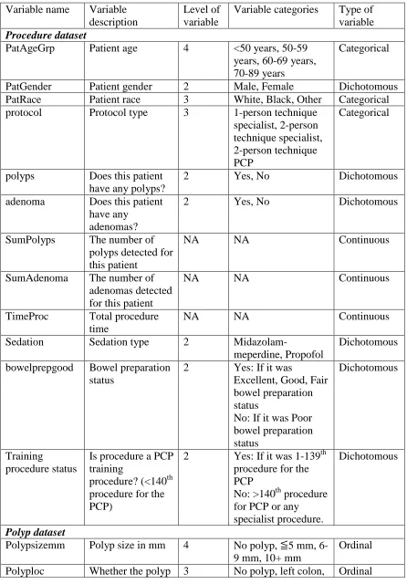
Table 3-1 variables of interest... 82
Table 3-2 Distribution of the number of polyps found per subject... 90
Table 3-3 Distribution of the number of polyps found per subject by protocol type ... 90
Table 3-4 Distribution of the number of polyps found per subject by sedation type ... 90
Table 3-5 Distribution of the number of adenomas found per subject ... 90
Table 3-6 distribution of the number of adenomas found per subject by protocol type ... 91
Table 3-7 Distribution of the number of adenomas found per subject by sedation type .. 91
Table 3-8 Distribution of study sample by key independent variables ... 91
Table 3-9 Breakdown of study sample by key independent variables with the number of polyps found... 92
Table 3-10 Breakdown of study sample by key independent variables with the number of adenomas found ... 93
Table 4-1: Demographic and procedure characteristics of the study sample ... 107
ix
Table 4-3: Adjusted estimates of colonoscopy performance and outcome quality
indicators by protocol type*** ... 112 Table 4-4: Adjusted estimates of colonoscopy performance and outcome quality
indicators by protocol type* ... 113 Table 4-5: Demographic and procedure characteristics of the study sample ... 126 Table 4-6: Indicators of colonoscopy quality by sedation type ... 128 Table 4-7: Adjusted estimates of colonoscopy performance and outcome quality
indicators by sedation type* ... 130 Table 4-8: Adjusted estimates of colonoscopy performance and outcome quality
x
List of Figures
Figure 2-1 Colorectal cancer incidences in the past decade ... 17
Figure 2-2 The trend line of colorectal cancer incidence in the past decade ... 18
Figure 2-3 Colorectal cancer mortalities in the past decade ... 19
Figure 2-4 The trend line of colorectal cancer mortality in the past decade ... 20
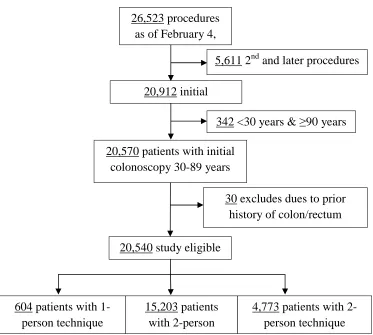
Figure 3-1 Sample Selection Flowchart ... 76
Figure 4-1: Study sample selection flowchart ... 104
1
Chapter 1 RESEARCH BACKGROUND AND SIGNIFICANCE
This chapter describes the background and the significance of the study topic.
1.1 Study Background
Colorectal cancer (CRC) is the third most prevalent cancer and second leading cause of cancer death in the U.S. (U.S. Cancer Statistics Working Group), with
almost140, 000 new cases and 55,000 deaths annually. This large number affected can be mostly prevented by screening tests both by removing pre-cancerous lesions and by early detection. In the past decade, age-adjusted CRC incidence decreased from 51.8 per 100,000 in 1999 to 44.7 per 100,000 in 2009; as well as age-adjusted CRC mortality, which decreased from 20.5 per 100,000 to 16.9 per 100,000 (SEER 1990-2010).
2
screenings until the age of 75. Currently, the USPSTF recommended screening tests and intervals are:
• Annual screening with high-sensitivity fecal occult blood testing
• Sigmoidoscopy every 5 years, with high-sensitivity fecal occult blood testing every 3 years
• Colonoscopy every 10 years
Colonoscopies have been recommended as the preferred screening method, including the American College of Obstetricians and Gynecologists, the American College of Gastroenterology (Rex 2000, Rex 2009) and the American Society for
Gastrointestinal Endoscopy (ASGE) (David 2006). Although colonoscopy every 10 years is considered to be the preferred screening method, it is imperfect because of variable quality of screening colonoscopy under community-based practice conditions.
As the use of colonoscopy screening increases, the need for measurement of colonoscopy quality is inevitable to ensure quality in the performance of colonoscopies. Generally, the cecal intubation rate is most commonly studied. However, this is a very limited measure of quality, and widespread poor quality of colonoscopy performance continues to limit the CRC prevention potential.
Adenoma detection and removal is the mechanism of conferring CRC prevention and is the main goal of colonoscopy, and should be a key indicator for assessing
3
primary care physicians (PCPs) with a total of 18,292 patients found an adenoma detection rate of 28.9%. The authors concluded that colonoscopy performance by PCPs can meet the professional, societies’ recommended standards (Wilkins 2009). The
association between colonoscope withdrawal times and adenoma detection rates was also studied. Barclay (2006) reported that physicians who had a mean withdrawal time less than 6 minutes (when no polyp was found) have an adenoma detection rate of 11.8%, while it was 28.3% for those with a mean withdrawal time of more than 6 minutes. This statistically significant difference persists in the next level of quality, the mean number of adenomas per subject, which were 0.17 vs. 0.61, respectively. Rex et al (2001)
videotaped 10 procedures performed by 2 colonoscopists and got them reviewed/scored by 4 experts based on four quality criteria related to colonoscopic withdrawal technique. They reported that technique does matter to the adenoma miss rate, which could be further associated with the potential cancer protection efficacy of colonoscopy screening. There is concern about colonoscopy quality because of the variable results and outcomes in terms of CRC prevention in the literature. Also, colonoscopy is a physician-dependent procedure. To make sure that physicians are doing a good job is more important than how many they have carried out.
4
1.2 Objectives
The aim of this study was to evaluate the quality of colonoscopies. Of the risk factors which impact colonoscopy performance, our variables of interest were the clinical procedure protocol type and the sedation type. We tested the impact of the protocol type and the sedation type on the polyp/adenoma detection rate, the mean number of
polyps/adenomas detected, polyp size, polyp location, and the procedure time.
We hypothesized that colonoscopy quality may be enhanced by applying the 2-person technique protocol relative to solo performance, and with deep sedation by propofol relative to the conventional Midazolam-meperidine combination.
The hypotheses tested are:
1. The screening colonoscopy quality of physicians using the 2-person technique yields more adenomas than with solo performers.
2. The screening colonoscopy quality of procedures with deep sedation by propofol yields more adenomas than with the conventional Midazolam-meperidine
combination.
1.3 Significance of the research and methodology
Although colonoscopy is considered to be the reference gold standard against which the sensitivity of other colorectal cancer screening tests is compared, it is not perfect. Most of the evidence about the sensitivity of a colonoscopy comes from experienced examiners conducting study colonoscopies in research settings without detailed documentation on the protocol followed.
hands-5
on 2-person technique, in which the endoscopy technician advances the colonoscope while the physician manipulates the scope tip for polyp search and removal, b) propofol sedation to substitute the conventional midazolam-meperidine (MM) combination
sedation starting in April 2006, and c) gradual insertion and withdrawal with polyp search and removal during both phases to maximize coverage of the colonic mucosal surface. The hands-on 2-person technique method avoids missing polyps due to physician's motor fatigue, confers the dexterity of two “right” hands for polyp search and removal, and ensures more persons (at least 3 persons, the third being the note taker ) watching the video screen for polyps (avoiding visual error). Of these elements, item (a) was not followed by experts and some specialists, which enables study of the contribution of the hands on 2-person technique. Item (b), propofol sedation was implemented from April 2006 onwards, enabling pre- and post-comparison to assess the role of propofol sedation. This research aims to contribute to the literature by:
1. Using clinical data for the analysis
2. Studying a state-of-the-art colonoscopy protocol, which has been applied for over 10 years
3. Identifying the effect of protocol elements on screening colonoscopy quality 4. Analyzing differences in adenoma detection rates by sedation type and number of
persons engaged in procedure performance.
1.4 Limitations
6
2. Due to the strict implementation of a uniform protocol at this center, the vast majority of the procedures were under state-of-the-art protocol. Only 2% of the procedures were performed using the conventional industry practice of a one-person technique, compared to 98% of procedures performed with the 2-person technique distributed across 57 physicians. Therefore the observed results may not generalize to all physicians using the 1-person technique.
1.5 Conclusions
This study finds that an innovation of a hands-on 2-person technique is highly associated with superior colonoscopy performance and lesion detection outcomes. By every sensitive measure, the results with the 2-person technique are superior and consistent across measures. Regarding sedation type, we find that while there is a suggestion of a positive association of propofol sedation with improved lesion detection and clearance as measured by sensitive indicators, the results did not attain statistical significance except in respect of one indicator, the advanced adenoma detection rate. Another important indicator for logistic reasons is the procedure time. Because propofol induces rapid and deep sedation, as anticipated the study showed a mean reduction in procedure time adjusted for all other variables that may impact procedure time. Our findings suggest that propofol sedation may contribute marginally to improved
7
8
Chapter 2 LITERATURE REVIEW
This chapter reviews the related colorectal cancer screening literature and makes the case for the significance of this study based on past research.
2.1 Overview of colorectal cancer in the United States
2.1.1 Incidence and mortality
Approximately 7.6 million people die of cancer each year. These deaths account for 13% of all deaths, and 64% occur in the developing countries. The burden of cancer is increasing both in the developed and developing countries due to the growth of
population, aging, and changes in lifestyle, especially for the cancer-associated behaviors, such as obesity, smoking, and adoption of Western-style diets. (Globocan (IARC) 2008, WHO 2008, Jemal 2011). Colorectal cancer (CRC) is one of the most common cancers and the leading cause of death in the U.S. since the late 1990s. The incidence of
9
steadily reduced except for a non-significant plateau during 1995-1998. In 2008, the incidence rate of CRC for men was 50.98 per 100,000 people and 39.64 per 100,000 people for women. The goal of Healthy People 2020 is to reduce CRC incidence to 38.6 per 100,000 people by 2020 (NCI 2012).
The significance of CRC is reflected both in the population affected and the rankings. Globally, in 2008, about 1.2 million new cases and 608,700 deaths occurred due to colorectal cancer (Globocan (IARC) 2008). It was found to be most prevalent in Oceania, Europe, and North America (Globocan (IARC) 2008). In 2010, an estimated 142,570 new CRC cases, 9.32% of all cancer new cases occurred in the United States. An estimated 51,370 people (9% of all cancer deaths) died from colorectal cancer (Jemal 2010, ACS 2010). The incidence and mortality rates of CRC rank as the 3rd most
frequent for both sexes (Jemal 2010, ACS 2010). The overall cost of cancer as estimated by the National Institutes of Health was $263.8 billion, 9% for CRC amounts to about $23.74 billion. Among these costs, $9.25 billion is for health expenditure, $1.88 billion is for lost productivity due to illness, $12.61 billion is for lost productivity due to premature death (ACS 2010).
10
early CRC diagnosis survive for 5 years. But it is predicated upon early detection. Furthermore CRC can be prevented. Reducing the colorectal cancer death rate is one of the objectives of Healthy People 2020, the target rate being 14.5 deaths per 100,000. In 2008, the worldwide age-standardized mortality was 8.2%, accounted for approximately 0.6 million individuals, and the age-standardized incidence was17.2%, accounted for 1.24 million persons (Globocan (IARC) 2008).
2.2 Colorectal cancer prevention and the role of polyps
While the role of hyperplastic polyps in colon cancer is debated, benign
adenomatous polyps have been documented to be the precursor for most cases of colon cancer, and polyps increase with age (Correa et al 1977, Rickert et al 1979). Detecting and removing adenomatous polyps is effective in reducing the incidence and mortality of CRC. Within polyps the proportion of villous architecture (showing rapid growth) is positively associated with the size of adenomatous polyps and, furthermore, the potential of having malignant characteristics (Rickert et al 1979). Evidence shows that
adenomatous polyps smaller than 10mm in diameter are rarely found to be cancer. The villous architecture component is more likely to be found in adenomatous polyps larger than 10mm (Enterline et al 1962, Morson 1974, Muto et al 1975, Spjut et al 1977).
11
invasive and in-situ CRC), followed by Hispanics (4.82% for invasive CRC and 5.08% for invasive and in-situ CRC), and highest in Asians/Pacific Islanders (5.58% for invasive CRC and 5.83% for invasive and in-situ CRC). The lifetime risk of dying from CRC is lowest in American Indians/Alaskan Natives at 1.86%, followed by Hispanics at 1.92%, and is highest in Blacks at 2.42% (Ries et al 2007). The pattern of lifetime risks between ethnics of being diagnosed with CRC mirrors the pattern of lifetime risk of dying from CRC.
12
rarely found to be cancer. The villous architecture component is more likely to be found in adenomatous polyps larger than 10mm (Enterline et al 1962, Morson 1974, Muto et al 1975, Spjut et al 1977).
According to Surveillance Epidemiology and End Results 17 (SEER-17), which captures cancer data in 17 metropolitan/rural regions (San Francisco, Connecticut, Detroit, Hawaii, Iowa, New Mexico, Seattle, Utah, Atlanta, San Jose-Monterey, Los Angeles, Alaska Native Registry, Rural Georgia, California excluding SF/SJM/LA, Kentucky, Louisiana and New Jersey) 2002-2004, the lifetime risk of being diagnosed with CRC is lowest in American Indians/Alaskan Natives (4.27% for invasive CRC and 4.45% for invasive and in-situ CRC), followed by Hispanics (4.82% for invasive CRC and 5.08% for invasive and in-situ CRC), and highest in Asians/Pacific Islanders (5.58% for invasive CRC and 5.83% for invasive and in-situ CRC). The lifetime risk of dying from CRC is lowest in American Indians/Alaskan Natives at 1.86%, followed by Hispanics at 1.92%, and is highest in Blacks at 2.42% (Ries et al 2007). The pattern of lifetime risks between ethnics of being diagnosed with CRC mirrors the pattern of lifetime risk of dying from CRC.
13
pedunculated adenomas had an increasing prevalence in the distal colon and were most prevalent in the sigmoid colon (Williams et al 1982) While the role of hyperplastic polyps in colon cancer is debated, benign adenomatous polyps have been documented to be the precursor for most cases of colon cancer, and polyps increase with age (Correa et al 1977, Rickert et al 1979). Detecting and removing adenomatous polyps is effective in reducing the incidence and mortality of CRC. Within polyps the proportion of villous architecture (showing rapid growth) is positively associated with the size of adenomatous polyps and, furthermore, the potential of having malignant characteristics (Rickert et al 1979). Evidence shows that adenomatous polyps smaller than 10mm in diameter are rarely found to be cancer. The villous architecture component is more likely to be found in adenomatous polyps larger than 10mm (Enterline et al 1962, Morson 1974, Muto et al 1975, Spjut et al 1977).
14
ethnics of being diagnosed with CRC mirrors the pattern of lifetime risk of dying from CRC.
Polyps in the colon are associated with different histology types. Some types of polyps are more likely to be found in the left colon while others were not. Hyperplastic polyps are most commonly found in the rectum – 86.1% of total polyps. Neoplastic adenomas are the second most common, and both increase with age and are more prevalent in men (Williams et al 1982). Although there is no consistent relationship between the size of adenomas and age, as age increases, adenomas larger than 1 cm in diameter are more prevalent. Sessile adenomas are more prevalent in the cecum, while pedunculated adenomas had an increasing prevalence in the distal colon and were most prevalent in the sigmoid colon (Williams et al 1982) While the role of hyperplastic polyps in colon cancer is debated, benign adenomatous polyps have been documented to be the precursor for most cases of colon cancer, and polyps increase with age (Correa et al 1977, Rickert et al 1979). Detecting and removing adenomatous polyps is effective in reducing the incidence and mortality of CRC. Within polyps the proportion of villous architecture (showing rapid growth) is positively associated with the size of adenomatous polyps and, furthermore, the potential of having malignant characteristics (Rickert et al 1979). Evidence shows that adenomatous polyps smaller than 10mm in diameter are rarely found to be cancer. The villous architecture component is more likely to be found in adenomatous polyps larger than 10mm (Enterline et al 1962, Morson 1974, Muto et al 1975, Spjut et al 1977).
15
Hawaii, Iowa, New Mexico, Seattle, Utah, Atlanta, San Jose-Monterey, Los Angeles, Alaska Native Registry, Rural Georgia, California excluding SF/SJM/LA, Kentucky, Louisiana and New Jersey) 2002-2004, the lifetime risk of being diagnosed with CRC is lowest in American Indians/Alaskan Natives (4.27% for invasive CRC and 4.45% for invasive and in-situ CRC), followed by Hispanics (4.82% for invasive CRC and 5.08% for invasive and in-situ CRC), and highest in Asians/Pacific Islanders (5.58% for invasive CRC and 5.83% for invasive and in-situ CRC). The lifetime risk of dying from CRC is lowest in American Indians/Alaskan Natives at 1.86%, followed by Hispanics at 1.92%, and is highest in Blacks at 2.42% (Ries et al 2007). The pattern of lifetime risks between ethnics of being diagnosed with CRC mirrors the pattern of lifetime risk of dying from CRC.
Polyps in the colon are associated with different histology types. Some types of polyps are more likely to be found in the left colon while others were not. Hyperplastic polyps are most commonly found in the rectum – 86.1% of total polyps. Neoplastic adenomas are the second most common, and both increase with age and are more prevalent in men (Williams et al 1982). Although there is no consistent relationship between the size of adenomas and age, as age increases, adenomas larger than 1 cm in diameter are more prevalent. Sessile adenomas are more prevalent in the cecum, while pedunculated adenomas had an increasing prevalence in the distal colon and were most prevalent in the sigmoid colon (Williams et al 1982).
2.3 Identifying at-risk population
16
17
Cancer Incidence: Full (Research) File
For South Carolina Residents
County: All Counties in South Carolina
Primary Cancer Sites: Colorectal (colon, rectum, and rectosigmoid) Age
35-39 40-44 45-49 50-54 55-59 60-64 65-69 70-74 75-79 80-84 85+ Year Rates Rates Rates Rates Rates Rates Rates Rates Rates Rates Rates
1996 10.5 19.1 36.1 69.6 100.7 156.2 190.8 275.5 316.5 366.7 429.6
1997 10.7 17.6 39.2 62.4 109.3 161.5 187.3 280.2 334.4 320.3 437.9
1998 13.8 18.2 38.7 64.4 110.6 161.9 214.0 302.2 344.0 418.1 402.5
1999 9.2 18.5 35.8 72.9 108.2 154.2 233.0 279.1 342.2 336.2 407.0
2000 8.6 21.8 45.6 65.2 108.1 161.2 210.6 274.9 321.3 414.1 402.8
2001 10.4 24.1 44.5 63.4 101.7 157.6 199.4 285.2 318.8 387.5 383.2
2002 11.3 23.5 45.1 70.9 105.4 161.2 233.1 283.1 326.6 362.5 364.0
2003 13.2 23.5 38.8 73.6 117.8 156.8 245.2 291.8 302.0 335.6 359.6
2004 16.2 27.3 34.5 79.2 110.1 138.0 216.7 259.4 323.1 346.1 370.4
2005 10.0 15.4 32.5 75.1 107.7 143.6 200.8 243.3 281.1 346.6 328.3
2006 14.3 21.2 41.9 66.0 91.0 131.1 195.3 221.9 259.8 280.2 256.0
2007 10.7 17.6 39.4 77.4 92.4 124.0 179.6 201.2 256.4 244.7 291.2
2008 8.9 20.1 39.4 68.5 84.4 115.1 159.0 186.4 228.8 260.2 277.4
2009 8.4 16.9 40.7 59.1 77.1 108.1 139.3 166.9 206.0 216.8 244.0 *Rate: Crude Rate calculated per 100,000 population
Figure 2-2 The trend line of colorectal cancer incidence in the past decade
18
19
Cancer Mortality
For South Carolina Residents
County: All Counties in South Carolina
Primary Cancer Sites: Colorectal (colon, rectum, and rectosigmoid) Age
35-39 40-44 45-49 50-54 55-59 60-64 65-69 70-74 75-79 80-84 85+
Year Rates Rates Rates Rates Rates Rates Rates Rates Rates Rates Rates
1996 # 6.6 10.9 14.5 34.2 42.9 66.4 106.8 108.8 182.4 270.9
1997 # 5.1 12.0 18.9 33.2 40.7 63.8 91.2 123.0 157.6 246.7
1998 # 5.3 8.0 19.2 30.6 37.8 58.1 87.0 124.9 178.0 230.3
1999 # 6.8 10.3 21.4 30.1 49.6 67.6 80.4 123.1 166.5 252.8
2000 # # 10.4 15.8 37.0 41.9 61.7 101.8 121.5 185.2 268.6
2001 # 8.6 11.5 16.9 23.8 39.5 75.7 79.3 129.9 153.5 229.2
2002 # 7.9 12.9 18.5 22.8 54.7 57.6 79.5 112.7 149.3 228.9
2003 # 7.3 13.4 14.7 26.7 46.8 57.5 84.4 102.6 161.6 222.7
2004 # 6.3 6.8 18.2 27.8 32.6 51.8 67.0 106.1 136.4 194.9
2005 # 5.7 12.7 15.1 26.1 37.5 59.2 60.6 106.5 145.3 231.0
2006 # 4.7 10.0 17.1 30.8 45.1 65.9 74.0 102.1 134.0 158.9
2007 # # 8.7 13.7 25.3 44.3 50.7 67.8 113.4 84.4 172.4
2008 # 5.5 11.7 18.2 24.6 27.9 49.6 78.1 82.4 110.2 195.0
2009 # # 10.9 17.9 26.2 35.4 40.4 58.8 81.0 118.3 167.0 *Rate: Crude Rate calculated per 100,000 population
Figure 2-4 The trend line of colorectal cancer mortality in the past decade
The U.S. Preventive Services Task Force (USPSTF), the American Cancer Society (ACS), the National Comprehensive Cancer Network (NCCN), and the U.S. Multi-Society Task Force (MSTF) recommend periodic screenings of the average
population to prevent CRC, which includes men and women aged over 50 years with no history of adenomas, colorectal cancer, inflammatory bowel disease, and family history (USPSTF 2008, Levin 2008, NCCN 2012). The USPSTF recommends routine CRC screenings with fecal occult blood test (FOBT), sigmoidoscopy, or colonoscopy for adults aged 50-75 years. Between 76 and 85 years old, routine screening is not recommended, but it can be provided a
considerations. For patients older than 85 years, CRC screening is not recommended (USPSTF, 2008).
2.3.2 High-risk persons
20
The trend line of colorectal cancer mortality in the past decade
The U.S. Preventive Services Task Force (USPSTF), the American Cancer Society (ACS), the National Comprehensive Cancer Network (NCCN), and the U.S.
ety Task Force (MSTF) recommend periodic screenings of the average
population to prevent CRC, which includes men and women aged over 50 years with no history of adenomas, colorectal cancer, inflammatory bowel disease, and family history
Levin 2008, NCCN 2012). The USPSTF recommends routine CRC screenings with fecal occult blood test (FOBT), sigmoidoscopy, or colonoscopy for
75 years. Between 76 and 85 years old, routine screening is not recommended, but it can be provided as required based on specific clinical
considerations. For patients older than 85 years, CRC screening is not recommended The U.S. Preventive Services Task Force (USPSTF), the American Cancer Society (ACS), the National Comprehensive Cancer Network (NCCN), and the U.S.
ety Task Force (MSTF) recommend periodic screenings of the average-risk population to prevent CRC, which includes men and women aged over 50 years with no history of adenomas, colorectal cancer, inflammatory bowel disease, and family history
Levin 2008, NCCN 2012). The USPSTF recommends routine CRC screenings with fecal occult blood test (FOBT), sigmoidoscopy, or colonoscopy for
75 years. Between 76 and 85 years old, routine screening is not
21
Asymptomatic individuals with a family history are categorized as high-risk for colorectal cancers. This group of persons is recommended to have screening at an earlier age than the average-risk group. The American College of Gastroenterology (ACG), the ACS and the NCCN have updated standards of the high-risk population and the
recommended screening guidelines (Levin 2008, Rex 2009, NCCN 2012):
Persons with single first-degree relative with CRC or advanced adenoma
diagnosed at age < 60 years or two first-degree relatives with colorectal cancer
or advanced adenomas will be considered as high risk population, and are
recommended to have colonoscopy screening every 5 years beginning at the age
of 40 years, or 10 years younger than the age at diagnosis of the youngest
affected relative. Or, with personal history of adenoma/sessile serrated polyps,
colon cancer, or inflammatory bowel disease (i.e., ulcerative colitis, Crohn’s
disease) additional screenings are recommended.
Other persons defined as high-risk have one of two hereditary syndromes (Byers 1997):
• Familial Adenomatous Polyposis syndromes (FAP)
People with this condition develop hundreds of colorectal polyps and will almost
certainly develop colorectal cancer unless the colon is removed.
• Hereditary Non-Polyposis Colorectal Cancer syndromes (HNPCC)
HNPCC has been classically defined as colorectal cancer in three or more family
members, two of whom are first-degree relatives of the third, involving people in at
least two generations, and with at least one person diagnosed with colorectal cancer
22
HNPCC, also known as the Lynch syndrome, accounts for approximately 5% of new cases of CRC each year (Winawar 1997).
Persons with a family history of hereditary syndromes, with relatives who received diagnoses of colorectal cancers at an early age, with two or more affected
relatives, or with persistent ulcerative colitis have a high risk of colon cancers. The risk is especially high among younger persons (40-59 years old), but not associated with
individuals after 60 (Fuchs et al 1994). Other principal risk factors include a history of colorectal cancers or adenomas in a first-degree relative, a personal history of large adenomatous polyps or colorectal cancers, and a prior diagnosis of endometrial, ovarian, or breast cancers (Rustogi 1994, USPSTF (Baltimore), 1996). Based on an ACS report in 1981, the high-risk population was described as (Eddy, 1981):
“Persons with familial polyposis, Gardner’s syndrome, ulcerative colitis, a
history of polyps or prior colon cancer, and a family history of cancer of the
colon or rectum”.
2.4 Early CRC detection and management
CRC is highly curable if detected in an early stage through routine screenings of the colon/rectum. When polyps/adenomas are detected and removed in the early
developmental course, the 5-year relative survival rate is 90% (CDC 2011). Among the screening methods, the effectiveness of the screening tool for
23
by flexible sigmoidoscopy, followed by fecal tests, Hemoccult SENSA being the best followed by fecal immunochemical test, and lowest for Hemoccult II (USPSTF 2008). Specificity of the screening tool is defined as the ability to identify true negatives among all negatives. In case of CRC, specificity is the percentage of people with no neoplastic lesions who are correctly identified as clear. Specificity is highest for colonoscopy as well, followed by flexible sigmoidoscopy, then Hemoccult II, which is also
approximately equal to fecal immunochemical test, and lowest for Hemoccult SENSA (USPSTF 2008).
One randomized clinical trial study in 1993 exploring the effect of FOBT screenings on CRC mortality for up to 13 years of follow-up reported that the annual FOBT group had the highest 13-year survival rate, with a 33% reduced mortality from colon cancers compared to the control group, and almost double the reduction observed in the biennial FOBT group (Mandel et al 1993). Although observational studies have reported incidence/mortality reductions associated with screening colonoscopy and polypectomy (Winawar et al 1993, Zauber et al 2012), less than half of the US screening-eligible population is covered by screening (Meissner et al 2006, Seeff et al 2004). Research has shown the effect of early detection and the removal of precancerous lesions through screening on CRC incidence and mortality reduction in the United States
(Edwards et al 2010, Center et al 2009, Chu et al 1994).
24
screening take up rate remains low and there is still a big gap to fill given that the target rate in the Healthy People 2020 is 70.5%.
Except for patients with bowel symptoms, the physician recommendation/referral is a required precondition for CRC screenings. Although Medicare coverage of
colonoscopies since 2001 reduced the racial disparity in colonoscopy screenings between older Whites and Blacks (Shih et al 2006), studies suggest that physician
recommendations are less frequent for Blacks than for Whites both in the general
population, and among Medicare beneficiaries (Klabunde et al 2006), which translates to a lower screening rate among Blacks. The low take up rate and the disparity in the access of screening could be a potential reason for Blacks having a relatively high CRC
incidence (Rex 2004, Daguise et al 2006).
2.5 History of CRC screening in the US
Before 1980, the American Cancer Society (ACS) recommended that people aged over 40 years should be screened with the annual sigmoidoscopy. The digital proctoscope and occult blood examinations were urged to be included in the regular health checkups for adults over age 40 by the ACS (Eddy 1980). Based on an ACS report in 1981, those at high risk were recommended to have more “frequent” and “intensive” examinations starting at an earlier age (Eddy, 1981). In a June 1992 meeting, revisions were made by the National Board of Directors of the American Cancer Society to the guidelines for asymptomatic individuals (Levin et al 1992):
25
2. “Fecal Occult Blood Test (FOBT)” substituted for “stool guaiac slide test” every year for individuals age 50 and over.
The ACS also made a revision to recommendations for the high-risk population at this meeting in June 1992 (Table 2-1) (Levin & Murphy 1992):
1. If first-degree relatives have a CRC diagnosis at an age less than 55 years, a
colonoscopy or a double-contrast barium enema (DCBE) was recommended every 5 years starting at age 35 – 40 years.
2. If family members have a history of familial adenomatous polyposis, early flexible sigmoidoscopy is required.
26
Table 2-1 American Cancer Society Recommendations for the Early Detection of Colon Cancer in Asymptomatic Persons (1992)
Test or Procedure Population
Sex Age Frequency Sigmoidoscopy, Perferably Flexible Male & Female 50 and over Every 3 to 5 years
Fecal Occult Blood Test Male & Female 50 and over Every year Digital Rectal Examination Male & Female 40 and over Every year
Source: Levin B, Murphy GP. Revision in American Cancer Society Recommendations for the Early Detection of Colorectal Cancer. CA Cancer J Clin. 1992; 42(5): 296-9.
27
Table 2-2 The 1997 ACS colorectal screening guidelines (Byers 1997) Average risk people
(Single first-degree relative diagnosed with CRC, or
adenomas after age 50, or no first-degree relative, or those without any personal or family history of CRC or adenomas)
Screening by either one of methods:
1. Annual FOBT with sigmoidoscopy every 5 years.
2. Annual FOBT with colonoscopy every 10 years.
3. Annual FOBT with DCBE every 5 to 10 years.
Moderate risk people
(People who are diagnosed as having adenomatous polyps)
1. Remove adenomatous polyp at the procedure, followed by surveillance in 3 years.
2. If the original polyp was smaller than 1cm/non-villous pathology and the 3-year surveillance is negative, then back to average risk pool.
3. If the original polyp was larger than 1cm or villous pathology, the surveillance should be repeated every 5 years.
High risk people 1. If FAP confirmed, consider colectomy. 2. If HNPCC confirmed, colonoscopy every 2
28
sigmoidoscopy, or colonoscopy, beginning at age 50 years and continuing until age 75 years (USPSTF 2008). In contrast to the 2002 USPSTF recommendation statements, the USPSTF updated the recommended screening eligibility from “individuals age 50 years and older” to “50 years and continuing to 75 years.” Also, high-sensitivity FOBTs,
sigmoidoscopies with interval FOBTs, or colonoscopies were recommended replacing the un-prioritized recommendations in 2002. For CT colongraphy and fecal DNA, the
USPSTF concluded to maintain that there is insufficient evidence for recommendation (USPSTF 2008).
2.6 CRC screening methods
Screening tests for CRC prevention basically are categorized into 2 types, the fecal tests (such as the Fecal Occult Blood Test (FOBT)), and the full or partial structure tests, such as Digital Rectal Examination (DRE), sigmoidoscopy, barium enema,
Computed Tomographic (CT) colonography, and colonoscopy.
29
the 3 methods is considered effective in detecting advanced adenomatous polyps and cancers at an early stage (USPSTF 2008).
The three CRC screening methods commonly used are – fecal occult blood test (FOBT), flexible sigmoidoscopy, and colonoscopy. All tests lead to colonoscopies if they are positive, which permits the visual detection of early stage cancers and removal of adenomatous polyps simultaneously during the procedure (Denis et al 2011).
2.6.1 Fecal occult blood test (FOBT) / Stool blood test
Stool blood tests are broadly known as guaiac fecal occult blood test (gFOBT) because the tests are designed to detect the occult blood in stool through guaiac method. CRC can be detected by finding occult blood in the stool, which is not readily visible. Also, the bleeding caused by cancers or advanced adenomatous polyps depends on the lesion size, friability, and location, and blood may might not be detected by the naked eye. But the problem with the stool blood test is its accuracy, because blood is unevenly distributed in the stool and the bleeding is intermittent. Further there are substances other than hemoglobin that can produce false positive results, such as iron in the diet (Eddy 1981). CRC screening cannot rely solely on FOBT results, but can use FOBT results as a preliminary test.
30
Positive reactions on guaiac-impregnated cards, the most common form of testing, signal the presence of bleeding from premalignant adenomas and early-stage colorectal cancers (USPSTF (Baltimore) 1996).The stool blood test is far less likely to help prevent cancers compared to invasive tests such as flexible sigmoidoscopy or colonoscopy. To be effective, FOBT must be repeated at a regular interval, otherwise the protection is nil. When the test is positive, an invasive test, such as a colonoscopy, is needed (Levin et al 2008). If patients are not willing to repeat the FOBT or take the invasive test when the FOBT is abnormal, FOBT is ineffective and should not be recommended (Levin et al 2008). FOBT has very low test sensitivity (especially a single test) for detecting adenomas, and has a reasonable sensitivity for detecting colorectal cancers. However, regarding the program sensitivity (serial tests over time in a program), it is relatively high. Therefore, repeating testing each year is a very important key for ensuring the quality of FOBT (Levin et al 2008). In a randomized clinical trial of FOBT screening with a 13-year follow-up period, the results showed 33% reduction in colorectal cancer mortality, and better 5-year survival for those with annual FOBT screening (Mandel et al 1993).
31
Due to the issue of intermittent bleeding of polyps and cancers, Hemoccult II Slides are designed so that patients can collect serial specimens at home from bowel movements over three days. This increases the probability of detecting hidden blood from polyps and cancer. After the patient prepares the Hemoccult II SENSA elite test, it may be returned in person or by mail to the laboratory, hospital or medical office for testing and interpretation. The test consists of two main components: 1) the test cards containing guaiac paper, 2) The Developer, a developing solution containing a stabilized mixture of less than 4.2% hydrogen peroxide and 80% denatured ethyl alcohol and enhancer in an aqueous solution. Research has shown that the rehydrated Hemocuult II slides show a high sensitivity in detecting CRC (92.2%) but disappointingly low specificity (90.4%), which causes too many colonoscopy referrals and produces high corresponding costs (Byers 1997, Mandel 1993).
A relatively new method for the fecal test is the fecal immunochemical test (FIT). FITs are highly specific in detecting human blood and in eliminating the dietary
restrictions. Research on the sensitivity and specificity of detecting the advanced adenomas, however, is disappointing. Allison’s (2007) study of three Northern Kaiser Permanente Medical Centers between April 1997 and October 1999 revealed that the sensitivity and specificity for advanced neoplasms in the left colon were only followed by colonoscopies within 2 years after the FOBT screening (29.5% for sensitivity and 97.3% for specificity).
2.6.2 Digital rectal examination (DRE)
32
rectal mucosa, which is 11cm in length, where about one-sixth of the colon cancers occur (Eddy 1981, USPSTF (Baltimore) 1996).
A digital (finger) rectal examination is used to check for problems with organs or other structures in the pelvis and the lower belly. During the examination, the doctor gently puts a lubricated, gloved finger of one hand into the rectum. The doctor may use the other hand to press on the lower belly or pelvic area (Healthwise 2010). The results for DRE can be normal, where there are no problems such as organ enlargements or growths are felt, and vice versa. Growths such as hemorrhoids, polyps, tumors, or abscesses may be found in the lower rectum. Breaks in the skin around the anus (anal fissures) may be found; problems of the bladder may also be felt (Healthwise 2010). The DRE alone is not effective to check for colorectal cancers. If problems are found during a DRE, more advanced tests are needed, such as a sigmoidoscopy or a colonoscopy
(Healthwise 2010).
There are no formal studies/reports for the effectiveness of DRE since the costs are small and its high safety feature, any benefits can be considered as worthwhile, the only potential harm is it might produce a false-positive results leading to other structure tests, such as sigmoidoscopies (Eddy 1980). Also, the DRE is not as effective as
sigmoidoscopy because neoplasms can be hidden by mucus or any residual substances even within the area reached by exploring fingers (Eddy 1981).
2.6.3 Sigmoidoscopy
33
(Baltimore) 1996). The effectiveness of risk reduction for sigmoidoscopy has been widely documented (Lang 1994, Selby 1992, Mewcomb 1992).
i. Rigid sigmoidoscopy
The 25-cm rigid sigmoidoscopy was introduced in the 19th century but no longer used since 1982 (Eddy 1980, Mandel et al 1993). Theoretically, it can cover up to 25 cm of the colon, where about one-half to two-thirds of cancers and adenomatous polyps grow (Eddy 1981). There are pros and cons of rigid sigmoidoscopes. It allows direct
visualization of the colon, biopsy, and removal of the suspicious lesions. Patient discomfort and the risk of perforation of the colon are problems to be considered,
however (Eddy 1981). Due to patient discomfort and the development of new technology, the rigid sigmoidoscopy is no longer used. A retrospective study in 1992 on the
effectiveness of rigid sigmoidoscopy showed that participants who had undergone one or more rigid sigmoidoscopy examinations within the past 10 years had only 30% of the risk of dying from distal colon or rectum cancers relative to those who did not (Selby 1992).
ii. Flexible sigmoidoscopy
34
flexible sigmoidoscopy of adults aged 55-64 years reduced the incidence of colorectal cancer by 33%, and mortality by 43% (Atkin et al 2010).
Sigmoidoscopies can also produce false-positive results, primarily from polyps detected that are unlikely to become malignant during the patients’ lifetime (USPSTF (Baltimore) 1996). It turns out that the majority of asymptomatic persons with colonic polyps discovered by routine sigmoidoscopic examinations will not develop into
clinically significant malignancies during their lifetime. For these patients, interventions typically followed (i.e., biopsies, polypectomies, and frequent colonoscopies). Costly procedures, anxiety provoking, and potential harms are unlikely to make up for the clinical benefits of sigmoidoscopies (USPSTF(Baltimore), 1996). Flexible
sigmoidoscopy was considered too expensive and specialized only for early detection usage (Eddy 1980).
Another disadvantage of sigmoidoscopy is the distribution of cancers in the colon. Studies indicated that the proximal colon cancer accounts for a significant portion of the colorectal cancers. The contribution of incident colon cancer in the proximal colon beyond the examination zone of sigmoidscopy is about 27% to 45% (Dinning 1994, Castiglione 1995, Lemmel 1996). The ratio of proximal cancer to total CRC is 0.338 among men and 0.421 among women (SEER CanQues 1973-2009).
35
aging population (65 years and older), the proportion of the population at risk of CRC is expanding with aging of the US population (Rabeneck 2003).
2.6.4 Barium enema
Barium enema is one variant of X-ray examination, and is of two types, single contrast barium enema (SCBE) and double contrast barium enema (DCBE).
Barium enema examinations have been shown to better identify most of the advanced, most likely incurable, carcinomas of the colon instead of the early, potentially curable carcinomas of the colon. Therefore, the routine use of barium enema
examinations is not reliable and is recommended for discontinuation in the diagnostic evaluation of carcinomas of the colon (Gilbertsen et al 1979).
i. Double Contrast Barium Enema (DCBE)
During this procedure, the physician inserts a tube into the rectum, fills in barium sulfate, and drains its out, leaving a thin layer of barium on the wall of colon, and then air is filled in to define the outline of the colon. Then X-ray images from various angles are taken to better view the whole colon and detect abnormal growths (Byers 1997). A positive result in DCBE should be followed by a colonoscopy or a sigmoidoscopy (Byers 1997).
ii. Single Contrast Barium Enema (SCBE)
The difference between SCBE and DCBE is that the former fills the barium in the colon to outline the colon for detecting abnormal growths. The procedure time for SCBE is shorter than DCBE. SCBE is mostly performed for specific medical reasons or for older people who may not be able to tolerate the more time-consuming and
36
The performance of barium enema varies by section of colon, and highest in the straight portions – descending colon (93%), transverse colon (89%), and ascending colon (88%); followed by the askew portions – the splenic flexure (86%), the hepatic flexure and the sigmoid flexure (83%). It is lowest in the globular portions – the rectum (77%) and the cecum (75%). The average sensitivity for cancer is 83%. In addition, the DCBE has better sensitivity than SCBE, with 0.78 being odds ratio of missed cancers (Rex, Rahmani, Haseman 1997).
Overall, barium enemas are less expensive than colonoscopies. But colonoscopies enable direct visualization of the colon and removal of the suspicious lesions directly for biopsy with a one-time procedure if the bowel prepared properly. Colonoscopies have a higher sensitivity and are least likely to miss cancers, with an odds ratio of 0.25 relative to barium enema (Byers 1997, Rex, Rahmani, Haseman, 1997).
2.6.5 CT colonography
37
the segment unblinded method, which declares the findings of each section of the colon after it is examined by colonoscopy. They reported a sensitivity of 93.8% and 96% specificity on polyps of 10 or more millimeters; however, when polyps more than 6 millimeters were included the sensitivity reduced to 88.7%, and specificity to 79.6%. Macari and colleagues (2005) using 2-dimensional views also reported the sensitivity of CT colongraphy reduced significantly with polyp size (>10 mm: 100%, 6-9 mm: 52.9%, 1-5 mm: 11.5%).
Besides the effectiveness of CT colonography in identifying a polyp, a study in 2008 of 2,531 asymptomatic individuals from 15 facilities using per-patient analysis showed that the sensitivity for polyps of 10 millimeters was 90% and 86% for specificity, which is consistent with the previous literature. However, the positive predictive value of CT colonography is 23% (with a high negative predictive value of 99%), which means the accuracy of diagnosis is extremely low, and the ability of radiologists identifying even the large polyps (>10mm) is an issue (Johnson 2008).
38
method for the prevention of CRC. Also, the use of CT colonography has not been recommended by USPSTF due to the insufficient evidence on its benefits and harms (USPSTF 2008).
2.6.6 Colonoscopy
Evidence has shown that the rate of colonoscopy screening is increasing concurrent with a decline in the uptake of other screening tests. In the 2005 National Health Interview Survey (NHIS) study, the colonoscopy take up rate exceeded FOBT and sigmoidoscopy (Meissner et al 2006). This study also concluded that a large proportion of the take-up rate increment since 2000 was due to the increase in colonoscopy. The
American College of Gastroenterology (2000) recommendation that colonoscopy was the preferred colorectal cancer screening test for average-risk individuals could have been the reason for the large increase in colonoscopy rate (Rex 2000).
Colonoscopy, which requires sedation and often involves the use of a hospital or surgery center suite, is more expensive than other screening tests and has a higher risk of sedation and procedural complications (USPSTF (Baltimore 1996)). Retrospective studies have reported the effectiveness of colonoscopy is superior to sigmoidoscopy because approximately 60% to 70% of proximal cancers are not accompanied by
neoplasms in the distal colon, which is the examination zone of sigmiodoscopy (Dinning 1994, Castiglione 1995, Lemmel 1996). Therefore a large proportion of test-negatives with sigmoidoscopy are not CRC-free. Proximal cancers account for 40% of the colon cancers. These cancers remain undetected despite preventive screening using
39
Research has shown that individuals with any distal adenomatous polyps were more likely to have advanced proximal neoplasia, the risk of advanced proximal neoplasia is significantly enhanced by increasing severity of distal adenomatous polyps (compared to patients without distal polyps). The relative risk of harboring distal hyperplastic polyps, distal tubular adenomas, and advanced distal polyps were 2.6, 4.0, and 6.7, respectively. Colonoscopy is irreplaceable among individuals without any distal adenomas, because 2.7% had advanced proximal neoplasias (Lieberman 2000; Imperial 2000).
The effectiveness of colonoscopy in reducing CRC incidence and mortality depends on how thoroughly visualization of the entire colon is achieved, the diligence in examining the mucosa, and patient acceptance of the procedure (Denis et al 2011).
Colonoscopy has an advantage over barium enemas because it can be both diagnostic and therapeutic. Its advantage over flexible sigmoidoscopy is that it can access the entire colon while flexible sigmoidoscopy can only access the distal colon (Robertson et al 2006). The use of colonoscopy in colorectal cancer screening has increased concurrent with the decline in barium enema and flexible sigmoidoscopy (Robertson 2006; Klbunde 2005).
40
because it enables both detection and removal of suspicious lesions in on step. Even with the price issue, screening with every 10 years makes it an attractive screening tool.
2.7 Quality of colonoscopy / effectiveness
Colonoscopy is currently regarded as the gold standard of colorectal cancer screening by removing polyps/adenomas (colonoscopic polypectomy) because there is strong evidence that polyps/adenomas are the precursor of colorectal cancer (Enterline et al 1967, Grinnell et al 1958, Kalus et al 1972, Muto et al, 1975). The effectiveness of colonoscopy screening in preventing the development of colorectal cancer depends on the quality of examination. A successful colonoscopy can be identified by the rate of
polyps/adenomas detected and the cecum intubated by the performer’s series of procedures. Achieving 95% cecal intubation rate is a recommended benchmark
(USPSTF). Missed polyp/adenoma rates are documented at 6% to 27% depending on the size of polyps/adenomas (Hixson et al 1990, Rex et al 1997, Leaper et al 2004). Evidence regarding the effectiveness of colonoscopy has caused other screening methods to
decline, and resulted in it being considered as the preferred method of CRC screening (Rex 2000, Rex 2009, Davila 2006, NCCN Clinical practice guidelines in oncology 2011).
With increasing use of colonoscopy, measuring colonoscopy quality is becoming inevitable to ensure its competent performance. Generally, the cecal intubation rate is the most commonly studied quality indicator, but is a very inadequate indicator. Poor quality colonoscopies limit its CRC protection potential.
41
quality. A wide range of ADR among endoscopists has been documented (Millan 2008, Barclay 2006, Wilkins 2009, Rex 2001). This implies that the quality of colonoscopies varies widely, which could severely undermine its efficacy in CRC protection. Another major issue is the specialist capacity to meet the demand for colonoscopy screening if it becomes the primary screening method. Some studies have explored the effectiveness of colonoscopies performed by trained PCPs.
A meta-analysis of 12 eligible studies of colonoscopies performed by PCPs with a total of 18,292 patients reported an adenoma detection rate of 28.9%. The authors
concluded that the performance of PCPs can meet the professional Societies’ recommended standards (Wilkins 2009). The association between colonoscope
withdrawal time and adenoma detection rate was also studied, Barclay (2006) indicated that physicians who had a mean withdrawal time of less than 6 minutes (among patients with no polyp removed) had an overall adenoma detection rate of 11.8%, while it was 28.3% for physicians whose mean withdrawal time was more than 6 minutes (without a polyp found). This statistically significant difference persists in the mean number of adenomas detected per subject, which were 0.17 vs. 0.61, respectively.
Rex et al (2001) videotaped 10 procedures performed by 2 colonoscopists and got them reviewed/scored by 4 experts based on four quality criteria related to colonoscopic withdrawal technique. They reported that technique does matter to the adenoma miss rate, which could be further associated with the potential cancer protection efficacy of
42
retrospective cohort study under an academic medical center setting reported a 100% of CRC protection over a mean follow-up of 5.34 years (Imperiale et al 2008). Zauber et al (2012) reported a 53% reduction in CRC mortality over a mean follow-up of 15.8 years per subject.
Owing to its potential of a high level of CRC prevention, high-quality
colonoscopy is recommended to be performed every 10 years for average risk population beginning at the age of 50 years, and 45 years for Blacks (Rex 2009). Colonoscopy every 5 years at the age of 40 years, or 10 years before the earliest age of CRC diagnosis in a first-degree relative (high risk population) is recommended.
2.7.1 Procedure quality indicators
The National Comprehensive Cancer Network (NCCN) guidelines for CRC screening indicate that to evaluate the quality of colonoscopy, the following indicators should be considered (NCCN 2011):
• Cecal intubation rate
• Withdrawal time
• Adenoma detection rate
• Appropriate intervals between endoscopic examinations based on family and personal history, and the number and histological type of polyps at last colonoscopy
• Minor and major complication rates
• Pre-procedure medical evaluations
43
The cecal intubation rate and withdrawal time can be signal indicators for the quality of colonoscopy screening. Cecum intubation is set as the standard for a completed colonoscopy since the cecum is considered as the beginning of the large bowel. Reaching the cecum implies that the colonoscope was inserted all the way through the colon and rectum starting from the anus. In addition, withdrawal time (an average of 6 minutes or more when no polyps are removed) can be the surrogate for the percentage of colonic mucosa inspected (Barclay et al 2006, Rex 2006).
i. Cecal intubation rate
Three types of cecal intubation rates are defined, the unadjusted rate, the MSTF-adjusted rate, and the circumstance-MSTF-adjusted rate. The unMSTF-adjusted rate measures the cecal intubation status for all study-eligible screenings. The MSTF-adjusted rate, according to the MSTF recommendations is calculated by excluding incomplete colonoscopies due to severe colitis or poor preparation (Rex 2002). The circumstance-adjusted rate further excludes procedures in which the endoscopist made a clinical decision not to attempt to reach the cecum because of severe diverticulosis, vital sign instability during the
procedure, obstruction or stricture, or because it was a therapeutic procedure without the goal of cecal intubation, such as colon decompression, treatment of active lower
gastrointestinal bleeding, removal of a previously discovered lesion, stent placement, etcetera (Aslinia 2006). The cecal intubation rate is the most commonly measured indicator and relatively easy to report as a preliminary quality indicator of colonoscopy (Aslinia 2006, Rex 2006, Rex 2002, Lieberman 2007).
44
Colonoscope insertion time and withdrawal time are considered as appropriate quality indicators (Lieberman 2007, Rex 2009), especially when the ADR is low (Rex 2006). Longer withdrawal time is shown to be associated with a better polyp detection rate (Rex 2000, Sanchez et al 2004). In contrast, shorter withdrawal time periods were associated with higher polyp miss rates (Rex 2000, Sanchez et al 2004). Also, the insertion time, which is the examination time period from insertion to the anus to the visualization of cecum, is documented to be associated with the adenoma detection rate (Benson et al 2005). In a series of 550 average-risk consecutive colonoscopy screenings performed by academic gastroenterologists, the ratio of insertion time to withdrawal time was found to be positively related to the adenoma detection rate (Benson et al 2005).
2.7.2 Outcome indicators
i. Polyps
A polyp is an overgrow tissue with part of its body attached to the site of origin. Studies suggest that about 1 in 4 colon cancers develop from polyps (Morson 1974, Muto et al 1975,Jass 1989), it takes averagely 10-15 years from a polyp to become a cancer by progression through the Stages of an adenoma (Morson 1974). Identifying and removing polyps during the screening exam has been considered the key to reduce the risk of developing colon cancers (Pabby 2005, Rex 2002, Rex 2006). When there are multiple polyps detected, they are most likely to be found in the ascending and transverse colon (Correa et al 1977).
ii. Adenomas
45
prevent the CRC (Rex 2006, Lieberman 2007, Rex 2009). A positive correlation is documented between withdrawal time and adenoma detection rate (Barclay 2006, Barclay 2008, Millan 2008). Barclay et al (2006) study reported wide variation in the mean number of adenomas per subject, in the adenoma detection rate, and in the mean withdrawal time. With a mean withdrawal time more than 6 minutes, the physician’s detection rates of adenoma in their patient panels are higher than physicians with less than 6 minutes by around two and half folds (11.8% vs. 28.3%). The mean number of adenomas detected per subject for physicians with a mean withdrawal time more than 6 minutes are nearly 4-fold larger than physicians with less than 6 minutes (0.17 vs. 0.61). In another study Barclay et al (2008) studied the association of a minimum of 8 minutes withdrawal time (mean for the endoscopist), and found that compared with endoscopists with a mean withdrawal time less than 8 minutes, those with at least 8 minutes have significantly higher rates of any neoplasia (37.8% vs 23.3%) and advanced neoplasms (6.6% vs 4.5%). More importantly, among the advanced neoplasms found by those with at least 8 minutes withdrawal time, 25% were 9 mm or less while for those with less than 8 minutes, only 10% were 9 mm or less. This indicates that the more gradual inspection is, a higher number of smaller, potentially deadly neoplasms are found, some of which are missed otherwise.
46
physicians with ADR less than 11% is 12.50, and about 11 for ADRs 11%-14.9% and 15.0–19.9% (Kaminski 2010).
2.8 Factors associated with CRC screening rates and adenoma rates
2.8.1 Patient factors
The factors associated with the uptake of CRC screening tests are widely
documented, and are similar to other prevention screenings, including race/ethnicity, age, education, income, having health insurance coverage, and having a usual source of care. One study evaluated the 1987, 1992, 1998, 2000, and 2003 National Health Interview Survey (NHIS) findings (Meissner 2006). Use of CRC screenings was higher among individuals with private health insurance, a usual source of care, and who were older, White, married, having higher annual household income, and higher education (Meissner 2006). The prevalence of adenomatous polyps was positively associated with age, but increasing age was not associated with an increase in polyp size (Rickert et al 1979, Hughes 1968). The incidence of colorectal cancer is higher in males than in females (Globocan (IARC) 2008, Jemal 2011, Meissner et al 2006). Nationally, the CRC
47
The CRC death rate remains highest among Blacks (Gargiullo et al 2002). This may due to lower screening rates in this group (Thornton et al 2007; Zimmerman et all 2006; James et al 2006; Vlahor et al 2005; Shokar et al 2007; Shokar et al 2008; Zhao et al 2006). At a population-level, reduction in CRC incidence rates are about 50% less among Blacks than among Whites (Gargiullo et al 2002). CRC incidence rates among Whites have been decreasing since 1985, and Blacks rates have remained relatively unchanged (Gargiulo et al 2002; Jemal et al 2005; Ries et al 2000). Further, Blacks are at higher risk of being diagnosed at an advanced or metastatic stage, where Whites are more likely to be diagnosed at a non-advanced or localized stage (Weir et al 2003; Daguise et al 2006).
Studies have demonstrated that Blacks suffered a higher proportion of adenomas in the proximal colon than Whites, which in other words, more proximal adenomas in Blacks that are missed by sigmoidoscopies (Thornton 2007, Johnson 1986, Ozick 1995, Mayberry 1995, Nelson 1997, Thomas 1992, Rex 2000). What makes worse is Blacks are documented to be less frequently screened by colonoscopy but sigmoidoscopy (Peterson 2008).
2.8.2 Physician factors in screening coverage
Although the effectiveness of colonoscopy screening in preventing CRC is documented, screening colonoscopy coverage in the US population remains low. This is partly due to the low endoscopy capacity due to a shortage of providers. Currently,
48
According to the American Board of Internal Medicine, there are only 13,968 certified gastroenterologists in the US, annually increasing by460 gastroenterologists (ABIM 2013). The colonoscopy capacity is only 63% (14.2 millions) of the estimated annual 22.4 million colonoscopies (Seeff 2004), and the increase of the aging population, including baby boomers, widens the gap. To cover 100% of all age-eligible
49
compared with non-gastroenterologists was the lack of information in the latters’ training in colonoscopy. Some of them were self-trained and others had variable (not
documented) levels of colonoscopy training.
Two Canadian studies reported that persons with colonoscopies performed by non-gastroenterologists had a higher risk of subsequent CRC compared to those performed by gastroenterologists. One retrospective cohort study of colonoscopies conducted in Canada during 1992-1997 with up to a 15-year follow-up of 110,000 patients reported higher risks of interval cancer after a negative colonoscopy when patients were provided the service in a hospital setting by non-gastroenterologists compared to gastroenterologists (40% higher for general surgeons and 30% higher for internists and family physicians), but no difference was found in physician office settings (Rabeneck 2010). Another study matched colonoscopies done in 1997-2002 with the Ontario Cancer Registry and also showed higher odds of CRC for non-gastroenterologists compared with gastroenterologists among both men (OR=1.77) and women (OR=1.85) (Bressler 2007).
In contrast, other research has shown that trained PCPs’ performance is
comparable or better than the current benchmarks of quality set by the US MultiSociety Task Force (USMSTF) for gastroenterologists, from both the patient safety and adenoma detection perspectives (Wilkins 2009, Xirasagar 2009). Pierzchajlo (1997) reported a 91.5% cecal intubation rate and 17.8% ADR among 751 colonoscopies performed by family physicians, which meets the USMSTF recommendation that ≥90% of
50
approaches the ADR standards of ≥15% for women and ≥25% for men (Rex, Bond, Winawer 2002).
Another series of 200 colonoscopies performed by family physicians documented highly competent performance by trained family physicians with 96.5% cecal intubation rate and 22.5% neoplastic polyp detection rate (Edwards 2004). A retrospective case review of 731 colonoscopies performed by two family physicians credentialed for sigmoidoscopy and initially supervised by their referral gastroenterologist reported that they had a ADR of 27.2% in men and 21.4% in women, though the cecal intubation rate was close but lower than the USMSTF standard (89.5%) in their starting phase (1996-1998) and improved to meet the USMSTF standard (94.6%) at a later phase (1999-2001) (Newman 2005).
51
unchanged. In comparison, black patients of black untrained PCPs showed a change over the same period from 10.4% to 38.7%, and the white patients from13.3% to 13.2% (Xirasagar 2011).
Comparing estimates of the number of gastroenterologists needed with the newly qualifying gastroenterologists each year, it will take approximately fifteen years to fill the colonoscopy capacity gap (Vijan 2004, ABIM 2013). Having primary care physicians perform screening colonoscopies may be a solution to cover the unscreened population, which is 60% of currently eligible population (Seeff 2004). However, the training of primary care physicians in colonoscopy remains very low. Only 4% of graduating family medicine residents applied for colonoscopy credentialing in 2002 even though about half of the residency programs offered colonoscopy training to family medicine residents, and only 18% of these programs had anyone registering and getting trained (Wilkins 2004). While research has documented that “trained” primary care physicians can provide competent and safe colonoscopy (Edwards 2004, Newman 2005, Wilkins 2009), there is no documentation of the training process or of the protocols used by high-performing PCPs. This study presents the protocol that was consistently used to train primary care physicians, which requires a 2-person technique, and includes other elements to maximize colon surface inspection and to minimize the likelihood of missing polyps.
2.8.3 Procedure protocol features in colonoscopy quality – 2-person technique