University of South Carolina
Scholar Commons
Theses and Dissertations
1-1-2013
Bioassay Guided Fractionation of American
Ginseng. A Hexane Fraction of American Ginseng
Suppresses Colitis and Associated Colon Cancer.
Mechanism of Action.
DEEPAK POUDYAL University of South Carolina
Follow this and additional works at:https://scholarcommons.sc.edu/etd Part of thePharmacy and Pharmaceutical Sciences Commons
This Open Access Dissertation is brought to you by Scholar Commons. It has been accepted for inclusion in Theses and Dissertations by an authorized administrator of Scholar Commons. For more information, please [email protected].
Recommended Citation
B
IOASSAYG
UIDEDF
RACTIONATIONO
FA
MERICANG
INSENG.
A
H
EXANEF
RACTIONO
FA
MERICANG
INSENGS
UPPRESSESC
OLITISA
NDA
SSOCIATEDC
OLONC
ANCER.
M
ECHANISMO
FA
CTION.
by
Deepak Poudyal
Bachelor of Science
North Dakota State University, 2008
Submitted in Partial Fulfillment of the Requirements
For the Degree of Doctor of Philosophy in
Pharmaceutical Science
South Carolina College of Pharmacy
University of South Carolina
2013
Accepted by:
Lorne J Hofseth, Major Professor
Theresa Smith, Committee Member
Campbell McInnes, Committee Member
Yuri Peterson, Committee Member
Taixing Cui, Committee Member
ii
D
EDICATIONiv
A
CKNOWLEDGEMENTSI would like to thank many people who have helped, supported and encouraged
me over the years without whom I would not have successfully accomplish my research
program and finish this dissertation.
Foremost, my mentor Dr. Hofseth is not only a conscientious advisor, but also has
become a good friend over the years. His knowledge and thoughtful insights in the
research project has been my source of inspiration that guided me for the successful
completion of the research program.
I am greatly thankful to Drs. Theresa Smith, Campbell McInnes, Yuri Peterson
and Taixing Cui for their insight, helpful instruction, criticism, assistance and suggestions
throughout the courses of work.
It has been a pleasure working with my colleagues, in particular, Anne Hofseth,
Dr. Yu Jin, Dr. Xiangli Cui, Dr. Alexander Chumanevich, Alena Chumanevich and Erin
Witalison; many of the ideas in my work originated in discussions with them.
Finally, I would also like to thank all the faculty, students and staff members in
my college, for who have offered invaluable help to me in the past four years. My thank
goes to Dr. Anthony Windust and Ms Phuong Mai Le for the collaborative support in the
project and also to Ms Tia Davis and Ms Diane Wise for their help. Moreover, I have
been fortunate to have many friends who always have generously supported me. I cannot
A
BSTRACTInflammatory Bowel Disease (IBD) [Ulcerative colitis (UC) and Crohn’s Disease
(CD)] is a group of chronic disorders of unknown etiology characterized by inflammation
in the gastrointestinal tract associated with a high colon cancer risk. UC is characterized
by periods of active disease (“flare-ups”), followed by periods when the disease is
inactive (“remission”). The end result is an abnormal immune response with repeated
episodes of colonic inflammation. Conventional treatment of UC by aminosalicylates,
TNFα inhibitors, and steroids have modest effects, and come with a high risk of side effects including dyspeptic symptoms, gastric ulceration and sometimes hospitalization
and deaths. Because of higher acceptance, efficacy, minimal side effects and relatively
low cost, patients (up to 50%) are using complementary and alternative medicines
(CAMs). In this light, because of the anti-inflammatory and anti-oxidant properties of
American Ginseng (AG), we tested the hypothesis that AG suppresses colitis and
prevents colon cancer in mice.
AG (Panax quinquefolius), an obligate shade perennial native of North America
has previously been shown by our lab to treat and prevent colitis and associated colon
cancer. Here, to further delineate the putative active components of AG against colitis
and colon cancer, we performed a bioassay-guided fractionation of AG using several
polar (Water, Butanol, Dicholoromethane, Ethylacetate) and non-polar (Hexane) solvent
anti-vi
oxidant and can drive inflammatory cell apoptosis and ameliorate colitis and associated
colon cancer in an experimental mouse model (Chapter 2).
Inflammation induced Reactive Oxygen Species (ROS) and Nitric Oxide (NO)
leads to p53 activation to eliminate damaged cells by apoptosis during colitis. HAG
showed increased anti-inflammatory and pro-apoptotic properties in a mouse model of
colitis (Chapter 2). From these observations, we tested the hypothesis that HAG might
prevent colitis through p53-mediated apoptosis of inflammatory cells. Results are
consistent with this hypothesis (Chapter 3).
MicroRNAs (miRNAs) have recently been shown to play a key role in
inflammation and cancer. Alternatively, inflammation can modulate miRNA expression,
which in turn regulates carcinogenesis. Because HAG suppresses colon inflammation
and prevents colon cancer, we examined the effects of HAG in miRNA expression.
miRNAs are the small non-coding RNA of approximately 22 nucleotides long that
post-transcriptionally regulates the gene expression in plants and animals. Dysregulated
microRNA (miR) expression has been observed in several disease conditions including
colon cancer. Using global miR expression profiling, we observed increased miR-29b in
colon cancer cells following exposure to HAG. Since miR-29b plays a role in regulating
the migration of cancer cells, we hypothesized that HAG induces miR-29b expression to
target matrix metalloproteinase-2 (MMP-2) thereby suppressing the migration and
invasion of colon cancer cells. Results are consistent with this hypothesis. Our study
supports the understanding that targeting MMP-2 by miR-29b is a mechanism by which
Finally to initiate further studies to identify the bioactive component/s present in
HAG, preparative reverse-phase HPLC subfractionation was performed (Chapter 5).
Subfractions F2 and F3 [both with a major polyacetylene content (Panaxynol, Panaxydol
and Panaxydiol)] exhibited anti-inflammatory property, thereby supporting the notion
that Polyacetylenes could be the bioactive compounds responsible for the
anti-inflammatory and anti-cancer property of HAG. Future studies will confirm
Polyacetylenes as bioactive compound of HAG in the suppression of colitis and
prevention of colon cancer. In conclusion, data presented here have identified key
components of AG and some mechanisms by which HAG suppresses colitis and prevents
colon cancer in mice. These results support the possibility of testing individual
viii
TABLE OF CONTENTS
DEDICATION... iii
ACKNOWLEDGEMENTS... iv
ABSTRACT...v
LIST OF TABLES...x
LIST OF FIGURES... xi
LIST OF ABBREVIATIONS... xiii
CHAPTER 1GENERAL INTRODUCTION...1
1.1 GENERAL OVERVIEW...1
1.2 IN VITRO STUDY MODELS...16
1.3 ANIMAL MODELS...17
1.4 OBJECTIVE OF THE RESEARCH...19
1.5 SPECIFIC AIMS...21
REFERENCES FOR CHAPTER 1 ...23
CHAPTER 2AHEXANE FRACTION OF AMERICAN GINSENG SUPPRESSES MOUSE COLITIS AND ASSOCIATED COLON CANCER:ANTI-INFLAMMATORY
AND PROAPOPTOTIC MECHANISMS...35
2.1 INTRODUCTION...37
2.2 MATERIAL AND METHODS...38
2.4 DISCUSSION...59
REFERENCES FOR CHAPTER 2 ...83
CHAPTER 3ALIMITED ROLE OF P53ON THE ABILITY OF AHEXANE FRACTION
OF AMERICAN GINSENG TO SUPPRESS MOUSE COLITIS...89
3.1 INTRODUCTION...90
3.2 MATERIAL AND METHODS...93
3.3 RESULTS...99
3.4 DISCUSSION...102
3.5 CONCLUSION...107
REFERENCES FOR CHAPTER 3 ...117
CHAPTER 4AHEXANE EXTRACT OF AMERICAN GINSENG SUPPRESSES COLON CANCER CELL MIGRATION AND INVASION THROUGH THE INDUCTION
OF MICRORNA-29B...122
4.1 INTRODUCTION...123
4.2 MATERIAL AND METHODS...125
4.3 RESULTS...129
4.4 DISCUSSION...132
REFERENCES FOR CHAPTER 4 ...144
CHAPTER 5BIOACTIVE COMPONENTS OF HAG,
CONCLUSION AND FUTURE DIRECTIONS...150
5.1 BIOACTIVE COMPONENTS OF HEXANE FRACTION OF AMERICAN GINSENG...150
5.2 CONCLUSION AND SUMMARY...157
5.3 FUTURE DIRECTIONS...159
REFERENCES FOR CHAPTER 5 ...165
x
L
IST OFT
ABLESTable 2.1 Percentage Of Early Apoptotic TK6 Cells Treated With HAG
And Whole AG Extract...67
Table 2.2 Percentage Of Early Apoptotic TK6 Cells Treated With Different
Fractions of AG...67
Table 2.3 Percentage Of Early Apoptotic CD4+/CD25- Effector T Cells
Isolated From The Spleens Of C57BL/6 Mice ...68
Table 2.4 Percentage Of Inflammatory and Ulcerative Lesions (A) And Of Precancerous and Cancerous Lesions (B) in Mice With
AOM/DSS ± AG ± HAG At Day 35 ...68
Table 2.5 Percentage Of Inflammatory and Ulcerative Lesions (A) And Of Precancerous and Cancerous Lesions (B) in Mice With
AOM/DSS ± AG ± HAG At Day 50 ...69
Table 2.6 Fatty-acid, Ginsenoside And Polyacetylene Content of HAG ...69
Table 2.7 Apoptosis of ANA-1 Cells Treated With 260 µg/ml of HAG,
Followed By 100 U/ml of IFNγ...70
Table 3.1 Apoptosis of CD4+/CD25- Effector T Cells From The Spleen Of
P53-/- and P53+/+ Mice By Increasing Concentration of HAG for 24h ...110
Table 3.2 Inflammation Score Based On The Inflammation Severity, Extent,
Crypt Damage and Percent Involvement ...110
Table 4.1.1 MicroRNA Expression Changes With Exposure to HAG.
Trend Change Analysis...137
Table 4.1.2 MicroRNA Expression Changes With Exposure to HAG.
Fold Change Analysis ...137
L
IST OFF
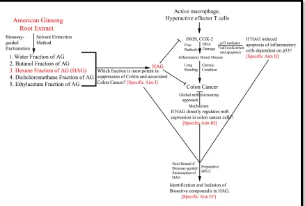
IGURESFigure 1.1 Schematic Overview Of The Project...22
Figure 2.1 HAG Suppresses The Induced Expression of iNOS And COX-2...71
Figure 2.2 Effect Of Whole AG Extract And Different Fractions Of AG On
IFNγ Induced iNOS Expression...73
Figure 2.3 Inflammatory Cells Exposed To The Whole AG Extract And The
HAG Undergo Apoptosis in vitro...74
Figure 2.4 Expression Of Apoptotic Markers in TK6 Cells Following Exposure
To HAG (Dose Response) ...75
Figure 2.5 Expression Of Apoptotic Markers in TK6 Cells Following Exposure
To HAG (Time Response) ...76
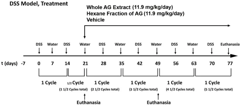
Figure 2.6 Experimental Protocol For The DSS Mouse Model Of UC...77
Figure 2.7 Effects Of Whole AG And The HAG On The Colon Histology
Score In The DSS Mouse Model Of Colitis ...78
Figure 2.8 Effects Of Whole AG And The HAG On Disease Activity Index
In The DSS Mouse Model Of Colitis ...78
Figure 2.9 iNOS, COX-2 And P53, Markers Of Inflammation And
Inflammatory Stress, Are Reduced In DSS + HAG Treated Mice...79
Figure 2.10 Effects Of HAG On Apoptosis In Cells Of Epithelium And
The Mesenteric Lymph Node ...80
Figure 2.11 Experimental Protocol For The AOM/DSS Mouse Model Of
Colon Cancer Associated With Colitis ...81
Figure 2.12 ANA-1 Murine Macrophage Cells Exposed To The HAG Undergo
Apoptosis in vitro...81
Figure 3.1 Representative Figure For The Purity Of CD4+/CD25- T Cells
Isolated From The Spleen Of The Mice ...112
Figure 3.2 Experimental Protocol For The DSS, Prevention Mouse Model
xii
Figure 3.3 HAG Drives Apoptosis (TUNEL+) Of Lymphoblasts Marginally
Better In P53+/+ Cells Compared With P53-/- Cells ...113
Figure 3.4 Effect Of The HAG On The Colon Histology Score Of The Acute DSS Colitis Model ...114
Figure 3.5 The HAG Induces G0/G1 Checkpoint In Colon Cancer Cells in Both P53+/+ And P53-/- Colon Cancer Cells...115
Figure 4.1 miR-29b Expression Increases in Colon Cancer Cells After Exposure To HAG ...138
Figure 4.2 HAG Suppresses MMP-2 Gene Expression...139
Figure 4.3 Suppression Of Endogenous miR-29b Using miR-VANA miR-29b Inhibitors...140
Figure 4.4 HAG Represses HCT-116 Colon Cancer Cell Migration in vitro...141
Figure 4.5 HAG Represses DLD-1 Colon Cancer Cell Migration in vitro...142
Figure 4.6 HAG Represses HCT-116 Colon Cancer Cell Invasion in vitro...143
Figure 5.1 Preparative HPLC Subfractionation Of HAG ...162
Figure 5.2 LC-UV/DAD Analysis Of HAG And Each Subfraction...163
L
IST OFA
BBREVIATIONSACN ... Acetonitrile
AG... American Ginseng
APC...Adenomatous Polyposis Coli
AML... Acute Myeloid Leukemia
AOM ... Azoxymethane
CAC ... Colon Cancer Associated with Colitis
CD ... Crohn’s Disease
CDK ...Cyclin Dependent Kinases
CLL ... Chronic Lymphocytic Leukemia
Con A ...Concanavalin A
COX ...Cyclo-oxygenase
CRC ...Colorectal Cancer
CYP2E1 ... Cytochrome P450 2E1
DAB ... Diamino benzidine
DAI ...Disease Activity Index
DMEM ...Dulbecco’s Modified Eagle Medium
DNA... Deoxyribonucleic Acid
DSS ...Dextran Sulfate Sodium
EMT ... Epithelial to Mesenchymal Transition
FID ...Flame Ionization Detector
xiv
HAG...Hexane Fraction Of American Ginseng
HPLC ...High Performance liquid Chromatography
H&E ...Hematoxylin and Eosin
IBD...Inflammatory Bowel Disease
IFNγ...gamma Interferon
IHC...ImmunoHistoChemistry
IL... Interleukin
IP ... Intraperitoneal
KDa ...Kilo Dalton
LC-UV ... Liquid Chromatography-Ultra Violet
LPL ...Lamina Propria Lymphocytes
LPS... Lipopolysaccharides
MAM ... Methyl Azoxymethanol
MDM2 ... Mouse Double Minute-2
MeOH ... Methanol
miR/miRNA...microRNA
miRNP ... micro Ribonucleoprotein
mRNA ... Messenger Ribonucleic Acid
MLN...Mesenteric Lymph Node
MMPs... Matrix Metalloproteinases
MMR...Mismatch Repair
MS...Mass Spectrometry
NF-κβ... Nuclear Factor Kappa-light-chain-enhancer of activated B cells
NO... Nitric Oxide
NRC ... National Research Council
NSAIDs... Non Steroidal Anti Inflammatory Drugs
nt/s... nucleotide/s
PARP ...Poly (ADP Ribose) Polymerase
PBS ...Phosphate Buffer Saline
PCR... Polymerase Chain Reaction
PI ... Propidium Iodide
P.O ... Oral
PUMA... P53 Upregulated Mediator of Apoptosis
ROS...Reactive Oxygen Species
RONS...Reactive Oxygen and Nitrogen Species
SFM ... Serum Free Medium
Th cells... T-helper cells
TNFα... Tumor Necrosis Factor- alpha
TP53... Tumor Protein 53
TUNEL ...Terminal Deoxynucleotidyl Transferase-Mediated dUTP Nick-End Labeling
UC ... Ulcerative Colitis
UTR...Untranslated region
Wip1 (PPM1D) ... Wild-type P53-induced Phosphatase-1
1
C
HAPTER1
G
ENERALI
NTRODUCTION1.1GENERAL OVERVIEW
Inflammation is the body’s first line of defense against injury and infection that could
lead to pain, heat, swelling and redness (due to the increased blood flow to the area) in
order to seal off the injured tissue, destroy damaged tissue and kill the invading bacteria.
Inflammation in general is an important mechanism that safeguards the body against
infection or injury by launching a well-coordinated immune response involving both
innate and adaptive immune systems [Reviewed in (1)]. Inflammation is both beneficial
and detrimental. When the body’s inflammatory response is normal, the body benefits
from the response. However if the response goes awry, the over-activation of an
inflammatory response could cause harm to the body by destroying the normal tissues in
addition to damaged tissues. Hence a balanced inflammatory response is necessary which
is maintained by the body’s immune system. An overactive immune system is associated
with autoimmune disorders.
The study of association between inflammation and cancer is long standing. In 1863,
Rudolf Virchow first suggested the connection between inflammation and cancer. Since
then, a tremendous amount of information has been obtained linking inflammation and
cancer. Chronic inflammation involves the generation of free radicals including Nitric
colon. Interesting, there is a correlation between NOS2 activity and p53 status in UC (1,
2).
Infection and chronic inflammation are implicated in about 25% of all human cancers
worldwide [Reviewed in (1)]. Several key molecules that are involved in
inflammation-driven carcinogenesis are nuclear factor κβ (NF-κβ); toll-like receptors; reactive oxygen
and nitrogen species (RONS); cyclooxygenases (COXs); nitric oxide synthases (NOSs);
pro and anti-inflammatory cytokines; metals; antioxidant enzymes; peroxisome
proliferator-activated receptor ligands; kinases; growth factors; and the tumor suppressor
proteins, p53 and retinoblastoma (pRb) proteins [Reviewed in (3)]. For the most part, in
IBD, neoplastic lesions arise within areas of the mucosa that have been involved with
colonic inflammation (4). This might explain that during inflammation, the healing of UC
by re-epithelization of colonic mucosa leads to abnormal cell growth resulting in
neoplastic lesions (4). Therefore targeting the key players that are involved in this
inflammation to cancer cascade has shown significant improvement in the therapeutics.
In the 1930s exploration of the role of diet in human cancers began and even at that
stage evidence emerged of the capacity of a higher intake of plant foods to reduce the risk
of cancer (5). Several classes of anti-inflammatory drugs, such as corticosteroids,
NSAIDs, and biologics possess several adverse effects and biologics are expensive (6).
Patients who received biologic therapies are also at higher risk for the development of
cardiac complications (7). Natural products or natural product derived compounds
represent great structural diversity, commonly not seen in synthetic compounds and plays
a dominant role in the discovery of leads for the development of drugs for the treatment
3
used for stress, to boost the immune system, and as a general tonic and stimulant. We
have recently reported the anti-inflammatory and anti-cancer properties of AG (9-11). To
further delineate the bioactive components of AG against colitis and colon cancer, we
performed a bioassay-guided fractionation of AG using various polar (water, butanol,
dicholoromethane, ethylacetate) and non-polar (hexane) solvent extraction methods. Of
these Hexane fraction of AG (HAG) possessed particularly potent anti-inflammatory and
anti-cancer properties. This thesis is aimed at further exploring the mechanisms of colitis
and colitis-associated colon cancer inhibition by HAG and identifying the bioactive
components present in HAG.
1.1.1 INFLAMMATORY BOWEL DISEASE
Ulcerative colitis (UC), Crohn’s disease (CD) and indeterminate colitis are defined by
a common term of inflammatory bowel disease (IBD) (12). Patients with UC and CD are
at an increased risk of developing the colorectal cancer (CRC). The risk of CRC in
patients with UC increases with the duration of the disease with a cumulative risk of 18%
after 30 years (1). A number of genetic alterations, including microsatellite instability and
mutation in TP53 tumor suppressor gene, are early events in UC-associated CRC (13,
14). The risk of CRC in UC patients is 2% after 10 years and 8% after 20 years and 18%
after 30 years of active disease (15). Current opinion regarding the pathogenesis of IBD
is that in genetically susceptible individuals, there is an overreaction of the immune
systems towards antigens of gut microbiota leading to chronic inflammation (16). The
pathogenesis of UC and CD is of multifactorial nature as genetic and immunologic
factors, alterations in the colonic barrier function, bacterial and viral infection, altered
UC is caused by an atypical T helper (Th)2-mediated immune response characterized by
high levels of IL-5 (but not IL-4) and IL-13: in CD there is a prevalent activation of Th1
cells with high expression of TNF-α and IFN-γ (18-21).
1.1.1.1CD4TCELLS AND IMMUNE RESPONSE IN IBD
T helper (Th) cells have important roles in combating infections and cancers;
however, dysregulation of their function can lead to chronic inflammatory diseases (22).
T cells are further divided into 2 types: i) CD4 T cells, which express the CD4 surface
glycoprotein and are termed T helper (Th) cells as they provide help to B and other T
cells in directing B- and T-cell responses; and ii) CD8 T cells, which express the CD8
surface glycoprotein and which are termed cytotoxic T cells owing to their high
expression of macrophage-activating interferon-γ (IFNγ) and granzymes. Dysfunctional
and aberrant immune responses in which the adaptive immune response attacks the host
tissues as if they were foreign is one of the main cause of chronic inflammatory disease
like IBD, Multiple Sclerosis, Systemic Lupus Erythematosus and Rheumatoid Arthritis
[Reviewed in (22)].
Animal studies have established that dysregulated effector T cell responses to the
commensal flora can be causative in IBD and likely represent a final, common
immunopathogenetic mechanism in most, if not all, forms of IBD, irrespective of the
inciting events that promote them (23). Th1, Th2 and Th17 are CD4+ effector T cell
lineages that are involved in some form of IBD. Adaptive transfer of CD4+CD45RBhigh T
cells from healthy wild-type (WT) mice into syngeneic recipients that lack T and B cells
induces colitis at 5–8 wk following T cell transfer (24, 25). Although breakdowns in
5
effector T cells of the adaptive immune system figure prominently in sustaining disease
and are probably essential to disease chronicity (23). IL-10-expressing CD4+ T cells
plays a dominant role in suppressing intestinal effector T cell development and function
and is consistent with the spontaneous development of colitis under conditions of genetic
deficiency of IL-10 production by CD4+ T cells (26), explain IL-10 as an
anti-inflammatory cytokine.
1.1.2 NON-STEROIDAL ANTI-INFLAMMATORY DRUGS (NSAIDS), STEROIDS AND
BIOLOGICS
NSAIDs are a group of drugs that are prescribed to reduce the pain and inflammation.
Some of these drugs require prescription, while some over the counter drugs do not
require prescription. Most commonly used NSAIDs are aspirin, sulindac, ibuprofen etc.
Acetaminophen and codeine are pure pain relievers that are not NSAIDs. NSAIDs
interfere with the cyclo-oxygenase (COX) pathways which lead to the production of
prostanoids (prostaglandins, prostacycline, and thromboxane) (27). Main problem with
NSAIDs is that there are two isoforms of COX enzymes (COX-1 and COX-2). COX-1
plays an important housekeeping role as it is constitutively expressed in most tissues,
including the stomach, duodenum, platelets, and kidneys; and involved in production of
prostaglandins which regulate important physiological processes such as gastrointestinal
cytoprotection (27). COX-2, on the other hand, is normally undetectable in most tissues,
but it can be induced rapidly, and in large quantities to 200–300-fold during inflammation
and other pathological processes (27). Most conventional NSAIDs are non-selective in
COX-2, but exert adverse effects (such as gastrointestinal mucosal damage and
nephrotoxicity) primarily due to inhibition of COX-1 (27).
Steroids are effective short term but fail long term in most patients and are associated
with numerous side effects and low rates of mucosal healing (28, 29). Another strategy is
the use of biologics, which are genetically engineered medications made from living
organisms and their products. The ideal biologic agent for treating IBD should target a
specific event of the inflammatory cascade, induce and maintain a sustained remission, be
well tolerated and induce no immunogenicity (30). Common biologics used against
inflammation that targets TNFα are adalimumab (Humira), certolizumab (Cimzia),
infliximab (Remicade); while natalizumab (Tysabri) targets cell adhesion molecule α
4-integrin. Because biologics are given either by intravenous infusions or subcutaneous
injections, it may produce redness, itching, bruising, pain, or swelling on the injection
site. Additionally, anti-TNFα (as biologics) are closely linked with mycobacterial
infections leading an incidence of tuberculosis (TB) (31) and sepsis, a life-threatening
blood infection is reported with biologics (32).
1.1.3 ROLE OF BIOASSAY-GUIDED FRACTIONATION OF HAG
American Ginseng (AG, Panax quinquefolius) is an obligate shade perennial native of
North America and its root is the commonly used part. AG has antioxidant properties,
and targets many key players involved in inflammation, including iNOS, COX-2 and
NF-κB (33). In a series of studies, we have recently shown that AG can treat colitis and
prevent colon cancer associated with colitis (9, 11, 33). In general, active or inactive
chemical entities obtained from ginseng species can be classified into five categories:
7
(34). Ginseng’s saponins (generally called ginsenosides, mostly Rb1, Rg1, Rg3, Re and
Rd), and acidic polysaccharides of AG have been the main focus of its pharmacological
activities (35-40) such as vasorelaxation, anti-inflammatory, and anti-cancer properties
(41). Other putative active components of AG, include polyacetylenes such as panaxynol
and panaxydol, which are non-polar compounds (42). In contrast to ginsenosides and
polysaccharides; polyynes, flavonoids, and volatile oils have been less studied, and
therefore less is known about their medicinal properties.
Many studies have also reported pro-apoptotic properties of some of the ingredients
we have determined to be in the Hexane Fraction of AG (HAG) (43-49). Mostly
polyacetylenes (panaxynol, panaxydol and panaxydiol) and fatty acids are present in the
HAG (50). Interestingly, others have found an n-Hexane extract of red ginseng is
particularly potent in inhibiting the growth of human lung tumor xenografts in nude mice
(51). However, many fatty acids detected in our HAG, can induce apoptosis in various
cell types (43-49), and conjugated linoleic acid and oleic acid has been shown to suppress
colitis in other studies (52-55). Thus the study of bioassay-guided fractionation of HAG
will provide a significant insight in the bioactive components present in AG against
colitis and colon cancer.
1.1.4 OXIDATIVE STRESS AND FREE RADICALS
As a result of photosynthesis, our earth’s atmosphere consists of 21% oxygen level.
Oxygen is toxic to biological organisms as it generates free radicals. Hence the aerobic
organism has developed antioxidant defense system to tolerate oxygen and protect from
oxidative damage caused by the oxygen-free radicals. It is estimated that in the aerobic
the reason why in aerobic organisms, certain amounts of ROS, including hydroxyl
radicals (OH), superoxide anions (O2) and hydrogen peroxide (H2O2) are constantly
generated (56). For every 1012 Oxygen molecules that enter a cell per day, it is estimated
that 1 in 100 of these molecules damages DNA and protein (57, 58). Oxidative stress is
defined as an imbalance between generation of ROS and decreased antioxidant defense
system (56). Oxidative stress develops particularly in inflammatory reactions because the
inflammatory cells, neutrophils, and macrophages produce large amounts of ROS (56).
Increased ROS lead to protein damage, lipid peroxidation, and DNA damage which
results in genetic and epigenetic alterations that accounts for increasing grades of
dysplasia and carcinoma [Modified (56)].
Oxidation of DNA by ROS can result in damage to all four bases and to the
deoxy-ribose-molecule. One of the most abundant molecules which is a product of this oxidation
is 8-oxo-7,8-dihydro-2,-deoxyguanosine (8-OH-dG), and has been established as a useful
biomarker of oxidative stress (59, 60). 8-OH-dG has a mutagenic effect in mammalian
cells, and can therefore be considered carcinogenic. Defense enzymes such as Superoxide
Dismutase (SOD) form a mutually supportive group of enzymes with peroxydases and
catalases to provide a defense against different ROS (61-64). Another enzyme
Glutathione-S-transferases (GSTs) protects macromolecules from attack of reactive
electrophiles and plays an important role in cellular detoxification (56). Nitric oxide, an
anorganic substance is an antioxidant defense factor that acts as a potent scavenger of
other free radicals and is a good inhibitor of lipid peroxidation (65, 66). However during
9
range) of NO by iNOS has been suggested to be cytotoxic and may contribute to cell
injury and is associated with pathogenesis of mucosal damage (67).
1.1.5 COLITIS ASSOCIATED COLON CANCER
Cancers in the setting of IBD are believed to occur by a progression from no
dysplasia to indefinite dysplasia to low-grade dysplasia (LGD) to high-grade dysplasia
(HGD) to carcinoma (4). Colitis-associated colorectal cancer (CAC) accounts for up to
5% of all colorectal cancers (68), and the incidence of CAC in the UC patients increases
with age. The frequency of CIN (85%) and MSI (15%) in CAC is roughly the same as in
SCC (13). For the most part, in IBD, neoplastic lesions arise within areas of the mucosa
that have been involved with colonic inflammation (4). This might explain that during
inflammation, the healing of UC by re-epithelization of colonic mucosa leads to
abnormal cell growth resulting in neoplastic lesions (4). Colorectal cancer is one of the
two major cancers, the risk of which is commonly agreed to be modified mainly by food
and nutrition (69).
1.1.6 MOLECULAR PLAYERS IN COLITIS ASSOCIATED COLON CANCER
Several key molecules that are involved in inflammation-driven carcinogenesis are
nuclear factor κβ (NF-κβ); toll-like receptors; reactive oxygen and nitrogen species
(RONS); cyclooxygenases (COXs); nitric oxide synthases (NOSs); pro and
anti-inflammatory cytokines; metals; antioxidant enzymes; peroxisome proliferator-activated
receptor ligands; kinases; growth factors; and the tumor suppressor proteins, p53 and
1.1.6.1NUCLEAR FACTOR-ΚΒ(NF-ΚΒ)
NF-κβ is a pleiotropic transcription factor with a key role in innate and adaptive
immunity and is required for the expression of various proinflammatory factors (70).
Activation of NF-κβ also supports carcinogenesis by increasing cell proliferation, and
angiogenesis, inhibiting cell death, promoting cell invasion and metastasis (71). NF-κβ is
a transcription factor consisting of closely related proteins that generally exist as dimers
and bind to a common DNA sequence within the promoter/enhancer of target genes
called the κβ site to promote transcription of target genes (such as COX-2, iNOS, Bcl-2,
Bcl-x(L), cyclin D1, MMP and VEGF) through the recruitment of coactivators and
corepressors (72). Inhibition of NF-κβ pathways by blocking one of the five members of
NF-κβ (p50, p52, p65(Rel A), c-Rel and Rel B) and upstream activators such as TNF-α
receptors and finally the target genes by nutraceuticals is one of the chemopreventive
aspects.
1.1.6.2P53FAMILY MEMBERS
The tumor suppressor, p53, plays a pivotal role in controlling cell cycle, apoptosis,
genomic integrity and DNA repair in response to various genotoxic stresses (72). After
activation, p53 can bind to regulatory DNA sequences and activate the expression of
target genes which can be grouped into four categories: cell cycle inhibition (p21,
reprimo, cyclin G1, GADD45, 14-3-3), apoptosis (PERP, NOXA, PUMA, p53AIP1,
ASPP1/2, Fas, BAX, PIDD), genetic stability (p21, DDB2, MSH2, XPC) and inhibition
of angiogenesis (TSP1, Maspin, BAI1, GD-AIF) [Reviewed in (72)]. Briefly, following
DNA damage, p53 levels rise and proliferating cells are arrested at G1-phase, allowing
11
stimulation of expression of p21CIP1, the cyclin kinase inhibitor (73). Increased p53 level
transactivates Bax (proapoptotic) for induction of apoptosis through caspase cascade
(74).
1.1.6.3P53 AND TUMOR FORMATION
The TP53 (Tumor Protein p53) gene provides instructions for making a protein called
tumor protein p53 which acts as a tumor suppressor by regulating cell division by
keeping the cells from growing and dividing too fast or in an uncontrolled way [Adapted
from NCBI]. The TP53 gene is located on chromosome 17p13.1. It directly binds to
DNA and if DNA is damaged (by toxic chemicals, radiation, UV light), p53 protein
determines whether DNA be repaired or the damaged cell be self destructed (undergo
apoptosis). p53 is regulated by MDM2 (Mouse Double Minute 2, a p53 specific ubiquitin
ligase) (75-77). Mdm2 inhibits p53 cell-cycle arrest and apoptotic functions (78, 79)
during a normal cell growth by binding and masking p53 transcriptional activation
domain leading to ubiquitlyation and degradation of p53 (76, 77). During cellular stress
or DNA damage, p53 gets activated (phosphorylated/ acetylated), which inhibits Mdm2
activity thereby facilitating the damaged cells to undergo apoptosis or cell cycle arrest.
Somatic mutation in TP53 gene is observed in about 50% of all human cancer (80, 81).
Many of these mutations change a single protein building block (Amino acid) in tumor
protein p53 leading to a production of non functional protein, that can’t regulate cell
growth and division, ultimately resulting in the unregulated growth and division of the
damaged cells forming a cancerous tumors [Adapted from NCBI]. When activated by
to the maintenance of the genomic stability by regulating cell cycle checkpoints, DNA
repair, apoptosis and senescence (82, 83).
1.1.6.4NITRIC OXIDE SYNTHASE
NOSs are the family of enzymes that catalyses the biosynthesis of NO. NOS are
dimeric enzymes with each monomer composed of two distinct catalytic domains:
NH2-terminal oxygenase domain and COOH-NH2-terminal reductase domain. N-NH2-terminal is the
binding site for heme 5,6,7,8-tetrahydrobiopterin (BH4), oxygen and l-arginine, whereas
NADPH, FMN and FAD bind on C-terminal (84). The catalytic mechanisms of NOS
involve flavin-mediated electron transport from NADPH to the heme centre, where
oxygen is reduced and incorporated into the guanidine nitrogen of l-arginine producing
NO and L-citrulline (85). There are 3 distinct isoforms of NOSs with 50% sequence
homology and can be divided into 2 broader classes:
1.1.6.4.1 CONSTITUTIVE NOSS: eNOS and nNOS isoforms are constitutive and
Calcium/Calmodulin dependent and generate NO in picomolar to nanomolar
range for short period of time [Reviewed in (86)]. cNOS is responsible for
physiological production of NO that has multiple beneficial effects including
modulation of platelet aggregation, inhibition of leukocyte adhesion and
control of vascular smooth muscle cell proliferation (87).
1.1.6.4.2 INDUCIBLE NOS: iNOS isoform is induced by cytokines and is independent
upon Calcium/Calmodulin for its enzymatic action [Reviewed in (86)]. iNOS
is expressed essentially in every cell type and can locally generate high output
quantities of NO at micromolar range for prolonged period of time [Reviewed
13
induced by exposure to cytokines and LPS in many cells (macrophages,
neurtrophils and the endothelium) (67) and is associated with NO production
in pathophysiological condition.
Thus NO serves as a dual role to mediate both physiological and
pathophysiological processes. In the Gastrointestinal tract, NO is important in protection
of mucosal damage and this is especially true in case of acute injury. NO produced in
such ways by cNOS influences virtually every component of mucosal defense by
reducing mast cell degranulation and release of pro-inflammatory substances from
macrophages, platelets and neutrophils (67). NO has been reported to be a free radical
scavanger (88) and its ability to scavange superoxide anion (O2-), has been well reported
both in vitro (89) and in vivo (90).
During chronic inflammatory conditions, sustained production of larger
concentration of NO (micromolar range) by iNOS has been suggested to be cytotoxic and
may contribute to cell injury (67). Increased level of NO has been associated with
experimental model of colonic inflammation induced by DSS (91) and increased activity
of iNOS has been measured in UC (92-95). This suggests that inhibition of iNOS may
have therapeutic implications against UC.
It is reported that NO induces p53 phosphorylation via ATM and ATR, which
leads to cell cycle checkpoint arrest at G2/M (96) during inflammation. NO also increases
cancer risk in chronically inflamed tissues. It has been shown that NO could target p53
tumor suppressor causing an increased p53 mutation load in inflamed colon tissue from
dual role of p53 stabilization and p53 mutation depending upon the condition of
inflammation.
1.1.6.5CYCLOOXYGENASES
COX-2 is an inducible prostaglandin G/H synthase, that is involved in prostaglandin
(PG) synthesis. Overproduction of COX-2 and PG production from free arachidonic acid
have been implicated in colon carcinogenesis (97). COX-2 mediated increased PGE2
levels have been believed to enhance tumor promotion by promoting cell proliferation,
angiogenesis and apoptotic evasion, stimulating tumor metastasis and decreasing immune
surveillance (98).
Cyclooxygenase-2 is a cytoplasmic protein that catalyzes the synthesis of lipid
inflammatory mediators (prostaglandins and prostacyclins) from arachidonic acid.
COX-2 expression is increased at the site of inflammation (99) and also in 80% of CRC and
40% of colorectal adenocarcinoma (100). Indeed, the COX-2 protein is found in the
cytoplasm of neoplastic colonic epithelial cells and to a lesser extent in stromal cells,
whereas normal epithelium is negative for COX-2 (101). COX-2 may contribute to tumor
development by modulating apoptosis, angiogenesis and tumor invasiveness, as
over-expression of COX-2 in rat intestinal epithelial cells had increased adherence to the
extracellular matrix, resistance to apoptosis-inducing agents and up-regulation of the
anti-apoptotic protein B-cell lymphoma-2 (Bcl-2) (102).
1.1.6.6WNT/Β-CATENIN PATHWAY
Abnormal activation of the Wnt/β-catenin pathway has been implicated in the
development of human colon cancer (97). In normal cells, Wild Type Adenomatous
15
for routine degradation (103, 104). Loss of WT APC function results in the translocation
of β-catenin to the nucleus, where it interacts with TCF-family transcription factors and
activates the transcription of several genes including those encoding cyclin D1, c-Myc
and anti-apoptotic protein survivin which is over-expressed by colorectal tumors (105).
1.1.6.7MATRIX METALLOPROTEINASES (MMPS)
MMPs are secreted by inactive zymogens and are activated extracellularly, and once
activated, they are able to degrade most of the extracellular matrix components (such as
Collagen, laminin, fibronectin, vitonectin, enactin and proteoglycans), thus enhancing the
metastatic potential of cancer cells (106). Degradation of Type IV-Collagen by MMP-2
and -9 often occurs in cancer (106).
1.1.6.8CDK/CYCLINS
Loss of the cell cycle regulation is a hallmark of cancer. The eukaryotic cell cycle is
regulated by the sequential activation and inactivation of cyclin-dependent kinases
(Cdks), that drive cell cyle progression by forming Cdk/Cyclin complex (assisted by Cdk
activating kinase, CAKs), and alternatively Cdk inhibitory subunits (CKIs, such as p21,
p27, p16INK4) inactivates the active Cdk/cylin complex [Reviewed in (107)]. Thus the
induction of cell cycle arrest and/or apoptosis is considered to be a promising
chemopreventive strategy.
1.1.7 MICRORNA
MicroRNAs (MiRs) are a group of small non-coding endogenous single stranded
RNAs of 18-25 nucleotide (approx. 22nts) length that negatively regulate gene expression
by translational inhibition or exonucleolytic messenger RNA (mRNA) decay (108).
inflammatory conditions (109-111). MiR-155 and miR-146a have been identified as
inflammatory response miRs, that are upregulated by NF-κβ (112-114). Additionally,
apoptosis of unwanted T cell clones is an important mechanism in resolution of
inflammation and miRs are clearly linked with regulation of apoptosis; indicates the
association of miR’s with inflammatory conditions. Several miRs have been found to be
either upregulated or downregulated in tumors (115-118). Besides the global microRNA
expression and signature identification in cancer, several individual microRNAs have
evolved as a key player in certain malignancies. The miRBase Version 16.0 has 1048
miRNA sequences annotated in the human genome, and miRNAs are believed to target
about one-third of human mRNAs; a single miR can target approximately 200 transcripts
simultaneously (119). There are different mechanisms to inhibit mutagenesis and
carcinogenesis and the modulation of miR as an epigenetic response to dietary agents is
one such mechanism that has recently evolved as an efficient tool in the cancer
chemoprevention study.
1.2IN VITRO STUDY MODELS
For the in vitro study of inflammation, several oncogene immortalized mouse
macrophage and lymphoblastoid cell lines are generated that emulate normal tissue
macrophage and lymphoblasts which avoid difficulties associated with the isolation of
large homogenous population of macrophage or lymphoblasts (120-123). Cell lines used
for the inflammatory studies are ANA-1 mouse macrophage, TK6, NH32 and Jurkat T
cells.
In vitro cellular models permit isolation of specific aspects of tumor biology such that
17
mediators and pharmacological compounds are faster and simpler than in the intact
organism (124). Various cell lines are used for the in vitro models such as HCT116,
LOVO, HT29, and DLD1 etc. The limitation of in vitro models of colon cancer is that it
reflects only a specific stage of tumor development depending on the stage, the cells
originated from and it also lacks the tissue context study of tumor growth and metastasis
in vivo (124). To address this issue, in vivo animal models similar to human CRC have
emerged to provide the opportunity to study tumor stages and metastatic processes due to
the reliability to induce and establish colon tumors and study chemopreventive features
1.3ANIMAL MODELS
Carcinogen induced CRC models are a highly reliable way to recapitulate the phases
of tumor growth and progression, that occurs in human and is frequently used to study the
chemopreventive compounds efficacy or their associated risk factors (124). An important
benefit of this model is that it is highly reproducible and can be readily tested on animals
with different genetic backgrounds.
1.3.1 DSS MOUSE MODEL OF COLITIS
DSS model of colitis is widely used model of experimental colitis because it has
similarities to human IBD in aetiology, pathology, pathogenesis and therapeutic response
(125). DSS is administered through the drinking water in cycles. For our experiments we
followed a protocol of each cycle of DSS as 7-days of DSS in drinking water followed by
7 days of normal drinking water. Acute colitis can be induced by single cycle of DSS
exposure (high concentration) to the mice whereas multiple cycles of DSS administration
(low concentration) will lead to chronic colitis (126). Exact mechanism of DSS induced
permeability where tight junction protein (Zona occludens-1) were directly reduced by
DSS leading to an increase permeability; changes that precede colonic inflammation
[Reviewed in (125)]. Another possible mechanism could be direct cytotoxicity of DSS on
the colonic mucosa which leads to alteration of integrin-α4 and M290 subunit level of
epithelial cells disrupting their interaction with γδ-intraepithelial T cells (127) thought to
be involved in mucosal protection and healing (128). The exact mechanism of how DSS
initiates colitis is unknown but the fact that crypt loss and increased permeability usually
precede inflammation is suggestive that DSS causes initial insult at the epithelial cell
level with inflammation developing secondarily (129).
1.3.2 AOM/DSS MOUSE MODEL OF COLITIS ASSOCIATED COLON CANCER
Single to double dose of AOM injections induce colon tumors with pathological
features similar to that seen in sporadic human diseases (130-132).Currently studies with
AOM are preferred because of its practical advantage such as reproducibility, high
potency, simple mode of application, excellent stability in solution and low price (131).
AOM-induced tumors share many of the histopathological characteristics of human CRC
and they carry frequent mutations in K-Ras and β-catenin and some show MSI indicative
of defective MMR system (124). APC and p53 mutations are rare and tendency to
metastasize is also low (133). The intraperitoneal (I.P.) injection of AOM (Tumor
initiating agent) with DSS (Dextran Sulfate Sodium: Tumor promoting agent) in drinking
water (periodic cycle-alternate week between water and DSS) (AOM/DSS-model) is
applicable for the study of tumor progression driven by colitis (131) which causes rapid
growth of multiple colon tumors within 10 weeks compared to 30 weeks (for AOM I.P.
19
AOM requires several metabolic activation steps (including N-oxidation and
hydroxylation) to induce DNA reactive adducts. Hydroxylation of AOM results in the
formation of reactive metabolite MAM which alkylate macromolecule in liver and colon
and operate the addition of methyl group at O6 and N7 positions of guanine
(O6-methyl-deoxyguanosine and N-7-methyl-(O6-methyl-deoxyguanosine) in the DNA molecule. Methylation at
the O6-position of guanine has been shown to be the primary promutagenic lesions
produced by AOM. MAM is activated by (mouse) colon alcoholdehydrogenase (134) and
alcohol inducible CYP2E1 (135).
1.4OBJECTIVES OF THE RESEARCH
UC is an idiopathic, chronic inflammatory disorder of the colonic mucosa, which
starts in the rectum and generally extends proximally in a continuous manner through
part of, or the entire colon (136). North American and Northern Europe have the highest
incidence rate (Varying from 9-20 cases/100000 person-years) and highest prevalence
rate (From 156-291 cases/100000 people) of UC (136). Incidence pattern of UC is
bimodal with main onset peak at 15-30 years (137) and second smaller peak at 50-70
years (136). The clinical features of UC are rectal bleeding, diarrhea, urgency, abdominal
pain, fever (severe cases); endoscopic features are loss of vascular pattern, erythema,
granularity, erosion and ulceration; and pathological features are distortion of crypt
architechture, crypt abscesses, Lamina propria cellular infiltrate, shortening of crypts,
lymphoid aggregates, erosion and ulceration [Reviewed in (136)]. Conventional NSAIDs
used for the treatment of UC have adverse side effects of gastrointestinal mucosal
damage (27). Anti-TNFα treatment have shown increased incidence of tuberculosis (31)
particularly in the form of herbal therapies, is widespread in the Western world as well as
in many Asian countries including China and India (138). The use of herbal medicines by
natives is dated for centuries because of its efficiency and lesser known side effects.
Ginseng has been used for centuries in eastern Asia as an immune suppressant. We have
reported the anti-inflammatory and anti-cancer effects of AG (9-11). To identify the
bioactive compound/s present in AG extract, we performed a bioassay guided
fractionation of AG using different solvents for extraction of AG components.
Increased iNOS activity and expression has been widely reported in active UC
(92, 94, 139-141). Increased COX-2 expression has also been widely reported in IBD
(142, 143). Stabilization of p53 leads to transactivation of series of proteins responsible
for cell cycle arrest, apoptosis and senescence (144). Thus, we tested the
anti-inflammatory property of the fractions of AG in vitro and in vivo for these end points of
inflammation. Since stabilization p53 is responsible for apoptosis and AG whole extract
has been reported to induce apoptosis of inflammatory cells through p53 pathway (9), in
Chapter 3 we assessed the ability of HAG to induce apoptosis in inflammatory cells and
tested if this HAG mediated apoptosis is p53 dependent. To further elucidate the
mechanism of action of HAG, we explored the approach of miR mechanism (Chapter 4).
To identify the differentially regulated miR after HAG treatment on colon cancer cells,
global miR microarray approach was utilized and significantly regulated miR (miR-29b)
was further studied. Functional assay was studied by performing a loss of miR-29b
functional analysis experiments (Chapter 4). Having explored the anti-inflammatory and
anti-cancer effects and mechanisms of HAG, we performed a next step of
21
the bioactive compound/s (Chapter 5). Suppression of iNOS expression in activated
mouse macrophages was assessed for each subfraction of HAG to initiate the
identification of bioactive compound/s.
Chapter 5 also includes the overall conclusion and summary of the project
followed by the future directions of this project.
1.5SPECIFIC AIMS
A proposed scheme summarizing this research project is shown in Figure 1.1. The
specific aims of this dissertation are addressed below:
Specific Aim One: To detect if HAG suppresses colitis and associate colon cancer in the
experimental mouse model. (Chapter 2)
Specific Aim Two: To determine if p53 is a key mediator of the protective effects of AG
against the experimental mouse model of colitis. (Chapter 3)
Specific Aim Three: To explore HAG mediated regulation of miR in colon cancer cells.
(Chapter 4)
Specific Aim Four: To perform a next step bioassay-guided fractionations to further
23 REFERENCES FOR CHAPTER 1
1. Hussain SP. Inflammation and cancer: is AID aiding? Gastroenterology. 2008;135:736-737.
2. Hussain SP, Amstad P, Raja K, Ambs S, Nagashima M, Bennett WP, et al. Increased p53 mutation load in noncancerous colon tissue from ulcerative colitis: a cancer-prone chronic inflammatory disease. Cancer Res. 2000;60:3333-3337.
3. Hofseth LJ, Wargovich MJ. Inflammation, cancer, and targets of ginseng. J Nutr. 2007;137:183S-185S.
4. Itzkowitz SH, Yio X. Inflammation and cancer IV. Colorectal cancer in inflammatory bowel disease: the role of inflammation. Am J Physiol Gastrointest Liver Physiol. 2004;287:G7-17.
5. Stocks P, Karn MN. A co-operative study of the habits, home life, dietary and family histories of 450 cancer patients and of an equal number of control patients. Ann Eugen Human Genet. 1933;5:30-33.
6. Gautam R, Jachak SM. Recent developments in anti-inflammatory natural products. Med Res Rev. 2009;29:767-820.
7. Strand V, Kimberly R, Isaacs JD. Biologic therapies in rheumatology: lessons learned, future directions. Nat Rev Drug Discov. 2007;6:75-92.
8. Newman DJ, Cragg GM. Natural products as sources of new drugs over the 30 years from 1981 to 2010. J Nat Prod. 2012;75:311-335.
9. Jin Y, Hofseth AB, Cui X, Windust AJ, Poudyal D, Chumanevich AA, et al. American ginseng suppresses colitis through p53-mediated apoptosis of inflammatory cells. Cancer Prev Res (Phila). 2010;3:339-347.
10. Jin Y, Kotakadi VS, Ying L, Hofseth AB, Cui X, Wood PA, et al. American ginseng suppresses inflammation and DNA damage associated with mouse colitis. Carcinogenesis. 2008;29:2351-2359.
11. Cui X, Jin Y, Poudyal D, Chumanevich AA, Davis T, Windust A, et al. Mechanistic insight into the ability of American ginseng to suppress colon cancer associated with colitis. Carcinogenesis. 2010;31:1734-1741.
13. Willenbucher RF, Aust DE, Chang CG, Zelman SJ, Ferrell LD, Moore DH, 2nd, et al. Genomic instability is an early event during the progression pathway of ulcerative-colitis-related neoplasia. Am J Pathol. 1999;154:1825-1830.
14. Brentnall TA, Crispin DA, Rabinovitch PS, Haggitt RC, Rubin CE, Stevens AC, et al. Mutations in the p53 gene: an early marker of neoplastic progression in ulcerative colitis. Gastroenterology. 1994;107:369-378.
15. Eaden JA, Abrams KR, Mayberry JF. The risk of colorectal cancer in ulcerative colitis: a meta-analysis. Gut. 2001;48:526-535.
16. Strober W, Fuss IJ, Blumberg RS. The immunology of mucosal models of inflammation. Annu Rev Immunol. 2002;20:495-549. Epub 2001 Oct 2004.
17. Head KA, Jurenka JS. Inflammatory bowel disease Part 1: ulcerative colitis--pathophysiology and conventional and alternative treatment options. Altern Med Rev. 2003;8:247-283.
18. Fais S, Capobianchi MR, Pallone F, Dimarco P, Boirivant M, Dianzani F, et al. Spontaneous Release of Interferon-Gamma by Intestinal Lamina Propria Lymphocytes in Crohns-Disease - Kinetics of Invitro Response to Interferon-Gamma Inducers. Gut. 1991;32:403-407.
19. Fuss IJ, Heller F, Boirivant M, Leon F, Yoshida M, Fichtner-Feigl S, et al. Nonclassical CD1d-restricted NK T cells that produce IL-13 characterize an atypical Th2 response in ulcerative colitis. J Clin Invest. 2004;113:1490-1497.
20. Fuss IJ, Neurath M, Boirivant M, Klein JS, de la Motte C, Strong SA, et al. Disparate CD4+ lamina propria (LP) lymphokine secretion profiles in inflammatory bowel disease. Crohn's disease LP cells manifest increased secretion of IFN-gamma, whereas ulcerative colitis LP cells manifest increased secretion of IL-5. J Immunol. 1996;157:1261-1270.
21. Monteleone G, Biancone L, Marasco R, Morrone G, Marasco O, Luzza F, et al. Interleukin 12 is expressed and actively released by Crohn's disease intestinal lamina propria mononuclear cells. Gastroenterology. 1997;112:1169-1178.
22. Zenewicz LA, Antov A, Flavell RA. CD4 T-cell differentiation and inflammatory bowel disease. Trends Mol Med. 2009;15:199-207.
23. Maynard CL, Weaver CT. Intestinal effector T cells in health and disease. Immunity. 2009;31:389-400.
25
25. Powrie F. T cells in inflammatory bowel disease: protective and pathogenic roles. Immunity. 1995;3:171-174.
26. Roers A, Siewe L, Strittmatter E, Deckert M, Schluter D, Stenzel W, et al. T cell-specific inactivation of the interleukin 10 gene in mice results in enhanced T cell responses but normal innate responses to lipopolysaccharide or skin irritation. J Exp Med. 2004;200:1289-1297.
27. Russell RI. Non-steroidal anti-inflammatory drugs and gastrointestinal damage-problems and solutions. Postgrad Med J. 2001;77:82-88.
28. D'Haens G, Baert F, van Assche G, Caenepeel P, Vergauwe P, Tuynman H, et al. Early combined immunosuppression or conventional management in patients with newly diagnosed Crohn's disease: an open randomised trial. Lancet. 2008;371:660-667.
29. Faubion WA, Jr., Loftus EV, Jr., Harmsen WS, Zinsmeister AR, Sandborn WJ. The natural history of corticosteroid therapy for inflammatory bowel disease: a population-based study. Gastroenterology. 2001;121:255-260.
30. Reenaers C, Louis E, Belaiche J. Current directions of biologic therapies in inflammatory bowel disease. Therap Adv Gastroenterol. 2010;3:99-106.
31. Keane J, Gershon S, Wise RP, Mirabile-Levens E, Kasznica J, Schwieterman WD, et al. Tuberculosis associated with infliximab, a tumor necrosis factor alpha-neutralizing agent. N Engl J Med. 2001;345:1098-1104.
32. Goode S, Tierney G, Deighton C. Life threatening intra-abdominal sepsis in patients on anti-TNF-alpha therapy. Gut. 2006;55:590-591.
33. Jin Y, Kotakadi VS, Ying L, Hofseth AB, Cui X, Wood PA, et al. American ginseng suppresses inflammation and DNA damage associated with mouse colitis. Carcinogenesis. 2008;29:2351-2359. Epub 2008 Sep 2318.
34. Jia L, Zhao Y. Current evaluation of the millennium phytomedicine--ginseng (I): etymology, pharmacognosy, phytochemistry, market and regulations. Curr Med Chem. 2009;16:2475-2484.
35. Kim KH, Lee YS, Jung IS, Park SY, Chung HY, Lee IR, et al. Acidic polysaccharide from Panax ginseng, ginsan, induces Th1 cell and macrophage cytokines and generates LAK cells in synergy with rIL-2. Planta Med. 1998;64:110-115.
37. Wang J, Li S, Fan Y, Chen Y, Liu D, Cheng H, et al. Anti-fatigue activity of the water-soluble polysaccharides isolated from Panax ginseng C. A. Meyer. J Ethnopharmacol. 2010;130:421-423.
38. Park E, Hwang I, Song JY, Jee Y. Acidic polysaccharide of Panax ginseng as a defense against small intestinal damage by whole-body gamma irradiation of mice. Acta Histochem. 2009.
39. Choi HS, Kim KH, Sohn E, Park JD, Kim BO, Moon EY, et al. Red ginseng acidic polysaccharide (RGAP) in combination with IFN-gamma results in enhanced macrophage function through activation of the NF-kappaB pathway. Biosci Biotechnol Biochem. 2008;72:1817-1825.
40. Sasaki T, Oh KB, Matsuoka H, Saito M. [Effect of Panax ginseng components on the differentiation of mouse embryonic stem cells into cardiac-like cells]. Yakugaku Zasshi. 2008;128:461-467.
41. Lu JM, Yao Q, Chen C. Ginseng compounds: an update on their molecular mechanisms and medical applications. Curr Vasc Pharmacol. 2009;7:293-302.
42. Liu JH, Lee CS, Leung KM, Yan ZK, Shen BH, Zhao ZZ, et al. Quantification of two polyacetylenes in Radix Ginseng and roots of related Panax species using a gas chromatography-mass spectrometric method. J Agric Food Chem. 2007;55:8830-8835.
43. Pan Z, Wang J, Tang H, Li L, Lv J, Xia L, et al. Effects of palmitic acid on lipid metabolism homeostasis and apoptosis in goose primary hepatocytes. Mol Cell Biochem. 2010.
44. Luo Y, Ling Y, Guo W, Pang J, Liu W, Fang Y, et al. Docetaxel loaded oleic acid-coated hydroxyapatite nanoparticles enhance the docetaxel-induced apoptosis through activation of caspase-2 in androgen independent prostate cancer cells. J Control Release. 2010;147:278-288.
45. Hsu YC, Meng X, Ou L, Ip MM. Activation of the AMP-activated protein kinase-p38 MAP kinase pathway mediates apoptosis induced by conjugated linoleic acid in p53-mutant mouse mammary tumor cells. Cell Signal. 2010;22:590-599.
46. Cho HJ, Kwon GT, Park JH. trans-10,cis-12 Conjugated linoleic acid induces depolarization of mitochondrial membranes in HT-29 human colon cancer cells: a possible mechanism for induction of apoptosis. J Med Food. 2009;12:952-958.
47. Cvjeticanin T, Stojanovic I, Timotijevic G, Stosic-Grujicic S, Miljkovic D. T cells cooperate with palmitic acid in induction of beta cell apoptosis. BMC Immunol. 2009;10:29.
27
49. Iguchi K, Okumura N, Usui S, Sajiki H, Hirota K, Hirano K. Myristoleic acid, a cytotoxic component in the extract from Serenoa repens, induces apoptosis and necrosis in human prostatic LNCaP cells. Prostate. 2001;47:59-65.
50. Poudyal D, Le PM, Davis T, Hofseth AB, Chumanevich AP, Chumanevich AA, et al. A hexane fraction of american ginseng suppresses mouse colitis and associated colon cancer: anti-inflammatory and pro-apoptotic mechanisms. Cancer Prev Res (Phila). 2012.
51. Lee SD, Park SK, Lee ES, Kim HM, Lee CW, Lee K, et al. A lipid-soluble red ginseng extract inhibits the growth of human lung tumor xenografts in nude mice. J Med Food. 2010;13:1-5.
52. Evans NP, Misyak SA, Schmelz EM, Guri AJ, Hontecillas R, Bassaganya-Riera J. Conjugated linoleic acid ameliorates inflammation-induced colorectal cancer in mice through activation of PPARgamma. J Nutr. 2010;140:515-521.
53. Bassaganya-Riera J, Hontecillas R. CLA and n-3 PUFA differentially modulate clinical activity and colonic PPAR-responsive gene expression in a pig model of experimental IBD. Clin Nutr. 2006;25:454-465.
54. Bassaganya-Riera J, Reynolds K, Martino-Catt S, Cui Y, Hennighausen L, Gonzalez F, et al. Activation of PPAR gamma and delta by conjugated linoleic acid mediates protection from experimental inflammatory bowel disease. Gastroenterology. 2004;127:777-791.
55. Borniquel S, Jansson EA, Cole MP, Freeman BA, Lundberg JO. Nitrated oleic acid up-regulates PPARgamma and attenuates experimental inflammatory bowel disease. Free Radic Biol Med. 2010;48:499-505.
56. Roessner A, Kuester D, Malfertheiner P, Schneider-Stock R. Oxidative stress in ulcerative colitis-associated carcinogenesis. Pathol Res Pract. 2008;204:511-524.
57. I.N. A, B. B. The Handbook of Oxidative Metabolism. Chelmsford, MA: ESA Inc; 1996.
58. Papa S, Skulachev VP. Reactive oxygen species, mitochondria, apoptosis and aging. Mol Cell Biochem. 1997;174:305-319.
59. Fortini P, Pascucci B, Parlanti E, D'Errico M, Simonelli V, Dogliotti E. 8-Oxoguanine DNA damage: at the crossroad of alternative repair pathways. Mutat Res. 2003;531:127-139.
61. Fridovich I. Superoxide anion radical (O2-.), superoxide dismutases, and related matters. J Biol Chem. 1997;272:18515-18517.
62. Ho YS, Gargano M, Cao J, Bronson RT, Heimler I, Hutz RJ. Reduced fertility in female mice lacking copper-zinc superoxide dismutase. J Biol Chem. 1998;273:7765-7769.
63. Lebovitz RM, Zhang H, Vogel H, Cartwright J, Jr., Dionne L, Lu N, et al. Neurodegeneration, myocardial injury, and perinatal death in mitochondrial superoxide dismutase-deficient mice. Proc Natl Acad Sci U S A. 1996;93:9782-9787.
64. Li Y, Huang TT, Carlson EJ, Melov S, Ursell PC, Olson JL, et al. Dilated cardiomyopathy and neonatal lethality in mutant mice lacking manganese superoxide dismutase. Nat Genet. 1995;11:376-381.
65. O'Donnell VB, Chumley PH, Hogg N, Bloodsworth A, Darley-Usmar VM, Freeman BA. Nitric oxide inhibition of lipid peroxidation: kinetics of reaction with lipid peroxyl radicals and comparison with alpha-tocopherol. Biochemistry. 1997;36:15216-15223.
66. Rubbo H, Radi R, Trujillo M, Telleri R, Kalyanaraman B, Barnes S, et al. Nitric oxide regulation of superoxide and peroxynitrite-dependent lipid peroxidation. Formation of novel nitrogen-containing oxidized lipid derivatives. J Biol Chem. 1994;269:26066-26075.
67. Elliott SN, Wallace JL. Nitric oxide: a regulator of mucosal defense and injury. J Gastroenterol. 1998;33:792-803.
68. Ishikawa TO, Herschman HR. Tumor formation in a mouse model of colitis-associated colon cancer does not require COX-1 or COX-2 expression. Carcinogenesis. 2010;31:729-736.
69. Food, Nutrition, Physical Activity, and the Prevention of Cancer: a Global Perspective. Washington DC: WCRF/AICR; 2007.
70. Hacker H, Karin M. Regulation and function of IKK and IKK-related kinases. Sci STKE. 2006;2006:re13.
71. Naugler WE, Karin M. NF-kappaB and cancer-identifying targets and mechanisms. Curr Opin Genet Dev. 2008;18:19-26.
72. Amin AR, Kucuk O, Khuri FR, Shin DM. Perspectives for cancer prevention with natural compounds. J Clin Oncol. 2009;27:2712-2725.
29
74. Miyashita T, Reed JC. Tumor suppressor p53 is a direct transcriptional activator of the human bax gene. Cell. 1995;80:293-299.
75. Barak Y, Oren M. Enhanced binding of a 95 kDa protein to p53 in cells undergoing p53-mediated growth arrest. Embo J. 1992;11:2115-2121.
76. Momand J, Zambetti GP, Olson DC, George D, Levine AJ. The mdm-2 oncogene product forms a complex with the p53 protein and inhibits p53-mediated transactivation. Cell. 1992;69:1237-1245.
77. Chen J, Marechal V, Levine AJ. Mapping of the p53 and mdm-2 interaction domains. Mol Cell Biol. 1993;13:4107-4114.
78. Haupt Y, Barak Y, Oren M. Cell type-specific inhibition of p53-mediated apoptosis by mdm2. Embo J. 1996;15:1596-1606.
79. Chen J, Wu X, Lin J, Levine AJ. mdm-2 inhibits the G1 arrest and apoptosis functions of the p53 tumor suppressor protein. Mol Cell Biol. 1996;16:2445-2452.
80. Hollstein M, Sidransky D, Vogelstein B, Harris CC. p53 mutations in human cancers. Science. 1991;253:49-53.
81. Levine AJ, Momand J, Finlay CA. The p53 tumour suppressor gene. Nature. 1991;351:453-456.
82. Vogelstein B, Lane D, Levine AJ. Surfing the p53 network. Nature. 2000;408:307-310.
83. Robles AI, Linke SP, Harris CC. The p53 network in lung carcinogenesis. Oncogene. 2002;21:6898-6907.
84. Knowles RG, Moncada S. Nitric oxide synthases in mammals. Biochem J. 1994;298 ( Pt 2):249-258.
85. Nathan C, Xie QW. Regulation of biosynthesis of nitric oxide. J Biol Chem. 1994;269:13725-13728.
86. Singh S, Gupta AK. Nitric oxide: role in tumour biology and iNOS/NO-based anticancer therapies. Cancer Chemother Pharmacol. 2011;67:1211-1224.
87. Moncada S, Palmer RM, Higgs EA. Nitric oxide: physiology, pathophysiology, and pharmacology. Pharmacol Rev. 1991;43:109-142.