University of South Carolina
Scholar Commons
Theses and Dissertations
1-1-2013
Modeling HIV-1-Associated Neurocognitive
Disorders and the Therapeutic Effects of the
Phytoestrogen Metabolite S-Equol in the HIV-1
Transgenic Rat
Landhing Mary Moran
University of South Carolina
Follow this and additional works at:https://scholarcommons.sc.edu/etd Part of thePsychology Commons
This Open Access Dissertation is brought to you by Scholar Commons. It has been accepted for inclusion in Theses and Dissertations by an authorized administrator of Scholar Commons. For more information, please [email protected].
Recommended Citation
Moran, L. M.(2013).Modeling HIV-1-Associated Neurocognitive Disorders and the Therapeutic Effects of the Phytoestrogen Metabolite
M
ODELINGHIV-1-A
SSOCIATEDN
EUROCOGNITIVED
ISORDERS AND THET
HERAPEUTICE
FFECTS OF THEP
HYTOESTROGENM
ETABOLITES-
EQUOL INTHE
HIV-1
T
RANSGENICR
ATby
Landhing M. Moran
Bachelor of Arts
College of Notre Dame of Maryland, 2005
Master of Arts
University of South Carolina, 2009
Submitted in Partial Fulfillment of the Requirements
For the Degree of Doctor of Philosophy in
Experimental Psychology
College of Arts and Sciences
University of South Carolina
2013
Accepted by:
Charles F. Mactutus, Major Professor
Rosemarie M. Booze, Committee Member
Jeffrey C. Schatz, Committee Member
James R. Fadel, Committee Member
D
EDICATIONTo my family: Mom, Dad, Steve, Kerri, Marty, and Lisa. Your love and support
A
CKNOWLEDGEMENTSI sincerely thank my mentor, Dr. Charles Mactutus, for his unwavering
commitment to my education and development as a scientist, as well as for his patience
and generosity in helping me to achieve my goals. I also thank Dr. Rosemarie Booze for
her mentorship and for providing opportunities for learning and career-building; Dr.
Jeffrey Schatz for his guidance as a member on my committees throughout my graduate
studies; and Dr. James Fadel for providing guidance as a member of my dissertation
committee. I also thank Dr. Steven Harrod for the use of his lab equipment, as well as for
his invaluable advice and support. Thank you to Robb Roscoe for helping in the
experiments with tissue collection and to Sarah Ezzell and Christian Meyers for help with
daily animal handling. Thanks also to lab members Dr. Marina Aksenova and Sarah
Bertrand for moral support.
This work was supported by National Institute of Child Health and Human
Development grant HD043680 awarded to Dr. Charles F. Mactutus, and by National
Institute of Drug Abuse grant DA013137 awarded to Dr. Rosemarie M. Booze. I would
like to thank the University of South Carolina College of Arts and Sciences for the
Dean’s Dissertation Fellowship, which allowed me to devote the last year to the
A
BSTRACTHIV-1 associated neurocognitive disorders (HAND) continue to afflict individuals
with HIV-1 in the combination antiretroviral treatment (cART) era, most notably
affecting executive function, as well as preattentive processing. Currently, there is no
effective treatment for HAND, with only adjunctive treatment targeting symptomatic
relief. There were two hypotheses in this dissertation: 1) that HIV-1 Tg rats will show
disruptions in measures of executive function and preattentive processing, as well as
neural network alterations in the prefrontal cortex (PFC), a brain region implicated in
executive function, and 2) that administration of the phytoestrogen metabolite S-equol
will improve performance as measured by executive function and attention, as well as
neuronal network complexity in the PFC.
In experiment 1, using prepulse inhibition of the auditory startle response and a series
of operant tasks, deficits were revealed in perceptual sharpening, sustained attention, and
core components of executive function. Daily oral S-equol treatments (0, 0.05, 0.1, and
0.2 mg), administered to the animals at 6-8 months of age, improved the performance of
the HIV-1 Tg animals on the sustained attention task. Assessment of neuronal networks
with diOlistic labeling of pyramidal neurons in the PFC suggested that the 0.2 mg dose of
S-equol ameliorated alterations in the HIV-1 Tg animals as well.
In experiment 2, treatment with S-equol (0.2 mg), begun at 2-3 months of age,
received S-equol also displayed enduring improvements in performance one month after
the treatment ended, an effect not detected in any of the other groups. However, further
assessments of increased demands on sustained attention as well as selective attention did
not further differentiate the HIV-1 Tg and control animals.
In summary, the HIV-1 Tg rats displayed impaired performance in preattentive
processing, attention, and executive function, prior to any clinical signs of wasting.
S-equol was effective in both ameliorating and preventing attentional deficits, suggesting its
T
ABLE OFC
ONTENTSDEDICATION ... iii
ACKNOWLEDGEMENTS ... iv
ABSTRACT ...v
LIST OF FIGURES ... ix
LIST OF ABBREVIATIONS ... xi
CHAPTER 1: INTRODUCTION ...1
HIV-1 ASSOCIATED NEUROCOGNITIVE DISORDERS ...1
EXECUTIVE FUNCTION AND HIV-1 ...3
PREATTENTIVE PROCESSING AND HIV-1 ...5
NEUROPATHOLOGY OF HIV-1 ...7
EFFECTS OF ESTROGENIC COMPOUNDS ON COGNITION AND NEUROANATOMY ...8
THE HIV-1 TRANSGENIC RAT ...11
EXPERIMENTAL GOALS ...13
EXPERIMENTAL QUESTIONS ...14
CHAPTER 2: EXPERIMENT 1 ...15
EXPERIMENT 1A: PREATTENTIVE PROCESSING ...16
EXPERIMENT 1B: EXECUTIVE FUNCTION ...29
EXPERIMENT 1C: AMELIORATION OF ATTENTIONAL DEFICITS WITH S-EQUOL ...42
EXPERIMENT 2A: PREATTENTIVE PROCESSING ...73
EXPERIMENT 2B: SUSTAINED AND SELECTIVE ATTENTION, AND PREVENTION OF
ATTENTIONAL DEFICITS WITH S-EQUOL TREATMENT ...78
CHAPTER 4: GENERAL DISCUSSION ...113
L
IST OFF
IGURESFigure 2.1 Body weight (Experiment 1) ...56
Figure 2.2 ASR Habituation (Experiment 1) ...57
Figure 2.3 PPI with an Auditory Prepulse (Experiment 1) ...58
Figure 2.4 PPI with an Auditory Prepulse Across Time (Experiment 1) ...59
Figure 2.5 PPI with a Visual Prepulse (Experiment 1) ...60
Figure 2.6 PPI with a Visual Prepulse Across Time (Experiment 1) ...61
Figure 2.7 Signal Detection Task (Experiment 1) ...62
Figure 2.8 Discrimination Task ...63
Figure 2.9 Reversal Task ...64
Figure 2.10 Extradimensional Set-shifting Task ...65
Figure 2.11 Signal Detection with Varying Signal Durations ...66
Figure 2.12 Signal Detection Post-S-Equol Treatment...67
Figure 2.13 Signal Detection Pre- and Post-S-Equol Treatment ...68
Figure 2.14 DiOlistically-labeled Varicosities ...69
Figure 2.15 Number of Varicosities ...70
Figure 2.15 Relative Uterine Weight ...71
Figure 3.1 Body weight (Experiment 2) ...99
Figure 3.2 ASR Habituation (Experiment 2) ...100
Figure 3.5 Signal Detection: Acquisition and Accuracy ...103
Figure 3.6 Signal Detection: Performance ...104
Figure 3.7 Signal Detection with Varying Signal Durations:Acquisition and Accuracy 105 Figure 3.8 Signal Detection with Varying Signal Durations: Hits and Misses One-Month Post-S-Equol Treatment ...106
Figure 3.9 Signal Detection with Varying Signal Durations: Performance One-Month Post-S-Equol Treatment ...107
Figure 3.10 Signal Detection with Higher Intertrial Interval Variability ...108
Figure 3.11 Signal Detection with a Visual Distractor: Signal Duration ...109
Figure 3.12 Signal Detection with a Visual Distractor ...110
Figure 3.13 Signal Detection with an Auditory Distractor: Signal Duration ...111
L
IST OFA
BBREVIATIONSAAN ... American Academy of Neurology
ANI ...Asymptomatic neurocognitive impairment
cART ... Combination antiretroviral therapy
HAD ... HIV-1-associated dementia
HAND ... HIV-1-associated neurocognitive disorders
HIV-1 ... Human immunodeficiency virus
HNRC ... HIV Neurobehavioral Research Center
MCMD ... Minor cognitive motor disorder
MND ... Mild neurocognitive disorder
C
HAPTER1
I
NTRODUCTIONHIV-1-Associated Neurocognitive Disorders
The current estimate of the number of people living with HIV-1 worldwide is 34
million (UNAIDS 2012). The annual rate of new HIV-1 infections has decreased in
several countries in the past decade, but many other regions, including the Middle East,
North Africa, Eastern Europe, and Central Asia, have seen a continued increase in yearly
incidence rates, contributing to an estimated 2.5 million new infections worldwide in
2011. Children represent a substantial proportion of these new cases; 330,000 children
under 15 years of age were newly infected in 2011. Combination antiretroviral therapy
(cART) has decreased the rate of mother-to-child-transmission (MTCT) of HIV-1 in the
U.S. and other western countries from approximately 25% to less than 2% (Connor et al.
1994; Taha et al. 2009), but in developing countries with limited access to cART and
other interventions, the MTCT rate is much higher, approximately 25-35% (Chen et al.
2009;Volmink and Marais, 2008). Although cART use substantially reduces the rate of
MTCT, other factors such as high maternal viral load and preterm delivery can increase
the risk of MTCT, even when cART is used (Townsend et al. 2008; Warszawski et al.
2008). Thus, HIV-1 remains a major health issue worldwide for both adults and children.
A significant and common consequence of HIV-1 infection, observed since the
Force 1991; Antinori et al. 2007), is neurocognitive impairment, ranging from mild
deficits to frank dementia. Prior to the introduction of cART, HIV-1-associated dementia
(HAD) occurred in up to 20% of individuals with HIV-1 (McArthur et al. 1993; Ances
and Ellis 2007). cART has dramatically reduced the prevalence of HAD to less than 5%
(Heaton et al. 2010). Nevertheless, HIV-1-associated neurocognitive disorders (HAND)
continue to afflict up to 50% of patients on cART (Ances and Ellis 2007; Heaton et al.
2011). Despite the increased life expectancy provided by cART, HAND is a significant
health problem for millions, disrupting normal functioning of daily activities and posing
an increased risk for early mortality (Vivithanaporn et al., 2010).
In the pre-cART era, neurological disorders related to HIV-1 were designated as
either HAD or minor cognitive motor disorder (MCMD) according to the criteria of the
American Academy of Neurology (1991). The criteria for HAD were an abnormality in at
least two cognitive areas causing difficulty in everyday activities, and an abnormality in
either motor function or in neuropsychiatric or psychosocial functions. MCMD was
defined as cognitive and motor impairment which causes difficulty in everyday activities,
which is not as severe as that meeting the criteria for HAD. The reduction in severity of
cognitive dysfunction in HIV-1+ individuals on cART, as well as the lack of clear
definitions of cognitive impairment in the AAN criteria, necessitated a revised nosology
for the changing face of HIV-1/AIDS. The HIV Neurobehavioral Research Center
(HNRC) at the University of California, San Diego, proposed diagnostic criteria of three
categories of neurocognitive disorders: HIV-1-associated asymptomatic neurocognitive
impairment (ANI), associated mild neurocognitive disorder (MND), and
neurocognitive impairment over motor impairment or psychosocial problems, and list the
following as cognitive domains that should be evaluated: verbal/language,
attention/working memory, abstraction/executive function, memory (learning/recall),
speed of information processing, sensory-perceptual ability, and motor skills. A diagnosis
of ANI is based on performance that is at least one standard deviation below the mean in
at least two cognitive areas. Individuals with neurocognitive impairment that falls into the
category of ANI do not experience difficulty in daily functioning as a result of the
impairment. MND is defined by performance that is at least one standard deviation below
the mean in at least two different cognitive domains, with the additional requirement
being some degree of difficulty in everyday functions as a result of neurocognitive
impairment. Lastly, HAD is diagnosed when the individual performs at least two standard
deviations below the mean in at least two different cognitive domains with difficulty in
performing everyday tasks. In each category, the impairment does not meet criteria for
delirium, and there are no comorbid conditions which may underlie the neurocognitive
impairment.
Currently, there is no effective treatment for HAND, despite a number of
strategies that have been attempted or that are presently being tested (Alfahad and Nath,
2013). cART is limited as a neurotherapeutic due to low penetration across the
blood-brain barrier (Thomas 2004), leaving only adjunctive treatments targeting symptomatic
relief (Chung et al. 2008; Dewhurst et al. 2007; Eilers et al. 2008; Zink et al. 2005).
Executive Function and HIV-1
Before the introduction of cART, HIV-1+ individuals with HAND showed
and language (Heaton et al. 1995; Heaton et al. 2011). Current studies on HAND, which
represent individuals on cART, demonstrate that attention, memory, and executive
function are the cognitive domains more readily affected in HAND (Cysique et al., 2004;
Garvey et al., 2009; Heaton et al., 2011). Executive function, in particular, shows the
greatest decline as a function of HIV-1 disease progression (Reger et al., 2002), which
can have significant effects on the ability to perform important daily activities (Scott et
al., 2011; Cattie et al., 2012). Executive function is a combination of higher-order
cognitive processes that include planning, decision-making, problem-solving, and
judgment (Spreen and Straus 1998). Attention, flexibility, response inhibition, and
set-shifting are core components forming the foundation of higher-order processes of
executive function (Keeler and Robbins, 2011).
A common clinical assessment of executive function is the Wisconsin Card
Sorting Test (WCST), in which the participant is instructed to match cards based on one
of several possible categories (e.g., color, number, shape), which is not made explicitly
known to the participant. The participant determines the sorting criterion, which changes
after a certain number of correct responses, based on feedback from the test administrator
or computer. With these task demands, the WCST targets flexibility, set-shifting or
shifting between sorting criterion, and inhibition of prepotent responses based on the
previously correct sorting criterion. It has been used extensively to characterize the
deficits in executive function in individuals with HIV-1 (Tozzi et al. 1993; Basso and
Bornstein, 2003; Heaton et al., 2011). Another typical test of executive function that has
been employed in studies of HAND is the Stroop Color Word Test (Chang et al., 2002;
presented, which, in the case of a word that is itself a color, requires inhibition of the
prepotent response to read the word aloud as the answer. The use of the Stroop Color
Word Test thus assesses inhibition, but not flexibility or set-shifting. Flexibility as an
executive function has been measured in terms of category fluency, revealing deficits in
switching subcategories while producing a list of words in a given category (Iudicello et
al., 2008).
In some cases, even when HIV-1+ individuals perform as well as HIV-1-
individuals on executive function tasks, significant differences may nevertheless be found
in brain function. Using fMRI, HIV-1+ participants have decreased BOLD signal change
in frontal regions, diminished functional frontostriatal connectivity, and a potential
compensatory activity increase in the parietal attentional networks (Chang et al. 2001;
Melrose et al., 2008). In conjunction with reduced activity, volume loss in the medial
frontal and premotor cortices, as well as thinning of the prefrontal cortex, are found in
HIV-1+ individuals, reflecting the worsening of executive function which is associated
with these brain regions (Thompson et al., 2005).
Preattentive Processing and HIV-1
One of the most consistent neurological impairments that has been observed in the
early stages of HIV-1 infection is an alteration in temporal processing, as assessed with
amplitude and latency measures of auditory evoked potentials (AEPs), both without
(Castello et al. 1998; Fein et al. 1995; Gil et al. 1992; Koralnik et al. 1990; Pagano et al.
1992; Schroeder et al. 1996; Vigliano et al. 2000), and after, cART initiation (Chao et al.
demonstrated alterations in evoked potentials (Phipps et al. 2000; Prospero-Garcia et al.
1999; Raymond et al. 1998; Riazi et al. 2009). Alterations in AEPs become more
apparent with disease progression in HIV-1+ patients (Goodwin et al. 1996; Lalwani and
Sooy 1992; Ollo et al. 1991; Schroeder et al. 1994), as is more generally observed in the
progression from mild cognitive impairment to dementia (Golob et al. 2007).
More recently, temporal processing in HIV-1+ individuals has been assessed
using prepulse inhibition (PPI) of the startle response. PPI is an operational measure of
sensorimotor gating, a preattentive process in which the startle reflex is reduced when the
startle stimulus is preceded 30–500 msec by a weak prepulse (Hoffman and Ison 1980).
In HIV-1+ individuals with HAND, PPI is significantly reduced compared to cognitively
unimpaired HIV-1+ individuals (Minassian et al. 2013). Alterations in PPI have also been
observed in 1 transgenic rats as well as in rats stereotaxically injected with the
HIV-1 viral proteins Tat and gpHIV-120. Specifically, female Sprague-Dawley HIV-HIV-1 transgenic
(Tg) rats have peak inhibition to an auditory prepulse at the 40 msec interstimulus
interval (ISI) when tested at 5, 6, and 7 months of age, which was a leftward shift from
the control group’s peak inhibition at the 80 msec ISI (Moran et al. 2013). A leftward
shift has also been demonstrated in the inhibition function in 30- and 60-day old male
Sprague–Dawley rats following neonatal Tat injection (Fitting et al. 2006a) and in
9-month old male and female Sprague–Dawley rats given neonatal gp120 injections
(Fitting et al. 2006b). Assessment of PPI in HIV-1 Tg rats prior to adulthood, which may
provide information regarding subtle early-onset neurological alterations analogous to
those reported in HIV-1+ humans, has not been conducted. Such information would be
Neuropathology of HIV-1
Central dopamine (DA) system dysfunction often results from HIV-1 infection
and is associated with subsequent cognitive deficits (Chang et al. 2008; diRocco et al.
2000; Kumar et al. 2011; Wang et al. 2004). HIV-1 entry to the brain occurs early in the
progression of the infection (An et al. 1999; Sinclair et al. 1994). The CNS is a main
target of HIV-1 due to the presence of the CD4 and CCR5 receptors on macrophages and
microglia of this system, to which the virus binds, subsequently allowing the virus to
enter the cell (Clapham and McKnight, 2001). Macrophages infected with HIV-1 cross
the blood-brain barrier, further infecting macrophages and microglia throughout the
brain. As a result, viral load in the brain increases and neurotoxic HIV-1 proteins, such as
Tat and gp120, are released, causing cell excitotoxicity and apoptosis (Ensoli et al. 1993;
Tardieu et al. 1992). Thus, HIV-1 does not directly infect neurons, yet it initiates a chain
of events resulting in neuronal damage and death that can persist even after viral
suppression (Nath et al. 1999).
The DA system is particularly vulnerable to neuronal damage caused by the
HIV-1 proteins, with DA-dependent signaling identified as one of the main mechanisms of its
neurotoxicity (Aksenova et al. 2006; Silvers et al. 2007; Wallace et al. 2006). The
dopamine transporter (DAT) is targeted by HIV-1 proteins Tat and gp120, resulting in
transporter impairment (Aksenov et al. 2008; Ferris et al. 2009; Midde et al. 2013; Zhu et
al. 2009; Zhu et al. 2011), as a function of direct protein-protein interactions (Zhu et al.
2009) involving an allosteric modulation of DAT by the Tat protein (Zhu et al. 2011).
Impairment of DAT subsequently elevates synaptic concentrations of dopamine (DA).
Dopamine (DA) thus binds to DA receptors on microglia, setting off several processes:
such as cytokines (TNF-alpha) and chemokines. The inflammatory mediators and HIV-1
proteins induce apoptosis of DA neurons, further impairing the DA systems (Purohit et
al. 2011).
Another distinguishing feature of HIV-1 neuropathology is the reduction of
neuronal network complexity, which may be evident in dendritic spine density and
morphology. Post-mortem neuropathological analyses on individuals with HIV-1 have
revealed dendritic abnormalities, including vacuolation (Wiley, 1991), dilation, reduced
branching density and dendritic length, and reduced dendritic spine density (Masliah et
al., 1997). Dendritic abnormalities found during autopsies have been correlated with poor
performance on neuropsychological tests, especially in tests of learning, perceptual-motor
skills, abstraction and verbal abilities (Masliah et al., 1997). An in vitro study of
prolonged exposure to lymphotropic HIV-1, macrophage-tropic HIV-1, or recombinant
gp120 from HIV-1 found a decrease in microtubule and neurofilament protein expression
in human fetal brain-derived neurospheres, inhibiting postmitotic neuronal dendrite
development (McCarthy, Vidaurre, & Geffin, 2006). HIV-1 protein Tat-treated neuronal
cultures from mice (Lu et al. 2011) and rats (Bertrand et al. 2013) exhibit significant loss
of dendrites and dendritic spine structures. Dendritic pathology including beading and
reduced spine density is also observed in the HIV-1 Tat transgenic mouse (Fitting et al.
2010).
Effects of Estrogenic Compounds on Cognition and Neuroanatomy
The effects of estrogen on cognition have long been studied, mostly to determine
those receiving estrogen treatments (Carlson and Sherwin, 1998; Duff and Hampson,
2000; Keenan et al. 2001; Smith et al. 2001). However, because of the potential side
effects of estrogen therapy, isoflavones have been explored as a safer, alternative
treatment for neurocognitive impairment; beneficial effects of isoflavones have been
reported (Casini et al. 2006; Duffy et al. 2003; File et al. 2005). Isoflavones, such as
genistein and diadzein, are nonsteroidal plant-derived compounds that bind to estrogen
receptors (Folman and Pope, 1966; 1969).
Findings from animal studies have also supported the use of isoflavones as a
therapy for neurocognitive impairment. Improved performance in rats has been observed
on hippocampal-based cognitive tasks such as the Morris water maze and the radial arm
maze, following daily oral soy isoflavone treatment (Monteiro et al. 2008; Pan et al.
2000; Sarkaki et al. 2008), although there are examples of isoflavone treatment either
having no effect or resulting in impairments (Neese et al. 2010; 2012). In the study of
HAND, improving performance on executive function tasks that engage the PFC is of
greater interest, primarily because executive function is more severely affected in HAND
than are other cognitive domains. In addition, soy isoflavones bind preferentially to the
estrogen receptor beta (ERβ) (Kuiper et al. 1997; Turner et al. 2007), which is highly
expressed in the PFC. If soy isoflavones can improve cognitive performance that relies on
the hippocampus, where a smaller concentration of ERβ is found, then the potential
improvements in executive function by soy isoflavones could possibly be even more
pronounced by their action in the PFC.
The beneficial effects of phytoestrogens on neurocognitive performance may
attenuate damage to DA neurons via inhibition of microglial activation and
proinflammatory factor generation, as occurs in Parkinson’s disease (Chen et al. 2007;
Wang et al., 2005). Rats administered daily oral genistein treatment show an increase in
DAT expression in the PFC (Neese et al. 2010). Stimulation of ERβ in ovariectomized
rats with the ERβ agonist diarylproprionitrile causes increases in norepinephrine (NE)
and DA activity in the PFC as indexed by their respective metabolites, as well as
enhanced recognition memory (Jacome et al. 2010).
Estrogen and estrogenic compounds also impact neuronal network complexity. In
female rats, spine density on pyramidal neurons of the primary sensory and motor
cortices are higher during proestrous, when estrogen levels are at their highest, than
during estrous or diestrous (Chen et al., 2009). Ovariohysterectomy reduces spine density
on these neurons, whereas treatment with an estrogen pellet implanted subcutaneously for
14 days increases spine density to levels approximating those of the estrous and diestrous
periods. Furthermore, ovariectomized rats that were given a phytoestrogen-enhanced diet
(810 micrograms/g) for 7-9 weeks showed a significantly higher spine density in
hippocampal and prefrontal cortex (PFC) pyramidal neurons (Luine et al., 2006). An
increase in dendritic spine density in the PFC has also been observed in monkeys
following intramuscular estradiol injections (Hao et al. 2006). Treatment of hippocampal
slices with 17beta-estradiol promoted new spine formation without altering the
pre-existing networks (Mendez et al., 2011). In addition to their benefits on neuronal growth,
estrogenic compounds including soy isoflavones improve performance in maze tasks in
ovariectomized female rats, suggesting an effect on memory processes (Monteiro et al.,
treatment significantly reduces neurotoxic effects of Tat in rat neuronal cultures (Adams
et al., 2010), and the phytoestrogens daidzein and liquiritigenin can both reduce
synaptodendritic damage as well as restore neuronal networks affected by Tat (Bertrand
et al. 2013). Clearly, the promise of phytoestrogen treatment for neurocognitive
impairment requires in vivo studies of the effect of such compounds on neurocognitive
function in an animal model of HIV-1, followed by measures of neuronal network
complexity.
Equol is a metabolite produced by intestinal bacteria following ingestion of the
soy isoflavone daidzein (Setchell et al. 1984). S-equol (the S-enantiomer) has selective
affinity for ERβ, and shows an even greater affinity for ERβ than 17β-estradiol. S-equol
is also the only enantiomer of equol that is produced by humans (Setchell et al. 2005).
The benefits of a soy food-based diet, such as a reduced risk for certain cancers and
increased bone density, are most typically found in adults who produce equol (Jackson et
al. 2011; Lampe 2009; Setchell et al. 2002), who represent only 25-35% of the Western
population (Rowland et al. 2000; Setchell and Cole, 2006). Therefore, equol, rather than
its precursor, may be a more efficient and effective potential therapeutic for
neurocognitive impairment of HAND.
The HIV-1 Transgenic Rat
The HIV-1 transgenic (Tg) rat is produced by microinjecting the HIV-1 provirus,
minus the gag-pol genes, into fertilized one-cell eggs. In this way, the rat expresses 7 of
the 9 genes for HIV-1, regulated by the viral promoter, Tat. It provides several
model, it is less expensive and more easily handled and housed than nonhuman primate
subjects; 2) with the deletion of the HIV-1 genes gag-pol, it is non-infectious; 3) it
expresses viral genes in lymph nodes, spleen, thymus and blood, similar to infected
humans; 4) it develops neurological abnormalities that resemble those observed in
humans with HIV-1, including proinflammatory responses and the expression of HIV
proteins in the brain (Royal et al. 2012). The use of the HIV-1 Tg rat provides the
opportunity to evaluate the progression of cognitive deficits over time, determine the
relationship between cognitive deficits and neuroanatomical alterations, and identify
appropriate treatments for neurocognitive impairment and the most effective time course
for these treatments.
In addition to the specific benefits of studying the HIV-1 Tg rat in terms of
modeling the expression of the virus, the rat can also provide a useful vehicle for the
study of cognitive function and its underlying neurobiology. Several complex behaviors
that rely on PFC function have been demonstrated in the rat. Homologous subregions of
the PFC in the rat, monkey, and human are associated with parallel cognitive functions as
well, suggesting that rat can be a useful and valid model of complex cognitive functions
(Kesner and Churchwell 2011). Thus, executive function and attention, as the most
affected domains in HAND, may be assessed with translatable results in the rat, with the
use of measures selected to model specific components of cognitive function such as
flexibility, inhibition, set-shifting, and sustained and selective attention.
Experimental Goals
HAND is a serious health issue that afflicts millions of people. The most notable
deficit observed in individuals with HAND is the deterioration of executive function,
which relies on several other core components of cognition, including attention. The
preattentive process of sensory gating is also disrupted in individuals with HAND, prior
to the development of deficits in higher level cognitive domains. Dopamine systems,
which are a major neural substrate for executive function and preattentive processing, are
particularly vulnerable to the neurotoxic effects of HIV-1. There is currently no effective
treatment for HAND, although cART can attenuate the severity of the disorder.
Estrogen-based compounds are a promising source of potential therapeutic use, given their
neuroprotective properties, even in the presence of the HIV-1 protein Tat. In order to
investigate potential therapeutics, the cognitive deficits and the underlying
neuroanatomical abnormalities of HAND need to be modeled in preclinical studies;
accordingly, the HIV-1 Tg rat was selected. The HIV-1 Tg rat provides an excellent
vehicle to study HIV-1, expressing 7 of the 9 genes for HIV-1, and further resembles
HIV-1+ humans with regard to the proinflammatory response and the presence of HIV-1
proteins in the brain. Little is known, however, of the cognitive deficits of the HIV-1 Tg
rat as they relate to the current profile of HAND.
Thus, the goals of the present experiments were twofold: 1) determine the profile
of cognitive deficits and neuroanatomical alterations present in the HIV-1 Tg rat; and 2)
assess the utility of the phytoestrogen metabolite S-equol as a therapeutic for cognitive
Experimental Questions
The major experimental questions of this dissertation are the following: How are
measures of attention, executive function and (pre)attentive processes, as well as neural
network complexity, altered in the HIV-1 Tg rat (i.e., Can we model neurocognitive
impairments across the progression of HAND?)? Can these cognitive, behavioral, and
neuroanatomical alterations be ameliorated (Experiment 1) or delayed (Experiment 2)
with the phytoestrogen metabolite S-equol?
The following hypotheses are proposed: 1) HIV-1 transgenic rats will exhibit
deficits in attention and executive function, as measured with signal detection, and in
preattentive processing, as measured with prepulse inhibition of the auditory startle
response, as well as neural network alterations in the medial prefrontal cortex, an area
implicated in executive function; and 2) S-equol will improve neurocognitive
C
HAPTER2
E
XPERIMENT1
The purpose of Experiment 1 was threefold: 1) to model the neurocognitive
impairments of HAND using measures of preattentive processing and executive function,
2) to determine if treatment with S-equol at doses comparable to human soy consumption
can ameliorate the neurocognitive impairments, and 3) to explore neural network
alterations in the PFC as a function of chronic expression of the HIV-1 transgene.
Executive function and preattentive processing are deficient in individuals with HIV-1,
and reduced neural network complexity is associated with symptoms of HAND. In vitro
studies show that HIV-1 viral proteins alter dendritic spine measures. Phytoestrogens
enhance spine density, which should reflect improved cognitive function. Phytoestrogens
have also been shown to improve cognition in preclinical studies (Monteiro et al. 2008;
Pan et al. 2000; Sarkaki et al. 2008), but in some cases, appear to be without effect or
impair it (Neese et al. 2010; 2012).
The prepulse inhibition (PPI) test and a series of operant tasks tapping attention
and core components of executive function were conducted on HIV-1 Tg and control rats
in experiments 1A and 1B. After assessing differences in performance between the two
groups, sucrose or S-equol pellets (90 mg) were administered to the animals for 45 days
while they continued to be tested on operant tasks, to determine if the S-equol treatment
dose groups, vehicle (sucrose) or one of three doses of S-equol (0.05, 0.1, and 0.2 mg),
which allowed for a between-groups comparison of the vehicle group, and low, medium,
and high dose groups, and a determination of the lowest dose to produce a detectable
effect for use in experiment 2. Brains were then extracted for assessment of alterations in
neuronal network complexity. Sample size for the experiment was determined by a power
analysis in previous studies with this procedure.
The following hypotheses were proposed: 1) HIV-1 transgenic rats will exhibit
deficits in attention and executive function, as measured with signal detection, and in
preattentive processing, as measured with prepulse inhibition of the auditory startle
response, as well as neural network alterations in the medial prefrontal cortex, an area
implicated in executive function; and 2) phytoestrogens will improve neurocognitive
performance as measured as by executive function and attention.
Experiment 1A: Preattentive Processing
Preattentive processing was assessed by measuring PPI of the auditory startle
response (ASR) using both auditory and visual prepulses. Alterations in PPI, as measured
by the amplitude of the ASR, have been demonstrated in adult HIV-1 Tg rats; in the
present experiment, animals were tested starting at 2 months of age to determine if
alterations in PPI can be observed early in the expression of the transgene, and were
subsequently tested at later time points to determine the development of PPI with age and
continued expression of the transgene. One of the readily apparent phenotypes of the
HIV-1 Tg rat (and mouse, see Mozes et al. 2002) is cataracts, which present potential
intensities, to determine if the HIV-1 Tg animals were capable of detecting a visual
prepulse, as evidenced by PPI of the ASR. If the HIV-1 Tg animals can display PPI with
a visual prepulse, then the generality of any potential alterations in PPI across prepulse
modality (visual and auditory) can be determined. It was hypothesized that the HIV-1 Tg
animals would display alterations in PPI, given the evidence that HIV-1+ individuals are
impaired in preattentive processing as measured with AEPs and PPI.
Animals
Two-month-old ovariectomized female Fischer (F344/N; Harlan Laboratories) rats
(HIV-1 Tg, n=4(HIV-1; control, n=42) were delivered to the facility in groups of 3-(HIV-14 animals every
week for 10 weeks. All animals were group- or pair-housed throughout the experiments.
Rodent food (2020X Teklad Global Extruded Rodent Diet (Soy Protein-Free)) and water
were available ad libitum throughout the first PPI test period. The animals were under
food restriction (85% body weight) beginning one week prior to and throughout operant
testing, which coincided with the last two PPI test periods. The animals were maintained
according to the National Institute of Health (NIH) guidelines in AAALAC-accredited
facilities. The animal facility was maintained at 21° ± 2°C, 50% ± 10% relative humidity
and had a 12-h light:12-h dark cycle with lights on at 0700 h (EST). Rats were handled
for one week prior to any behavioral testing procedures. The Institutional Animal Care
and Use Committee (IACUC) of the University of South Carolina approved the project
Apparatus
The startle platform (SR-Lab Startle Reflex System, San Diego Instruments, Inc.,
San Diego, CA) was enclosed in a 10 cm-thick double-walled, 81x81x116-cm isolation
cabinet (external dimensions) (Industrial Acoustic Company, INC., Bronx, NY), rather
than the 1.9 cm thick ABS plastic or laminate cabinets offered with this system. This
double-walled isolation chamber provides over 30dB(A) of sound attenuation relative to
the external environment. The ambient sound level in the chamber without any stimuli
presented is 22dB(A). The high-frequency loudspeaker of the SR-Lab system (Radio
Shack model#40-1278B) that was used to deliver all auditory stimuli (frequency range of
5k-16k Hz) was mounted inside the chamber 30 cm above the Plexiglas animal test
cylinder. Three white LED lights were mounted inside the chamber, one on the wall
behind the test cylinder (22 lux) presented as the visual prepulse at each of the three test
periods, and two on opposite walls on either side of the cylinder (100 lux combined)
presented together as the visual prepulse during a session in the first test period. The
animal’s whole body startle response to the auditory stimulus produced deflection of the
test cylinder, which was converted into analog signals by a piezoelectric accelerometer
integral to the bottom of the cylinder. The response signals were digitized (12 bit A to D)
and saved to a hard disk. Response sensitivities were calibrated using a SR-LAB Startle
Calibration System. Sound levels were measured and calibrated with a sound level meter
(Bruel & Kjaer 2203) with the microphone placed inside the Plexiglas cylinder.
All animals were tested for PPI starting at 2 months of age, approximately one week after
ASR Habituation
One week after arrival, the animals were administered a 36-trial auditory startle
test session to habituate them to the auditory stimulus and test procedures. Each session
began with a 5-min acclimation period of 70dB(A) background white noise, followed by
36 trials of a 100dB(A) white noise stimulus with a 20-msec duration and a 10-sec
intertrial interval (ITI). All test sessions were conducted in the dark.
Prepulse Inhibition Test
One day after ASR habituation, animals were tested for PPI of the ASR with both
visual and auditory prepulse stimuli. All animals were administered a 30-min test session,
which began with a 5-min acclimation period in the dark with 70dB(A) background white
noise, followed by 6 pulse-only ASR trials with a 10-sec ITI. An equal number of visual
and auditory prepulse trials [72 trials total; Interstimulus intervals (ISI) of 0, 8, 40, 80,
120, and 4000 msec, 6-trial blocks, Latin square design] were interdigitated in an ABBA
order of presentation. The 0 and 4000 msec ISI trials were control trials used to calculate
percent inhibition on PPI trials. The pulse stimulus intensity was 100dB(A) (20 msec
duration) measured inside the test cylinder. A variable 20-sec ITI was used (range=15-25
sec). All animals were tested on two consecutive days. On the first day, one LED light
(22 lux at the level of the test cylinder) was presented as the visual prepulse; on the
second day, two LED lights (100 lux at the level of the test cylinder) were presented. On
both test days, the auditory prepulse was an 85dB(A) white noise stimulus (20 msec
duration) measured inside the test cylinder. Mean peak ASR amplitude values were
collected for analysis. Percent PPI was derived from mean peak ASR amplitude data as
which peak inhibition was observed, divided by average peak amplitude at 0 and 4000
msec ISIs, multiplied by 100.
Animals were tested similarly a second and third time with a minimum of 2
months of age separating each of the 3 tests for each animal. Only the 22 lux visual
prepulse was used in the second and third test sessions, as it was determined after the first
test that this stimulus was sufficient to cause robust inhibition of the ASR.
Statistics
All data were analyzed using analysis of variance (ANOVA) techniques (IBM
SPSS Statistics 20). For each prepulse type (auditory and visual), a four-way
mixed-factor ANOVA was performed on mean peak ASR amplitude for the 0-4000 msec ISIs,
with genotype (HIV-1 Tg vs. control) as the between-subjects factor, and age, ISI, and
trial as the within-subjects factors. A four-way mixed-factor ANOVA was also performed
on mean peak ASR amplitude during visual prepulse trials at 2 months of age, with
genotype as the between-subjects factor, and light intensity, ISI, and trial as the
within-subjects factors. A similar analysis was conducted on mean peak ASR amplitude during
auditory prepulse trials at 2 months of age, to determine if the intensity of the light
stimulus used in the interdigitated visual prepulse trials differentially affected auditory
PPI. Mean peak ASR amplitude during the habituation session was also analyzed with
linear regression analyses. An alpha level of p≤0.05 was considered significant for all
statistical tests. Sample sizes were chosen with the goal of sufficient statistical power (>
0.80) to maximize the likelihood of detecting subtle early alterations of expression of the
Experiment 1A: Results
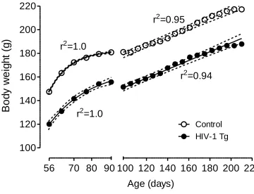
Body Weight.
The HIV-1 Tg group weighed significantly less than the control group across the
6-month period during which they were tested, F(1, 80)=92.4, p≤0.001 (Figure 2.1). Both
groups increased significantly in body weight across this period [HIV-1 Tg: F(21, 798)=
128.0, p≤0.001; control: F(21, 882)=121.6, p≤0.001]. Through PD 91, both groups
increased in weight according to a one-phase association curve fit. After attaining
adulthood, and with the implementation of food restriction, both groups increased in
weight in a linear function. There was no significant difference in the slope of these lines,
indicating that the groups did not differ in their rates of growth.
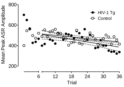
ASR Intrasession Habituation.
Mean peak ASR amplitude data from the habituation session are illustrated in
Figure 2.2. With the exception of the first 6 trials (during which the HIV-1 Tg group
showed a sharp decrease in the ASR), linear regression analysis revealed that there was
no difference in the rate of habituation across the test session for the HIV-1 Tg and
control groups (Regression line slopes: HIV-1 Tg, -4.24+/-1.24; Control: -2.7+/-0.91),
with no difference in overall ASR between groups, F<1.0.
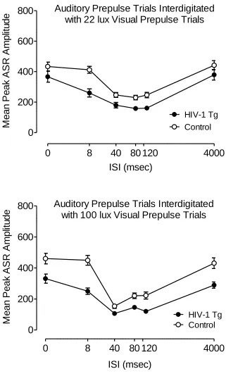
PPI with an Auditory Prepulse (in Both 22 and 100 lux Visual Prepulse Contexts).
Mean peak ASR amplitude during auditory prepulse trials assessed at 2 months of
age is illustrated in Figure 2.3. This first test period included a comparison of auditory
prepulse trials that were interdigitated with 22 lux visual prepulse trials, and auditory
prepulse trials that were interdigitated with 100 lux visual prepulse trials. Most
nor an interaction between genotype and light intensity, suggesting that the brightness of
the visual prepulse did not differentially affect PPI of either group during auditory
prepulse trials. In fact, the ISI functions of the HIV-1 Tg and control groups changed in a
similar manner with the increased light intensity of the prepulse, reflected by a significant
light intensity x ISI interaction in each group [Control: F(5,205)=5.0, p≤0.001; HIV-1
Tg: F(5,200)=3.1, p≤0.05]; with both groups, the ISI functions sharpened and the point of
peak inhibition shifted from the 80 msec ISI to the 40 msec ISI. The HIV-1 Tg and
control groups also both displayed quadratic trends for ISI during auditory prepulse trials
in each visual prepulse context, characteristic of the fundamental temporal domain of PPI
[22 lux visual prepulse: Control, F(1,41)=76.3, p≤0.002; HIV-1 Tg, F(1,40)=69.6,
p≤0.001; 100 lux visual prepulse: Control: F(1,41)=122.0, p≤0.001; HIV-1 Tg:
F(1,40)=110.7, p≤0.001]. However, there was a genotype x ISI interaction for mean peak
ASR amplitude during auditory prepulse trials that were interdigitated with the 100 lux
visual prepulse trials, F(5,405)=6.3, p≤0.001, indicating a relative insensitivity to
manipulation of ISI duration in the HIV-1 Tg group. There was no genotype condition x
ISI interaction during trials interdigitated with 22 lux visual prepulse trials.
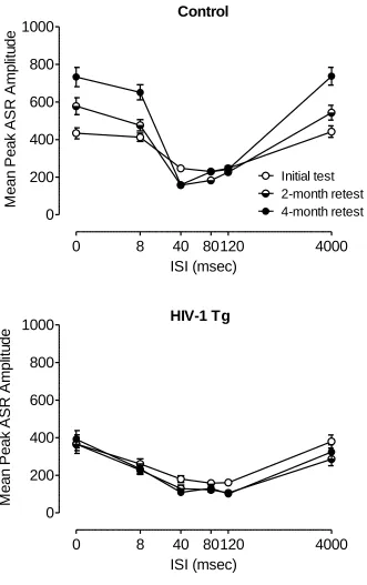
PPI with an Auditory Prepulse Across Age (in the 22 lux Visual Prepulse
Context).
The ANOVA conducted on mean peak amplitude during auditory prepulse trials
interdigitated with 22 lux visual prepulse trials across all three test periods revealed a
significant genotype x age x ISI interaction, F(10, 790)=8.1, p≤0.001, as well as a
790)=14.3, p≤0.001, and a genotype x age interaction, F(2,158)=16.6, p≤0.001.
Additional analyses were conducted to identify the locus of these interactions.
Separate analyses of each group revealed a main effect of age, F(2,82)=21.3,
p≤0.001, and an age x ISI interaction, F(10,410)=18.4, p≤0.001, in the control group,
illustrated in Figure 2.4. These effects were not observed in the HIV-1 Tg group,
suggesting that the expression of the HIV-1 transgene interfered with the age-dependent
development of perceptual sharpening.
Complementary results were obtained after separate analyses at each age, which
revealed significant genotype x ISI interactions during the 2-month retest [F(5,395)=10.5,
p≤0.001] and the 4-month retest [F(5,395)=26.1, p≤0.001], but not during the initial test
(2 months old). The genotype x ISI interactions at the later ages indicate, as does the age
x ISI interaction in the control group but not the HIV-1 Tg group, altered development of
the ISI function in the HIV-1 Tg group; they did not exhibit the same sharpening of the
ISI function with age that is apparent with the control group. Changes in percent PPI
across age also reflect the differential development of the ISI function. The HIV-1 Tg
group increased in percent PPI from 30.0%±5.5 during the initial test to 52.8%±4.4
during the 4-month retest, whereas the control group showed a much greater relative
increase, from 15.2%±7.8 to 64.0%±3.9.
The overall genotype x ISI interaction reflects not only the relative insensitivity of
the HIV-1 Tg group to manipulation of ISI duration, but also the differential peak
inhibition of the two groups, observed at the 80 msec ISI during the initial test and the
120 msec ISI during the 2-month and 4-month retests for the HIV-1 Tg animals, and at
4-month retests for the control animals. Both groups showed significant quadratic trends for
ISI, characteristic of the fundamental temporal domain of PPI [Control: F(1,41)=261.9,
p≤0.001; HIV-1 Tg: F(1,38)=108.6, p≤0.001].
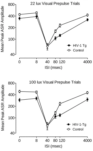
PPI with a Visual Prepulse (22 and 100 lux).
The ANOVA conducted on mean peak ASR amplitude during 22 and 100 lux
visual prepulse trials at 2 months of age revealed that there was no interaction between
genotype, light intensity, and ISI. There was also no genotype x light intensity or light
intensity x ISI interaction, nor a significant main effect of light intensity. The effect of the
22 lux light on the ASR was robust and sufficient to demonstrate visual PPI in the HIV-1
Tg as well as the controls (percent PPI at the 40 msec ISI: Control, 72.2±2.6%;
HIV-1-Tg, 74.9%±2.5); therefore, only the 22 lux visual prepulse was used for the subsequent
test periods. Both groups displayed quadratic trends for ISI in each visual prepulse
condition [22 lux: Control, F(1,41)=148.3, p≤0.001; HIV-1 Tg, F(1,40)=110.7, p≤0.001;
100 lux: Control, F(1, 41)=138.4, p≤0.001, HIV-1-Tg: F(1,40)=54.9, p≤0.001], with
comparable peak inhibition at the 40 msec ISI (100 lux visual prepulse trials, percent PPI:
Control, 73.9%±3.1; HIV-1-Tg, 66.2%±3.8). However, there was a significant genotype
x ISI interaction during 22 lux visual prepulse trials, F(5,405)=2.4, p≤0.05, as well as
during 100 lux visual prepulse trials, F(5,405)=9.0, p≤0.001, reflecting the relative
insensitivity of the HIV-1 Tg group to manipulation of ISI duration (see Figure 2.5).
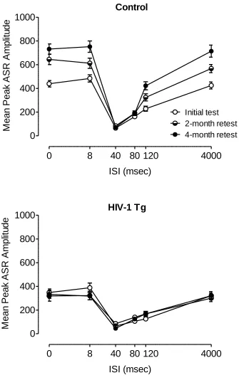
PPI with a Visual Prepulse Across Age (22 lux only).
Mean peak amplitude during 22 lux visual prepulse trials at each test period is
illustrated in Figure 2.6. There was a significant genotype x age x ISI interaction, F(10,
age x ISI interaction, F(10, 790)=5.5, p≤0.001, and a genotype x age interaction,
F(2,158)=15.7, p≤0.001. Additional analyses were conducted to identify the locus of
these interactions.
Separate analyses of each group revealed a main effect of age, F(2, 82)=24.2,
p≤0.001, and an age x ISI interaction in the control group, F(10,410)=10.4, p≤0.001, but
neither effect was present in the HIV-1 Tg group, suggesting that the expression of the
HIV-1 transgene interfered with the age-dependent development of perceptual
sharpening.
Significant genotype x ISI interactions were detected at each test period [initial
test: F(5,405)=2.4, p≤0.05; 2-month retest: F(5,400)=18.7, p≤0.001; 4-month retest:
F(5,400)=29.3, p≤0.001]. Each group showed a significant quadratic trend for ISI
[Control: F(1,41)=381.3, p≤0.001; HIV-1 Tg: F(1,38)=184.7, p≤0.001], but the genotype
x ISI interactions indicated that the HIV-1 Tg group was relatively insensitive to
manipulation of ISI duration at each test period, providing further evidence that the
HIV-1 Tg group did not develop normal perceptual sharpening.
The HIV-1 Tg and control groups each displayed peak inhibition at the 40 msec
ISI at each test period. The HIV-1 Tg group showed a quadratic trend for percent PPI
across age, F(1,38)=13.0, p≤0.005, whereas the control animals displayed similar percent
PPI at each age. The quadratic trend reflects the reduction in percent PPI during the
2-month retest in the HIV-1 Tg group (58.2%±4.2, compared to 74.9%±2.5 during the
initial test period and 72.1%±2.7 during the 4-month retest), which can be attributed to
inhibition, 40 msec, was almost identical for the two groups (HIV-1 Tg = 85.0±15.3;
Control = 83.7±8.2).
Experiment 1A: Discussion
The present experiment demonstrated alterations in preattentive processing as
assessed with PPI of the ASR in the HIV-1 Tg rat early in the expression of the HIV-1
transgene and prior to any documented neurological symptoms or signs of wasting. In the
absence of any difference in overall ASR or rate of habituation to the startle stimulus, the
HIV-1 Tg group exhibited a flatter ISI function during PPI trials, which did not sharpen
with age, as it did with control animals. Furthermore, the flatter ISI function was
observed in both auditory and visual prepulse conditions, demonstrating the generality of
sensorimotor gating deficits across prepulse modality. Over time, auditory prepulses
precipitated a temporal shift in peak inhibition in HIV-1 Tg animals relative to controls,
whereas with visual prepulses, both groups displayed peak inhibition at the 40 msec ISI.
The observed alterations in PPI indicate a lack of perceptual sharpening with age and a
relative insensitivity to the temporal dimension of sensorimotor gating in the HIV-1 Tg
rat, resembling the temporal processing deficits reported in HIV-1+ individuals early in
the disease course.
Perceptual sharpening is a developmental process in which responses are evoked
by more specific stimuli, i.e., stimulus discrimination (Ganz 1968; Gibson 1969; Tees
1976; Werner 1948). Younger subjects (Rubel and Rosenthal 1975) and sensory-deprived
subjects (Kerr et al. 1979) exhibit significantly flatter stimulus generalization gradients.
sharpening. In rats, the heart rate orienting response has been used to measure the
ontogeny of perceptual sharpening. Rats at 16-17 days of age that have been habituated to
an auditory stimulus will generalize the habituated response to a wide range of auditory
stimuli; by day 20, stimulus discrimination is apparent with a much sharper
generalization gradient (Campbell and Haroutunian 1983). We have previously observed
perceptual sharpening of PPI with auditory, visual, and tactile prepulses in Long-Evans
rats, at PD 18, 35, and 90 (Hord et al. 2008). These animals showed gradually sharper ISI
functions for PPI with an auditory or tactile prepulse across age. As the visual system is
the last system to develop in the rat (Gottlieb 1971), PPI with a visual prepulse was not
apparent until PD 90, when peak inhibition was exhibited at the 40 msec ISI. At PD 18
and 35, the ISI functions were flat, reflecting the immaturity of the visual sensory system.
The HIV-1 Tg and control rats in the present study exhibited robust PPI with a visual
prepulse at two months of age, which suggests that the visual system and its afferents to
PPI circuitry are well-developed at this age. However, the point of peak inhibition in the
ISI functions of the HIV-1 Tg group under both prepulse conditions did not become
clearly defined across age as was observed in the control group. Although the HIV-1 Tg
rats appear to have functional auditory and visual systems, a more specific deficit in the
development, or perceptual sharpening, of temporal sensitivity in the context of PPI was
exhibited.
In the present experiment, the ISI functions of the HIV-1 Tg and control groups
were most similar at two months of age, and then changed in different ways across age.
For PPI with an auditory prepulse, both groups had peak inhibition at the 80 msec ISI at
inhibition at the 120 msec ISI, representing a rightward shift from the control group’s
peak of inhibition at the 40 msec ISI. Differences in the point of peak inhibition have
previously been observed in female Sprague-Dawley HIV-1 Tg and control rats as well,
between 5-7 months of age (Moran et al. 2013). Shifts in peak inhibition have also been
demonstrated in rats administered HIV-1 viral protein injections. Leftward shifts were
observed in 30- and 60-day old male Sprague-Dawley rats following neonatal Tat
injection (Fitting et al. 2006a) and in 9-month old male and female Sprague-Dawley rats
given neonatal gp120 injections (Fitting et al. 2006b).
Despite the differences in sensorimotor gating observed between the HIV-1 Tg
and control groups, they displayed similar inhibition to a visual prepulse, which is
particularly notable given the presence of cataracts in the HIV-1 Tg animals. It is clear
from this finding that the HIV-1 Tg group could detect the 20 msec visual stimulus
despite their cataracts, and thus, a visual stimulus greater than or equal to the intensity
and duration used in this experiment would have utility in other experimental paradigms
with HIV-1 Tg rats, especially measures of executive function and other cognitive
domains relevant to the study of HAND. The use of visual stimuli permits utilization of a
variety of methods to test cognition in HIV-1 Tg rats.
The utilization of a visual prepulse in the PPI paradigm also affords the
opportunity to determine the generality of any alterations in sensorimotor gating
independent of prepulse stimulus modality. Comparable alterations in auditory and visual
PPI, as observed in the present experiment, are consistent with a deficit in sensorimotor
gating. Differential alterations in auditory versus visual PPI would, alternatively, be
In summary, the present experiment demonstrates that HIV-1 Tg rats exhibit
neurological deficits early in the expression of the HIV-1 transgene, prior to clinical signs
of wasting, which progress with age, bearing a marked resemblance to the temporal
processing deficits observed in individuals with HIV-1. Both the relative insensitivity to
the temporal dimension of sensorimotor gating and the lack of development of perceptual
sharpening with age suggest clear evidence of temporal processing deficits in the HIV-1
Tg rat.
Experiment 1B: Executive Function
The animals were trained on a series of operant tasks tapping attention and core
components of executive function, modeling the analogous functions in humans,
specifically the fundamental components of flexibility, inhibition, and set-shifting. It was
hypothesized that the HIV-1 Tg animals would exhibit deficits in attention and the core
components of executive function, which are the cognitive domains that are most
typically impaired in individuals with HAND. Parallel components of executive function
supported by homologous subregions of the PFC in the rat and human have been widely
demonstrated (Kesner and Churchwell, 2011). Thus, for the present experiment,
executive function and attention may be assessed with translatable results in the rat, with
the use of measures selected to model specific cognitive functions.
Apparatus
Behavioral training and testing was conducted in 22 operant chambers located
inside sound-attenuating chambers (Med Associates). One wall of each chamber
consisted of two retractable levers, a pellet dispenser (45 mg) between the two levers, and
light was located on the rear wall. Signal presentation, lever operation, reinforcement
delivery, and data collection were controlled by a PC and Med-PC for Windows software
(V 4.1.3; Med Associates).
Signal Detection Task
Starting at 3-months of age, animals were initially trained to press both levers on
an FR-1 schedule of reinforcement for sucrose pellets (45 mg). To prevent side-bias,
subjects were not rewarded for more than five consecutive presses on a single lever. After
the animals achieved at least 40 reinforcers during the 42-min sessions for three
consecutive days, with less than 20% variance across days, they were trained on the
signal detection task.
Each signal detection session began with a 5-min habituation period. The house light was
off for the duration of the session. The presentation of signals (central panel light
illumination) and non-signals (no illumination) was randomized over the 160
trial-session, with intertrial intervals of 9 ± 3 sec, during which time the levers remained
retracted. Levers were extended 2 seconds after each trial (signal or non-signal) began
and remained extended for 6 seconds for the animal to make a response. During signal
trials, the light stimulus remained illuminated until the animal made a response, or 6
seconds elapsed, whichever occurred first. For half of the animals, lever presses on the
left lever during signal trials and on the right lever during non-signal trials were rewarded
(hits and correct rejections, respectively). The reverse set of rules was used for the other
half of the subjects. Incorrect responses during signal trials (misses) and non-signal trials
repetitions of the trial. Finally, failure to respond appropriately to the correction trials
resulted in a forced-choice trial in which the same stimulus type was repeated (signal or
non-signal) but only the correct lever was extended and remained extended until a
response was made or 2 minutes elapsed, whichever occurred first. Each animal was
trained on the task each day until it achieved 70% or greater accuracy on three
consecutive sessions. Accuracy was calculated as the total number of hits and correct
rejections divided by the total number of correct and incorrect responses in a session.
Discrimination Task
Each discrimination task session began with a 5-min habituation period. The
house light was off for the duration of the session. Left panel light and right panel light
trials were presented randomly in a 160-trial session, with intertrial intervals of 9 ± 3 sec,
during which time the levers remained retracted. The light stimulus during each trial was
presented for 1 second, followed by the presentation of both levers for 6 seconds or until
the animal made a response, whichever occurred first. Animals were rewarded for
pressing the lever underneath the light stimulus. Following an incorrect response, animals
were presented with three correction trials. If the animal again responded incorrectly, it
was given a forced trial. Each animal was trained on the task until it achieved 70% or
greater accuracy on three (non-consecutive) sessions. Accuracy was calculated as the
total number of correct responses divided by the total number of correct and incorrect
Reversal Task
Each discrimination reversal task session began with a 5-min habituation period.
The houselight was off for the duration of the session. Left panel light and right panel
light stimulus trials were presented randomly in a 160-trial session, with intertrial
intervals of 9 ± 3 sec, during which time the levers remained retracted. The light stimulus
during each trial was presented for 1 second, followed by the presentation of both levers
for 6 seconds or until the animal made a response, whichever occurred first. The opposite
set of response rules from the discrimination task was used in the reversal task; the
animals were rewarded for pressing the lever on the opposite side of the light stimulus.
Following an incorrect response, animals were presented with three correction trials. If
the animal again responded incorrectly, it was given a forced trial. Each animal was
trained on the task until it achieved 70% or greater accuracy on three (non-consecutive)
sessions. Accuracy was calculated as the total number of correct responses divided by the
total number of correct and incorrect responses in a session.
Extradimensional Set-Shifting Task
Each extradimensional set-shifting task session began with a 5-min habituation
period. The houselight was off for the duration of the session. Left panel light and right
panel light stimulus trials were presented randomly in a 160-trial session, with intertrial
intervals of 9 ± 3 sec, during which time the levers remained retracted. The light stimulus
during each trial was presented for 1 second, followed by the presentation of both levers
animals were rewarded for pressing the left lever, and the other half were rewarded for
pressing the right lever, regardless of the position of the light stimulus. Following an
incorrect response, animals were presented with three correction trials. If the animal
again responded incorrectly, it was given a forced trial. Each animal was trained on the
task until it achieved 70% or greater accuracy on five consecutive sessions. Accuracy was
calculated as the total number of correct responses divided by the total number of correct
and incorrect responses in a session.
Statistics
All data were analyzed using SPSS Statistics 20 (IBM Corp., Somers, NY). For
the signal detection task, percent accuracy, and hits, misses, correct rejections, and false
alarms were analyzed with independent samples T-tests. For the discrimination, reversal,
and extradimensional set-shifting task, a two-way mixed-factor analysis of variance
(ANOVA) was used to analyze correct responses and errors, with genotype (HIV-1 Tg or
control) as the between-groups factor, and response type (correct responses vs. errors) as
a within-subjects factor. Each animal’s performance on each measure was averaged
across the first three days at which the animal performed with 70% accuracy, to provide
the data for the analyses. Trials and errors to criterion were analyzed with independent
sample T-tests, andsessions to criterion were analyzed with curve-fitting to assess the
temporal process of acquisition. An alpha level of p≤0.05 was considered significant for
all statistical tests. Sample sizes were chosen with the goal of sufficient statistical power
(> 0.80) to maximize the likelihood of detecting subtle early alterations of expression of
Experiment 1B: Results
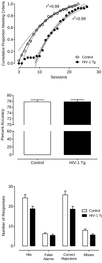
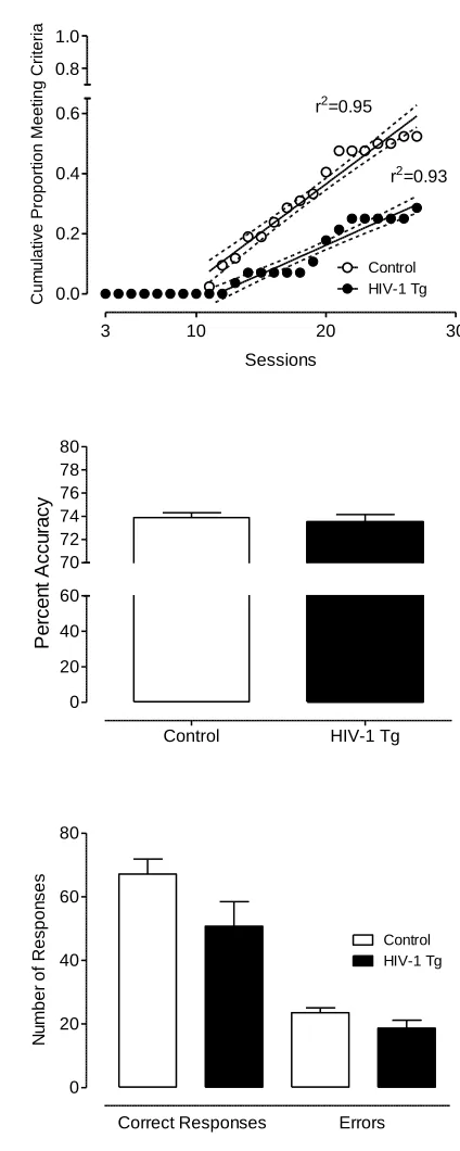
Signal Detection Task.
All controls and all but 2 HIV-1 Tg animals met the criterion within one month of
daily testing. The HIV-1 Tg and control groups followed similar rates in achieving the
criterion, but the HIV-1 Tg group showed an initial 7-day lag before any of the animals
met the criterion, reflected by their significantly greater number of trials [t(80)=-2.1,
p≤0.05] and errors to criterion [t(80)=-2.4, p≤0.05] compared to the control group. In
addition, a one-phase association regression analysis on the cumulative proportion of
animals meeting the criterion over sessions revealed that a different curve was best fit to
each group, F(3,27)=245.2, p≤0.01, further illustrating the gap between groups in the
number of sessions needed to reach criterion (see Figure 2.7). Once the animals met the
criterion, percent accuracy was not significantly different between groups (3-day average:
HIV-1 Tg: 77.8+/-0.43; Control: 77.8+/-0.42). However, the HIV-1 Tg animals
responded significantly less than the control animals during the signal detection task,
F(1,80)=8.2, p≤0.01. A significant group x response type (correct vs. error) interaction
[F(1,80)=8.6, p≤0.01] suggested that the HIV-1 Tg animals displayed a different
response profile than the control group. The HIV-1 Tg animals had fewer hits than the
control animals, t(80)=2.6, p≤0.05, misses, t(80)=2.5, p≤0.05, and correct rejections,
t(80)=2.9, p≤0.005, than the control group, consistent with a lapse of attention. There was
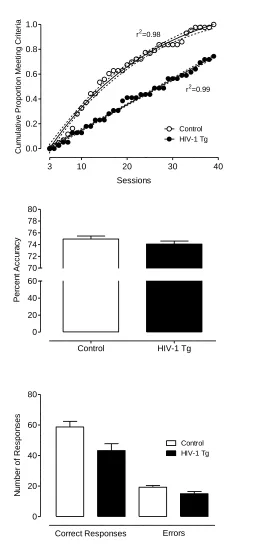
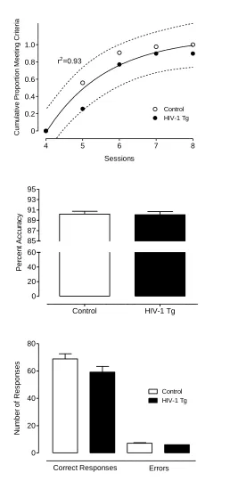
Discrimination Task.
All controls and 29 HIV-1 Tg animals met the criterion within 40 days of daily
testing. There was no significant difference between HIV-1 Tg and control animals in
errors or trials to criterion (t≤1, p>0.8). However, regression analyses revealed that the
rate at which each group achieved the criterion followed different functions; the HIV-1
Tg animals exhibited a linear function (r2=0.99) whereas the control group showed a
curvilinear one-phase association function (r2=0.98), illustrating the slower acquisition of
the task by the HIV-1 Tg animals over sessions (see Figure 2.8). Of the animals that met
the criterion, there was no significant difference in percent accuracy (3-day average:
HIV-1 Tg: 74.9+/-0.52; Control: 74.1+/-0.51). Despite attaining the same accuracy, the
HIV-1 Tg animals that met the criterion made significantly fewer overall responses,
F(1,70)=6.8, p≤0.05. A significant interaction between group and response type,
F(1,70)=6.8, p≤0.05, indicates that there was a smaller difference between the number of
correct responses and the number of errors in HIV-1 Tg group compared to control
group.
Reversal Task.
Of the subjects that met the criterion for the discrimination task and continued to
the discrimination-reversal task, 22 control animals and 8 HIV-1 Tg animals met the
discrimination-reversal task criterion within one month of daily testing. There was no
significant difference between HIV-1 Tg and control animals in errors or trials to
criterion (t≤1, p>0.4). However, linear regression analysis on the cumulative proportion