0095-1137/07/$08.00⫹0 doi:10.1128/JCM.01344-06
Copyright © 2007, American Society for Microbiology. All Rights Reserved.
Development of Novel Real-Time PCR Assays for Detection and
Differentiation of Eleven Medically Important
Aspergillus
and
Candida
Species in Clinical Specimens
䌤
Claudia Schabereiter-Gurtner,* Brigitte Selitsch, Manfred L. Rotter,
Alexander M. Hirschl, and Birgit Willinger
Department of Clinical Microbiology, Institute of Hygiene and Medical Microbiology, Medical University of Vienna, Vienna, Austria
Received 30 June 2006/Returned for modification 8 August 2006/Accepted 13 January 2007
In the present study, novel real-time PCR assays targeting the fungal ITS2 region were developed for the
detection and differentiation of medically importantAspergillusspecies (Aspergillus fumigatus,Aspergillus
flavus,Aspergillus nidulans,Aspergillus niger, andAspergillus terreus) andCandidaspecies (Candida albicans,
Candida dubliniensis,Candida glabrata,Candida krusei,Candida parapsilosis, andCandida tropicalis) using
a LightCycler instrument. The combination of a group-specific and a universal primer with fiveAspergillus
or sixCandida species-specific biprobes in one reaction mixture facilitated rapid screening and species
differentiation by the characteristic peak melting temperatures of the biprobes. Both assays can be performed either as single assays or simultaneously in the same LightCycler run. The analytical sensitivity using pure cultures and EDTA-anticoagulated blood, cerebrospinal fluid (CSF), and tissue samples spiked withA. fumigatusandC. albicanscell suspensions was shown to be at least 1 CFU per PCR, corresponding
to 5 to 10 CFU/ml blood and 10 CFU/200l CSF or 0.02 g tissue. To assess the clinical applicability, 26
respiratory samples, 4 tissue samples from the maxillary sinus, and 1 blood sample were retrospectively tested and real-time PCR results were compared with results from culture, histology, or a galactomannan enzyme-linked immunosorbent assay (ELISA). Twenty samples (64.5%) were both culture positive and positive by real-time PCR. Six samples (19.4%) showed no growth of fungi but were positive by real-time PCR. However, all of the tissue samples were positive by both PCR and histology. The blood sample
showed no growth of Aspergillus, but aspergillosis was confirmed by positive galactomannan ELISA,
histology, and PCR results. The remaining samples (16.1%) were culture and PCR negative; also, no other
signs indicating fungal infection were observed. Our data suggest that theAspergillusandCandidaassays
may be appropriate for use in clinical laboratories as simple and rapid screening tests for the most
frequently encounteredAspergillusandCandida species and might become an important tool in the early
diagnosis of fungal infections in the future.
Invasive fungal infections are increasingly recognized as a primary cause of morbidity and mortality, especially in immu-nocompromised patients. The frequency of nosocomial candi-demia has increased 10-fold during the past two decades (3).
Candida albicans, formerly the most important species, is still the one which most often causes disease. However, other spe-cies thanC. albicans, in particular,Candida glabrata, but also
Candida tropicalis, Candida krusei, and Candida parapsilosis, have gained greater significance and must not be overlooked (15, 31). The prevalence of candidiasis and the increase in
Candidabeing resistant to polyene and azole drugs have made rapid species differentiation mandatory (29, 31). There has been a less striking, though also substantial, increase in the incidence of invasive aspergillosis (IA) (27). Invasive aspergil-losis is mainly caused by Aspergillus fumigatus, followed by
Aspergillus flavusandAspergillus terreus. Other species, such as
Aspergillus nidulans,Aspergillus niger, andAspergillus ustus, are rarely found in diagnosing IA.
The definite and rapid diagnosis of invasive fungal infections
is difficult due to the lack of sensitive test methods. Therefore, efforts to improve diagnosis are ongoing and need to be further intensified. Although proving the presence of infection by his-tology and culture remains the cornerstone of diagnosis, non-culture-based methods are being developed to allow early de-tection. Among the most promising approaches are the detection of fungal antigens and PCR (2, 4, 42). The results of some studies suggest that a combination of theAspergillus galacto-mannan enzyme-linked immunosorbent assay (GM-ELISA) and real-time PCR may provide improved diagnosis of invasive aspergillosis (5, 7, 20, 35).
Nevertheless, not only the detection of fungi, but also their identification, which can be obtained only by PCR or culture, is important for the optimal choice of antifungals and duration of therapy. A variety of PCR assays based on the detection of fungal DNA in sterile human body fluids or tissue samples to allow early diagnosis of fungal infections and to improve the survival rate of patients suffering from invasive infections has been described. However, in contrast to the GM-ELISA, none of the developed PCR assays have been standardized, resulting in diverging results. In general, the introduction of real-time PCR technology in the detection of fungal infections has in-creased the reliability of PCR results compared to results ob-tained by conventional PCR methods. Real-time PCR sharply
* Corresponding author. Mailing address: Department of Clinical Microbiology, University Hospital of Vienna, Wa¨hringer Gu¨rtel 18-20, 1090 Vienna, Austria. Phone: 43 (1) 5975. Fax: 43 (1) 40400-5162. E-mail: [email protected].
䌤Published ahead of print on 24 January 2007.
906
on May 16, 2020 by guest
http://jcm.asm.org/
decreases the risk of false-positive results due to PCR product carryover during gel electrophoresis with subsequent Southern blot hybridization or enzyme-linked immunoassays to check the specificity of the PCR product. The identification of species via melting curve analysis with species-specific hybridization probes further increases specificity, as one mismatch in the probe binding site would lead to an altered melting tempera-ture (Tm). The fast turnaround time of less than 2 h is another
advantage of the real-time PCR technology (4, 13, 14). Although infections withCandidaandAspergillusspecies are the ones most encountered by high-risk patients, there are only a few reports of PCR assays specifically targetingCandidaspp. and Aspergillusspp. at the same time (21, 32). Furthermore, the majority of Aspergillus-specific real-time PCR assays are restricted to the detection ofA. fumigatusalone. Therefore, we have decided to design a real-time PCR approach which en-ables culture-independent screening forCandidaand Aspergil-lusinfections within only a few hours. The aim of this study was to develop assays specific for the fungal ITS2 region, allowing the simultaneous detection and differentiation of 11 medically important species ofAspergillus(Aspergillus fumigatus, Aspergil-lus flavus, Aspergillus nidulans, Aspergillus niger, and Asper-gillus terreus) and Candida (Candida albicans, Candida dubliniensis, Candida glabrata, Candida krusei, Candida parapsilosis, andCandida tropicalis) with species-specific bi-probes on the LightCycler instrument (Roche Diagnostics GmbH, Mannheim, Germany). Biprobes were chosen as they guarantee high specificity and have not yet been ap-plied to the species differentiation of fungi.
MATERIALS AND METHODS
Fungal strains and DNA extraction.Fungal strains were obtained from the American Type Culture Collection (ATCC), Manassas, VA; the Centraalbureau voor Schimmelcultures (CBS), Utrecht, The Netherlands; or the Institute of Hygiene and Medical Microbiology, Medical University of Vienna. The identi-fication of all clinical isolates was confirmed by conventional morphological and physiological methods (1, 40). DNA was extracted from the following isolates:C. glabrata(ATCC 90030),C. tropicalis(ATCC 750),C. albicans(ATCC 44374 and 10 clinical isolates),C. parapsilosis(ATCC 22019),C. krusei(ATCC 6258 and 2 clinical isolates),C. dubliniensis(4 clinical isolates),Candida kefyr(clinical isolate),
Cryptococcus neoformans(ATCC 62006),Malassezia furfur(clinical isolate), Malasse-zia pachydermatis(clinical isolate),Trichosporonsp. (clinical isolate),Saccharomyces cerevisiae(ATCC 9763),A. flavus(ATCC 64025),A. niger(ATCC 10578 and 9 clinical isolates),A. fumigatus(ATCC 14110 and 3 clinical isolates),A. terreus(CBS 116.46 and 5 clinical isolates),A. nidulans(3 isolates obtained from an external quality control program; UK NEQAS, Central Public Health Laboratory, London, United Kingdom), and clinical isolates ofAspergillus sydowii,Aspergillus versicolor,
Aspergillus ustus,Penicilliumspp.,Rhizopus oryzae,Mucorsp.,Rhizomucor pusillus,
Absidia corymbifera,Cunninghamellasp.,Syncephalastrumsp.,Scedosporium apio-spermum,Fusariumsp.,Verticilliumsp., andPhialophorasp.
Fungal isolates were cultured by standard cultivation methods. Cell suspen-sions were prepared with 0.9% saline and adjusted to a 3 McFarland standard. For quantification of theCandidaandAspergillussuspensions, 10-fold serial dilutions were plated and CFU were counted. The fungal suspensions were centrifuged, resuspended in 200l 0.9% NaCl, and incubated with 20 U recom-binant Lyticase (Sigma-Aldrich, Austria) at 37°C for 30 min. DNA was extracted with a High Pure PCR template preparation kit (Roche Molecular Biochemicals, Mannheim, Germany) by following the instructions of the manufacturer. DNA was eluted with 100l elution buffer provided with the kit.
Clinical samples.Samples obtained for routine microbiology diagnostic pro-cedures from patients suspected and not suspected of having fungal infections were retained for evaluation of the PCR assays. Bronchoalveolar lavage (BAL) (n⫽19), bronchial secretion (n⫽5), and tracheal secretion (n⫽2) samples and an EDTA-anticoagulated blood sample (n⫽1) from lung transplant recipients and from patients in the intensive care unit were used for further investigations. Tissue samples from the maxillary sinus (n⫽4) were obtained from patients with
histologically proven fungus balls. Part of the material was cultured using stan-dard cultivation methods. The remaining material (1 to 2 ml) was stored at ⫺20°C until it was used for DNA extraction.
DNA extraction from 3 ml EDTA-blood sample.A modification of a protocol described by Loeffler et al. (25) was used. For red cell lysis, 3 ml EDTA-blood was mixed with 15 ml lysis buffer (LB; 10 mM Tris [pH 7.6], 5 mM MgCl2, 10 mM
NaCl), incubated for 15 min on ice, and then centrifuged for 10 min at 3,000 rpm. The pellet was resuspended in 15 ml LB, incubated again for 15 min on ice, and then centrifuged for 10 min at 3,000 rpm. For white cell lysis, the pellet was then resuspended in 1 ml LB containing 200g/ml protease (QIAGEN, Hilden, Germany), incubated at 65°C for 45 min, and then centrifuged at 13,000 rpm for 10 min. In order to obtain spheroplasts, the pellet was resuspended in 500l Lyticase solution (50 mM Tris [pH 7.6], 1 mM EDTA [pH 8.0], 0.2% 2-mercap-toethanol) containing 20 U recombinant Lyticase, incubated at 37°C for 30 min, and then centrifuged at 13,000 rpm for 10 min. Finally, DNA was extracted with a High Pure PCR template preparation kit by following the instructions of the manufacturer. DNA was eluted with 100l elution buffer.
DNA extraction from 200l cerebrospinal fluid (CSF), BAL, bronchial, or tracheal secretion samples.For concentration of the fungi, 1 to 2 ml of the specimen was centrifuged for 5 min at 13,000 rpm. In order to obtain sphero-plasts, the pellet plus 200l supernatant was incubated with 20 U recombinant Lyticase at 37°C for 30 min. Finally, DNA was extracted with a High Pure PCR template preparation kit by following the instructions of the manufacturer. DNA was eluted with 100l elution buffer.
DNA extraction from tissue samples.Tissue (0.02 g) was incubated in 200l elution buffer and 200l binding buffer from the High Pure PCR template preparation kit with 900g protease at 55°C until the tissue was completely digested. After inactivation of the protease (95°C for 5 min), the sample was treated with 20 U recombinant Lyticase at 37°C for 30 min to obtain sphero-plasts. Finally, DNA was extracted with a High Pure PCR template preparation kit by following the instructions of the manufacturer. DNA was eluted with 100 l elution buffer.
In order to avoid contamination, all steps were performed with aerosol-resis-tant tips. DNA extraction, preparation of the master mix, and addition of the template were carried out in two separate rooms. For each extraction, a reagent blank was carried out to exclude false-positive PCR results due to contamination.
Aspergillus-andCandida-specific biprobe assays.GenBank was searched for sequences of the ITS2 regions ofCandidaandAspergillusspecies and phyloge-netically related fungi. The published sequences were aligned using ClustalW (http://www.ebi.ac.uk/clustalw/), and primers and probes were designed. A BLAST search (http://www.ncbi.nlm.nih.gov/BLAST/) was performed to check the specificity of the DNA sequences of the primers and probes. The sequences of the primers and probes are shown in Table 1. Primer Asp-F is mainly specific for members of theHyphomycetes, whereas primer Cand-F is mainly specific for members of the yeasts. Both primers anneal within the ITS2 region. Primer ITS-R anneals to a highly conserved region of the 28S rRNA gene. For the detection and differentiation of the fiveAspergillusspecies and the sixCandida
species, 11 different species-specific biprobes were designed, allowing species identification by specific melting peaks. Sequence alignments of the correspond-ingAspergillusandCandidaspecies are shown in Fig. 1 and 2, respectively.
To facilitate rapid screening for the presence ofCandidaorAspergillusin a sample, the sixCandida-specific biprobes or the fiveAspergillus-specific biprobes were used together in one reaction mixture. The 20-l real-time PCR mixtures were prepared with 2l of LightCycler-FastStart DNA master SYBR green I (Roche Molecular Biochemicals, Mannheim, Germany), 4 mM MgCl2, primers
and probes as shown in Table 1, and 3l of DNA extract made up to 20l with water. PCRs were performed in a LightCycler instrument with preliminary de-naturation for 10 min at 95°C, followed by 60 amplification cycles (with a temperature transition rate of 20°C/s) of denaturation at 95°C for 8 s, annealing at 55°C for 10 s, and primer extension at 72°C for 10 s, with a single fluorescence acquisition step at the end of the extension. This was followed by a melting analysis of the probe-PCR product duplex consisting of 95°C for 30 s and then cooling to 35°C for 60 s before the temperature was raised to 98°C at a rate of 0.2°C/s with continuous fluorescence acquisition. A final cooling step was per-formed at 40°C for 10 s.
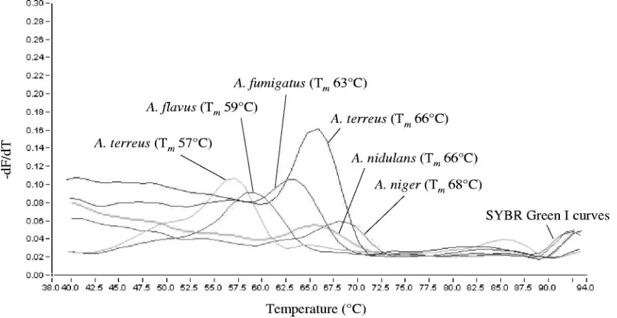
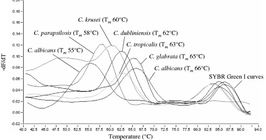
Light emission due to SYBR green was monitored in the F1 channel, whereas biprobe-specific melting peaks were analyzed in the F3 channel of the instru-ment. Samples were considered positive for anAspergillusorCandidaspecies upon the presence of a biprobe-specific melting peak. In this case, the species was confirmed by repeating the analysis with the appropriate specific biprobe in the reaction mixture. SpecificTmvalues of the biprobes are shown in Table 1. Melting peaks obtained with theAspergillusandCandidaspecies-specific bi-probes are shown in Fig. 3 and 4, respectively.
on May 16, 2020 by guest
http://jcm.asm.org/
The detection of galactomannan in the blood sample (patient 26) was per-formed by means of the PlateliaAspergillusenzyme immunoassay (EIA) kit (Bio-Rad Laboratories Central Europe, Vienna, Austria), according to the man-ufacturer’s instructions.
Evaluation of analytical sensitivity and specificity.In order to evaluate the analytical sensitivity of the assays, dilutions of DNA of the 11 species in the range between 105and 10⫺1CFU per PCR were used as template DNA. For
evaluation of the DNA extraction protocols, 3 ml EDTA-blood, 200l CSF, and 0.02 g tissue (aorta) of noninfected patients or healthy volunteers were spiked with dilutions ofC. albicansandA. fumigatusin the range between 104
and 100CFU/ml. DNA was extracted and analyzed. To confirm the absence
of PCR inhibitors, one additional reaction mixture containing the DNA extract was spiked with 4 CFU ofC. albicansorA. fumigatus to exclude inhibition. The analytical specificity of the assays was evaluated with DNA extracted fromAspergillus,Candida, and all the other fungal isolates de-scribed. Furthermore, BLAST analyses of primers and probes were per-formed.
RESULTS
Specificity of the Aspergillus-specific biprobes and melting
temperature values.A BLAST search was performed to check
the specificity of theAspergillus-specific biprobes. Biprobes spe-cific for A. flavus, A. fumigatus, A. nidulans, andA. niger had 100% sequence similarity to each of the respective Aspergillus
strains in the database. They further revealed 100% similarity to some otherAspergillus,Emericella, andNeosartoryaspecies, which may lead to cross-reactions (Table 2). The biprobe specific forA. terreusshowed mismatches with threeA. terreusstrains (Fig. 1). To further prove the specificity, DNA of all fungal isolates men-tioned under “Fungal strains and DNA extraction” was tested with each of theAspergillusspecies-specific biprobes. As expected, cross-reactivity of biprobe A.nid-S was observed withA. sydowii,
A. ustus, andA. versicolor. No cross-reactivity was observed with the other isolates. TheTmvalues of the biprobe-specific melting
peaks are shown in Table 1 and Fig. 3. A. terreusstrain CBS 116.46 revealed a biprobe-specific melting peak with aTmvalue of
[image:3.585.43.549.79.310.2]57°C instead of 66°C. The PCR product of this strain was se-quenced and revealed the same sequence variation as the two strains with accession numbers AJ001333 and AJ001331 found in
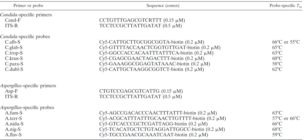
TABLE 1. Sequences and concentrations of primers and probes and corresponding probe-specificTmin real-time PCR assays
Primer or probe Sequence (concn) Probe-specificTm
Candida-specific primers
Cand-F CCTGTTTGAGCGTCRTTT (0.15M)
ITS-R TCCTCCGCTTATTGATAT (0.5M)
Candida-specific probes
C.alb-S Cy5-CATTGCTTGCGGCGGTA-biotin (0.2M) 66°C or 55°C
C.glab-S Cy5-GTTTTACCAACTCGGTGTTGAT-biotin (0.2M) 65°C
C.trop-S Cy5-GGCCACCACAATTTATTTCA-biotin (0.2M) 63°C
C.krus-S Cy5-CGAGCGAACTAGACTTT-biotin (0.2M) 60°C
C.para-S Cy5-GAAAGGCGGAGTATAAAC-biotin (0.2M) 58°C
C.dubl-S Cy5-CATTGCTAAGGCGGTCT-biotin (0.2M) 62°C
Aspergillus-specific primers
Asp-F CTGTCCGAGCGTCATTG (0.15M)
ITS-R TCCTCCGCTTATTGATAT (0.5M)
Aspergillus-specific probes
A.fum-S Cy5-AGCCGACACCCAACTTTATTT-biotin (0.2M) 63°C
A.terr-S Cy5-ACGCATTTATTTGCAACTTGTTTT-biotin (0.2M) 57°C or 66°C
A.nidu-S Cy5-GTCACCCGCTCGATTAGG-biotin (0.2M) 66°C
A.nig-S Cy5-TCACATGCTCTGTAGGATTGGCC-biotin (0.2M) 68°C
A.flav-S Cy5-TGCCGAACGCAAATCAAT-biotin (0.2M) 59°C
FIG. 1. ClustalW multiple-sequence alignment of the ITS2 region amplified with primers Asp-F and ITS-R fromA. fumigatus(AY373851),A. flavus(AJ876522),A. nidulans(AY373888),A. niger(AY373852), andA. terreus(A. terreus1, AY373871;A. terreus2, AJ001333; andA. terreus3, AJ001368) (designations in parentheses are GenBank accession numbers). Incomplete sequences of the ITS2 regions ofA. terreus2 andA. terreus
3 were found in the database. Nucleotides corresponding to the species-specific biprobes are bold, in italics, and underlined.
on May 16, 2020 by guest
http://jcm.asm.org/
[image:3.585.44.541.569.685.2]the database. Occasionally, an indistinct and broad peak at the 52°CTmwas observed when the fiveAspergillusspecies-specific
biprobes were used together for screening (Table 3).
Specificity of the Candida-specific biprobes and melting
temperature values.A BLAST search was performed to check
the specificity of theCandida-specific biprobes. Each biprobe had 100% sequence similarity to the respectiveCandidastrains in the database. None of those revealed 100% similarity to any other sequence (Table 2). To further prove the specificity, DNA of the fungal isolates described above was tested with each of theCandidaspecies-specific biprobes. Table 1 and Fig.
4 show theTmvalues of the biprobe-specific melting peaks. One clinical isolate ofC. albicansrevealed a biprobe-specific peakTmof 55°C instead of 66°C. The PCR product of this strain was sequenced and revealed one mismatch in the bi-probe-specific annealing site (Fig. 2). Furthermore, C.alb-S showed cross-reactivity withC. dubliniensis(Tmvalue of 47°C),
C.dubl-S showed cross-reactivity withC. albicans(Tmvalue of 52°C), and C.glab-S showed cross-reactivity withS. cerevisiae
(Tmvalue of 50°C).
Analytical sensitivity. The lowest Candida or Aspergillus
[image:4.585.42.544.73.259.2]concentration delivering a positive PCR result was 1 CFU per
FIG. 2. ClustalW multiple-sequence alignment of the ITS2 region amplified with primers Cand-F and ITS-R fromC. albicans(C. albicans1, AY139782, and C. albicans 2, clinical isolate), C. dubliniensis (AY382338), C. glabrata (AY139784), C. krusei (AY235807),C. parapsilosis
(AY217022), andC. tropicalis(AF321539) (designations in parentheses are GenBank accession numbers). Nucleotides corresponding to the species-specific biprobes are bold, in italics and underlined.
FIG. 3. Melting peaks obtained fromA. fumigatus,A. flavus, A. nidulans,A. niger, and A. terreuswith the corresponding species-specific biprobes. The values on theyaxis are the first negative derivative of the change in fluorescence (dF) divided by the change in temperature (dT) (⫺dF/dT).
on May 16, 2020 by guest
http://jcm.asm.org/
[image:4.585.75.513.470.696.2]PCR. After DNA extraction, the lowest concentration deliver-ing a positive PCR result was 15 to 30 CFU in 3 ml blood (0.45 to 0.9 CFU per PCR) and 10 CFU in 200l CSF or 0.02 g tissue (0.3 CFU per PCR). No change in analytical sensitivity was observed when the fiveAspergillus-specific or the six Can-dida-specific biprobes were used together in one reaction mix-ture.
Detection of Aspergillus spp. and Candida spp. in clinical
samples.Thirty-one clinical samples derived from patients
ir-respective of the presence of possible fungal infections were examined by standard cultivation and by real-time PCR (Table 3). In 25 samples (80.6%), fungi were detected by either
[image:5.585.74.512.72.302.2]cul-ture (n⫽20), histology (n⫽4), or a GM-ELISA (n⫽1). For the remaining six samples (19.4%), both culture and histology were negative. When the results of culture were compared with those of real-time PCR, 20 samples (64.5%) (patients 1 to 20) were both culture positive and positive by real-time PCR. In samples 1 to 8, concordant results were obtained by both culture and real-time PCR. In samples 9 to 16, real-time PCR detected not only the fungus grown in culture, but also additional Candida species such as C. albicans and/or C. glabrata. In sample 17, real-time PCR detectedC. glabrata, whereasC. lusitaniaewas grown by culture. In samples 18 to 20, showing the growth of more than one species, not all of
FIG. 4. Melting peaks obtained fromC. albicans,C. dubliniensis,C. glabrata,C. krusei,C. parapsilosis, andC. tropicaliswith the corresponding species-specific biprobes. The values on theyaxis are the first negative derivative of the change in fluorescence (dF) divided by the change in temperature (dT) (⫺dF/dT).
TABLE 2. Results of BLAST analyses of theAspergillusandCandidaspecies-specific biprobes
Biprobe
No. with 100% sequence similarity to biprobe/total no.
in GenBank database
Other species with 100% sequence similarity
A.flav-S 29/29A. flavusstrains A. oryzae,A. parasiticus, andAureobasidium mansoniia
A.fum-S 25/25A. fumigatusstrains Aspergillus fumisynnematus,Aspergillus lentulus,Aspergillus viridinutans,Neosartorya aureola,
Neosartorya botucatensis,Neosartorya fischeri,Neosartorya glabra,Neosartorya spinosa, andNeosartorya udagawae
A.nid-S 14/14A. nidulansstrains Emericella astellata,Emericella indica,Emericella quadrilineata,Emericella rugulosa,
Emericella variecolor,A. sydowii,Aspergillus unguis,A. ustus,A. versicolor,Aspergillus granulosus, andGigaspora margarita
A.nig-S 12/12A. nigerstrains A. awamori,Aspergillus carbonarius,Aspergillus coreanus,A. foetidus,Aspergillus ibericus,A. phoenicis,Aspergillus wentii,Gliocladium cibottii,aandVerticillium bulbillosuma
A.terr-S 17/20A. terreusstrainsb None
C.alb-S 32/32C. albicansstrains None C.dubl-S 14/14C. dubliniensisstrains None C.glab-S 15/15C. glabratastrains None C.krus-S 9/9C. kruseistrains None C.para-S 79/79C. parapsilosisstrains None C.trop-S 12/12C. tropicalisstrains None
aProbably a mislabeled GenBank sequence (17).
bThere were mismatches with threeA. terreusstrains (accession numbers AJ001368, AJ001333, and AJ001331).
on May 16, 2020 by guest
http://jcm.asm.org/
[image:5.585.45.545.526.709.2]the fungi grown by culture were also detected by real-time PCR. Six samples (19.4%) showed no growth of fungi (sam-ples 21 to 26) but were positive by real-time PCR. Conven-tional PCR with subsequent sequencing and BLAST analysis performed for the maxillary sinus tissue samples (samples 21 to 24) as described elsewhere (45) confirmed the results of the new real-time PCR assays (data not shown). Furthermore, in samples 21 to 24, the presence of hyphae was proven by his-tology. In sample 26, infection withAspergilluswas confirmed
by GM-ELISA and histology. For the remaining five samples (16.1%), culture, PCR, and histology were concordantly neg-ative.
DISCUSSION
[image:6.585.52.541.80.573.2]The detection of noncultivable/nonviable cells and circulat-ing free fungal DNA by PCR has been described as an impor-tant tool in the early diagnosis of aspergillosis and candidiasis
TABLE 3. Culture and real-time PCR results for clinical specimens
Patient Specimen type Culture result(s)
Real-time PCR
Result or peakTm(s) in:
Species confirmedc(peakT m[s])
Aspergillus
screeninga Candida screeningb
1 BAL C. glabrata Negative 65°C C. glabrata(65°C)
2 BAL C. albicans Negative 56°C and 66°C C. albicans(56°C and 66°C)
3 BAL C. albicans 52°C 66°C C. albicans(66°C)
4 BAL C. albicansandC. tropicalis Negative 63°C C. albicans(66°C) andC. tropicalis(63°C) 5 BAL C. albicansandC. tropicalis Negative 64°C C. albicans(66°C) andC.
tropicalis(63°C) 6 BAL C. dubliniensisandC. glabrata Negative 64°C C. dubliniensis(63°C) and
C. glabrata(65°C) 7 Bronchial secretion A. fumigatusandC. albicans 63°C 66°C A. fumigatus(63°C) andC.
albicans(66°C) 8 Tracheal secretion A. fumigatusandC. albicans 63°C 66°C A. fumigatus(63°C) andC.
albicans(66°C)
9 BAL C. glabrata Negative 65°C C. glabrata(65°C) andC.
albicans(66°C) 10 BAL C. glabrata Negative 64°C C. glabrata(65°C) andC.
albicans(66°C)
11 BAL C. tropicalis 52°C 65°C C. tropicalis(63°C) andC.
albicans(66°C)
12 Bronchial secretion A. fumigatus 63°C 66°C A. fumigatus(63°C) andC.
albicans(66°C)
13 Bronchial secretion A. fumigatus 63°C 66°C A. fumigatus(63°C) andC.
albicans(66°C)
14 Bronchial secretion A. fumigatus 63°C 66°C A. fumigatus(63°C) andC.
albicans(66°C) 15 BAL A. fumigatusandA. flavus 59°C 66°C A. fumigatus(63°C),A.
flavus(59°C), andC. albicans(66°C)
16 Bronchial secretion A. fumigatus 63°C 65°C A. fumigatus(63°C),C.
albicans(66°C), andC. glabrata(65°C)
17 BAL C. lusitaniae Negative 65°C C. glabrata(65°C)
18 BAL A. nigerandC. albicans 68°C Negative A. niger(68°C) 19 BAL C. dubliniensis,C. glabrata,C.
albicans, andC. tropicalis
Negative 63°C C. dubliniensis(63°C) and
C. glabrata(65°C) 20 BAL A. flavusandA. fumigatus 59°C Negative A. flavus(59°C)
21 Fungus balld Negative 63°C Negative A. fumigatus(63°C)
22 Fungus balld Negative 63°C Negative A. fumigatus(63°C)
23 Fungus balld Negative 63°C Negative A. terreus(63°C)
24 Fungus balld Negative 63°C 63°C A. fumigatus(63°C) andC.
tropicalis(63°C)
25 Tracheal secretion Negative Negative 55°C and 66°C C. albicans(55°C and 66°C)
26 Blood Negative 59°C Negative A. flavus(59°C)
27 BAL Negative Negative Negative Negative
28 BAL Negative 52°C Negative Negative
29 BAL Negative Negative Negative Negative
30 BAL Negative Negative Negative Negative
31 BAL Negative Negative Negative Negative
aScreening was performed with fiveAspergillus-specific biprobes in one PCR mixture. bScreening was performed with sixCandida-specific biprobes in one PCR mixture.
cSpecies confirmation was performed with only the appropriate species-specific biprobe in the PCR mixture. dThe identification was proven by histology.
on May 16, 2020 by guest
http://jcm.asm.org/
(4). As a rapid diagnosis would improve the survival rate in high-risk patients, we decided to design a real-time PCR ap-proach which enables screening for Aspergillus and Candida
infections within a few hours. To our knowledge, no protocol using biprobes to detect and distinguish eitherAspergillusor
Candidaspecies exists. So far, real-time PCR with biprobes has been restricted to a few studies for the detection of Helico-bacter pylori,Campylobacter spp.,Mycobacterium tuberculosis, and coagulase-negative staphylococci and for the detection, but not the differentiation, of sevenCandidaspecies (10, 11, 28, 36, 43). In this study, the application of the biprobe tech-nology facilitated a rapid screening for and simultaneous dif-ferentiation of 11 medically importantAspergillusandCandida
species in only two individual PCR mixtures and simulta-neously in the same LightCycler run. Biprobes are sequence-specific hybridization probes labeled with the fluorophore Cy5, which is excited by SYBR green I when the probe hybridizes to the target sequence. High specificity of the assay is guaranteed by the right peakTm, as a probe-specific melting peak with the appropriateTmcan be considered to be a positive result. In
both assays, a group-specific and a universal primer were com-bined with species-specific probes. This enabled a higher de-gree of specificity than that obtained with genus-specific probes and minimized the risk of false-positive results due to exoge-nous environmental fungal DNA, such as that ofPenicillium,
Alternaria, andSaccharomycesspecies. Nevertheless, one of the drawbacks of the biprobe technology may be the fact that it does not allow for quantification of the pathogen. As a conse-quence, differentiating between colonization and infection when investigating specimens from sterile body sites may be-come more difficult. At present, all PCR results of these spec-imens must be collated with other clinical evidence, such as radiology, culture, histopathology, patient history, and other diagnostic assays.
OurAspergillusassay includes not onlyA. fumigatus, but also
A. flavus,A. nidulans,A. niger, andA. terreus, which in general extends the diagnostic range of Aspergillus-specific real-time PCR. TaqMan- and HybProbe-based real-time PCR tests for the diagnosis of aspergillosis have so far been targeted to the detection ofA. fumigatusonly, with the notable exception of an assay described by Costa et al. (7) designed for the detection of
A. fumigatusandA. flavus. A PCR-EIA for the detection and differentiation of seven medically importantAspergillusspecies was designed by de Aguirre et al. (9). Nevertheless, although aspergillosis is mainly due toA. fumigatus, species such asA. flavus, A. terreus, and others are also of clinical importance. The ability to detect and distinguish between the various clin-ically relevantAspergillusspecies is of great diagnostic value, as certain species vary in their resistance to antifungal therapy and are associated with increased virulence and higher mor-tality. This is mainly the case forA. terreusand A. nidulans, which are frequently resistant to amphotericin B (22, 23). Var-ious PCR targets have been evaluated, but the most commonly used are sequence areas within the ribosomal DNA gene com-plex (4–8, 19, 20, 26, 30, 32, 34, 35, 39). However, several studies have shown that the ITS1 or ITS2 regions are the most promising targets for refined discrimination between Aspergil-lusspecies (9, 17, 33). Our BLAST analyses and alignments of the ITS region revealed that someAspergillusspecies are phy-logenetically very closely related and show high sequence
iden-tity to other fungi, such asPenicilliumandVerticillium. In fact, some species cannot be identified to the species level due to identical ITS2 sequences. For example,Aspergillus phoenicis,
Aspergillus awamori,Aspergillus foetidus, and Aspergillus tubig-ensisare molecular siblings ofA. niger;Aspergillus oryzaeand
Aspergillus parasiticusofA. flavus; andEmericella rugulosaand
Emericella quadrilineataofA. nidulans. However, this should not be of major concern, as therapy would not be altered substantially for these species. On the other hand, some in-traspecies sequence diversity is found in the ITS2 region. Thus, for eachAspergillusspecies, probes with a 100% intraspecies sequence similarity had to be designed. BLAST analyses and PCR revealed no strain-to-strain variability in the probe bind-ing regions, except forA. terreus. As expected, the intraspecies sequence diversity and the presence of molecular siblings re-sulted in some potential for cross-reactivity only with very closely related species, as shown in Table 2.
As yet, published Candida-specific real-time PCR assays have been based on species-specific TaqMan probes or hybrid-ization probes (8, 16, 26, 37, 38, 43, 44). Most of these assays facilitated the specific detection of medically important Can-didaspecies and were tested on DNA extracted from blood cultures and whole-blood samples. The ability to distinguish between the various clinically relevantCandida species is of eminent importance for guidance on the specific therapy. Al-thoughC. kruseiis intrinsically resistant to fluconazole andC. glabrata and C. tropicalis may become resistant very quickly during therapy, fluconazole remains the drug of choice to treat invasive candidiasis (41) and the identification of the species is rated as indispensable in critical specimens. ForCandida, it is much easier to develop species-specific real-time protocols, since this genus is phylogenetically more diverse than Aspergil-lus. The Candida-specific assays introduced in the present study were highly specific. Cross-reactions of probes designed forC. albicans,C. dubliniensis, andC. glabratacould easily be detected by a lower (minimum of more than 8°C difference) peakTm. The ITS2 region showed some intraspecies sequence
diversity for severalCandidaspecies. However, except for the probe specific forC. albicans, no strain-to-strain variability was observed in the other probe binding regions. Our results are in agreement with those of others suggesting that the ITS2 region is a proper target for the differentiation of Candida species (37).
Both real-time PCR assays proved to be highly sensitive (5 to 10 CFU/ml). For a preliminary clinical evaluation, samples from the respiratory tract and tissue samples were examined by culture and real-time PCR. The combination of a group-spe-cific and a universal primer with fiveAspergillusor sixCandida
species-specific probes in one reaction mixture facilitates rapid screening. However, results showed that the differentiation of species with a peakTmdifference of 1°C is difficult. In addition,
with clinical samples, the peakTmof hybridization probes can
vary slightly. This was observed with patients 6 and 19. Here, theC. dubliniensis-specific biprobe was detected at a peakTm
of 63°C instead of 62°C. As a consequence, species confirma-tion should be performed with the corresponding biprobes whenever theCandidaassay reveals a peakTmbetween 62°C and 66°C. This is true forC. albicans,C. dubliniensis,C. gla-brata, andC. tropicalis. Concerning the Aspergillus assay, A.
on May 16, 2020 by guest
http://jcm.asm.org/
fumigatusand A. nidulansshow the same peakTmof probes and should also be confirmed in a second PCR.
In the present study, real-time PCR showed mostly a higher sensitivity than culture. Since different forward primers were used forAspergillusandCandida, simultaneous occurrence of
AspergillusandCandidacould be properly determined in the majority of cases. In some samples, PCR even detected a greater number of different Aspergillus and Candida species than culture. In these cases, patients had received antifungal therapy before sampling, which may be a reason for these discrepancies. Furthermore, whenever C. albicans was de-tected in addition to the culturedCandida(C. glabrataandC. tropicalis) orAspergillus(A. fumigatusandA. flavus) species,C. albicansmight have been overgrown by the other fungi and therefore missed by culture. On the other hand, culture de-tected a greater number of different species than real-time PCR in three samples, which may be due to an outcompetition of a species present in lower numbers. The higher sensitivity of the real-time PCR was clearly indicated by the results obtained from the fungus balls, all of them being PCR positive and culture negative. Discrepancies in the results of culture and PCR are well known and have already been observed in pre-vious studies (45). In addition, culture of the blood sample from patient 26, with histology-proven aspergillosis, was neg-ative, whereas real-time PCR detected A. flavus. In general, blood culture is considered to be an important tool for the detection of systemic infection, but it has been shown to be positive in less than 50% of patients with chronic disseminated candidiasis (12). Blood cultures of patients suffering from in-vasive aspergillosis remain negative in almost all cases (18). Also, the sensitivity of cultures from BAL fluid is low. As has been described by Levy et al. (24),Aspergillusspp. are isolated in only 50 to 57% of all cases.
However, it has to be considered that the newly developed assays cover the range of the most important, but not every,
AspergillusandCandidaspecies. This is illustrated by a sample for which real-time PCR did not detectC. lusitaniae, since the
Candida-specific assay does not include a biprobe for this spe-cies. PCR detectedC. glabratainstead. A cross-reaction ofC. lusitaniaewithC. glabratacan be excluded, as primer Cand-F shows 2 mismatches at the 3⬘ end with C. lusitaniae, which would most probably prevent the amplification ofC. lusitaniae
in a clinical sample. Furthermore, the biprobe C.glab-S reveals 10 mismatches with the ITS2 region ofC. lusitaniae.
In summary, this is the first real-time PCR assay which allows the simultaneous detection and identification of various
Candidaand Aspergillusspecies. The results of the analytical and clinical evaluations show that both assays are highly sen-sitive and can be used in clinical laboratories as simple screen-ing tests for the most commonly encounteredAspergillusand
Candidaspecies. As our results have been quite promising, it is planned to evaluate the clinical impact of these assays in com-bination with other tests, such as the detection of GM, in a prospective study which will start in the near future.
ACKNOWLEDGMENT
We thank A. Makristathis for helpful discussion on the manuscript.
REFERENCES
1.Ajello, L., and R. J. Hay (ed.).1998. Medical mycology, p. 1–711. Edward Arnold, London, United Kingdom.
2.Ascioglu, S., J. H. Rex, B. de Pauw, J. E. Bennett, J. Bille, F. Crokaert, D. W. Denning, J. P. Donnelly, J. E. Edwards, Z. Erjavec, D. Fiere, O. Lortholary, J. Maertens, J. F. Meis, T. F. Patterson, J. Ritter, D. Selleslag, P. M. Shah, D. A. Stevens, and T. J. Walsh.2002. Defining opportunistic invasive fungal infections in immunocompromised patients with cancer and hematopoietic stem cell transplants: an international consensus. Clin. Infect. Dis.34:7–14. 3.Beck-Sague, C. M., W. R. Jarvis, et al.1993. Secular trends in the epidemi-ology of nosocomial fungal infections in the United States, 1980–1990. J. In-fect. Dis.167:1247–1251.
4.Bretagne, S., and J.-M. Costa. 2005. Towards a molecular diagnosis of invasive aspergillosis and disseminated candidosis. FEMS Immunol. Med. Microbiol.45:361–368.
5.Challier, S., S. Boyer, E. Abachin, and P. Berche.2004. Development of a serum-based TaqMan real-time PCR assay for diagnosis of invasive aspergil-losis. J. Clin. Microbiol.42:844–846.
6.Costa, C., D. Vidaud, M. Olivi, E. Bart-Delabesse, M. Vidaud, and S. Bretagne.
2001. Development of two real-time quantitative TaqMan PCR assays to detect circulatingAspergillus fumigatusDNA in serum. J. Microbiol. Methods44:263– 269.
7.Costa, C., J. M. Costa, C. Desterke, F. Botterel, C. Cordonnier, and S. Bretagne.2002. Real-time PCR coupled with automated DNA extraction and detection of galactomannan antigen in serum by enzyme-linked immu-nosorbent assay for diagnosis of invasive aspergillosis. J. Clin. Microbiol.
40:2224–2227.
8.Cruz-Perez, P., M. P. Buttner, and L. D. Stetzenbach.2001. Detection and quantitation ofAspergillus fumigatusin pure culture using polymerase chain reaction. Mol. Cell. Probes15:81–88.
9.de Aguirre, L., S. F. Hurst, J. S. Choi, J. H. Shin, H. P. Hinrikson, and C. J. Morrison.2004. Rapid differentiation ofAspergillusspecies from other med-ically important opportunistic molds and yeasts by PCR-enzyme immunoas-say. J. Clin. Microbiol.42:3495–3504.
10.Edwards, K. J., M. E. Kaufmann, and N. A. Saunders.2001. Rapid and accurate identification of coagulase-negative staphylococci by real-time PCR. J. Clin. Microbiol.39:3047–3051.
11.Edwards, K. J., L. A. Metherell, M. Yates, and N. A. Saunders. 2001. Detection ofrpoBmutations inMycobacterium tuberculosisby biprobe anal-ysis. J. Clin. Microbiol.39:3350–3352.
12.Einsele, H., H. Hebart, G. Roller, J. Loffler, I. Rothenhofer, C. A. Muller, R. A. Bowden, J. van Burik, D. Engelhard, L. Kanz, and U. Schumacher.
1997. Detection and identification of fungal pathogens in blood by using molecular probes. J. Clin. Microbiol.35:1353–1360.
13.Espy, M. J., J. R. Uhl, L. M. Sloan, S. P. Buckwalter, M. F. Jones, E. A. Vetter, J. D. Yao, N. L. Wengenack, J. E. Rosenblatt, F. R. Cockerill III, and T. F. Smith.2006. Real-time PCR in clinical microbiology: applications for routine laboratory testing. Clin. Microbiol. Rev.19:165–256.
14.Ferns, R. B.2006. Evaluation of the role of real-time PCR in the diagnosis of invasive aspergillosis. Leuk. Lymphoma47:15–20.
15.Fidel, P. L., J. A. Vazquez, and J. D. Sobel.1999.Candida glabrata: review of epidemiology, pathogenesis, and clinical disease with comparison toC. albicans. Clin. Microbiol. Rev.12:80–96.
16.Guiver, M., K. Levi, and B. A. Oppenheim.2001. Rapid identification of
Candidaspecies by TaqMan PCR. J. Clin. Pathol.54:362–366.
17.Hinrikson, H. P., S. F. Hurst, T. J. Lott, D. W. Warnock, and C. J. Morrison.
2005. Assessment of ribosomal large-subunit D1–D2, internal transcribed spacer 1, and internal transcribed spacer 2 regions as targets for molecular identification of medically importantAspergillusspecies. J. Clin. Microbiol.
43:2092–2103.
18.Hopfer, R. L.1997. Contemporary techniques for molecular diagnoses of mycoses. Clin. Microbiol. Newsl.19:169–173.
19.Imhof, A., C. Schaer, G. Schoedon, D. J. Schaer, R. B. Walter, A. Schaffner, and M. Schneemann.2003. Rapid detection of pathogenic fungi from clinical specimens using LightCycler real-time fluorescence PCR. Eur. J. Clin. Mi-crobiol. Infect. Dis.22:558–560.
20.Kami, M., T. Fukui, S. Ogawa, Y. Kazuyama, U. Machida, Y. Tanaka, Y. Kanda, T. Kashima, Y. Yamazaki, T. Hamaki, S. Mori, H. Akiyama, Y. Mutou, H. Sakamaki, K. Osumi, S. Kimura, and H. Hirai.2001. Use of real-time PCR on blood samples for diagnosis of invasive aspergillosis. Clin. Infect. Dis.33:1504–1512.
21.Klingspor, L., and S. Jalal.2006. Molecular detection and identification of
CandidaandAspergillusspp. from clinical samples using real-time PCR. Clin. Microbiol. Infect.12:745–753.
22.Kontoyiannis, D. P., R. E. Lewis, G. S. May, N. Osherov, and M. G. Rinaldi.
2002.Aspergillus nidulansis frequently resistant to amphotericin B. Mycoses
45:406–407.
23.Lass-Flo¨rl, C., G. Kofler, G. Kropshofer, J. Hermans, A. Kreczy, M. P. Dierich, and D. Niederwieser.1998. In-vitro testing of susceptibility to am-photericin B is a reliable predictor of clinical outcome in invasive aspergil-losis. J. Antimicrob. Chemother.42:497–502.
24.Levy, H., D. A. Horak, B. R. Tegtmeier, S. B. Yokota, and S. J. Forman.1992. The value of bronchoalveolar lavage and bronchial washings in the diagnosis of invasive pulmonary aspergillosis. Respir. Med.86:243–248.
25.Loeffler, J., H. Hebart, U. Schumacher, H. Reitze, and H. Einsele.1997.
on May 16, 2020 by guest
http://jcm.asm.org/
Comparison of different methods for extraction of DNA of fungal pathogens from cultures and blood. J. Clin. Microbiol.35:3311–3312.
26.Loeffler, J., N. Henke, H. Hebart, D. Schmidt, L. Hagmeyer, U. Schumacher, and H. Einsele.2000. Quantification of fungal DNA by using fluorescence resonance energy transfer and the LightCycler system. J. Clin. Microbiol.
38:586–590.
27.Marr, K. A., R. Carter, F. Crippa, A. Wald, and L. Corey.2002. Epidemi-ology and outcome of mould infections in hematopoetic stem cell transplant recipients. Clin. Infect. Dis.34:909–917.
28.Menard, A., F. Dachet, V. Prouzet-Mauleon, M. Oleastro, and F. Megraud.
2005. Development of a real-time fluorescence resonance energy transfer PCR to identify the main pathogenicCampylobacterspp. Clin. Microbiol. Infect.11:281–287.
29.Ostrosky-Zeichner, L., J. H. Rex, P. G. Pappas, R. J. Hamill, R. A. Larsen, H. W. Horowitz, W. G. Powderly, N. Hyslop, C. A. Kauffman, J. Cleary, J. E. Mangino, and J. Lee.2003. Antifungal susceptibility survey of 2,000 blood-stream Candidaisolates in the United States. Antimicrob. Agents Che-mother.47:3149–3154.
30.O’Sullivan, C. E., M. Kasai, A. Francesconi, V. Petraitis, R. Petraitiene, A. M. Kelaher, A. A. Sarafandi, and T. J. Walsh.2003. Development and validation of a quantitative real-time PCR assay using fluorescence reso-nance energy transfer technology for detection ofAspergillus fumigatusin experimental invasive pulmonary aspergillosis. J. Clin. Microbiol.41:5676– 5682.
31.Pfaller, M. A., D. J. Diekema, S. A. Messer, L. Boyken, and R. J. Hollis.2003. Activities of fluconazole and voriconazole against 1,586 recent clinical iso-lates ofCandidaspecies determined by broth microdilution, disk diffusion, and Etest methods: report from the ARTEMIS Global Antifungal Suscep-tibility Program, 2001. J. Clin. Microbiol.41:1440–1446.
32.Pryce, T. M., I. D. Kay, S. Palladino, and C. H. Heath.2003. Real-time automated polymerase chain reaction (PCR) to detectCandida albicansand
Aspergillus fumigatusDNA in whole blood from high-risk patients. Diagn. Microbiol. Infect. Dis.47:487–496.
33.Rakeman, J. L., U. Bui, K. Lafe, Y. C. Chen, R. J. Honeycutt, and B. T. Cookson. 2005. Multilocus DNA sequence comparisons rapidly identify pathogenic molds. J. Clin. Microbiol.43:3324–3333.
34.Rantakokko-Jalava, K., S. Laaksonen, J. Issakainen, J. Vauras, J. Nikoske-lainen, M. K. Viljanen, and J. Salonen.2003. Semiquantitative detection by real-time PCR ofAspergillus fumigatusin bronchoalveolar lavage fluids and tissue biopsy specimens from patients with invasive aspergillosis. J. Clin. Microbiol.41:4304–4311.
35.Sanguinetti, M., B. Posteraro, L. Pagano, G. Pagliari, L. Fianchi, L. Mele, M. La Sorda, A. Franco, and G. Fadda.2003. Comparison of real-time PCR, conventional PCR, and galactomannan antigen detection by enzyme-linked immunosorbent assay using bronchoalveolar lavage fluid samples from he-matology patients for diagnosis of invasive pulmonary aspergillosis. J. Clin. Microbiol.41:3922–3925.
36.Schabereiter-Gurtner, C., A. M. Hirschl, B. Dragosics, P. Hufnagl, S. Puz, S. Kova´ch, M. Rotter, and A. Makristathis.2004. Novel real-time PCR assay for detection ofHelicobacter pyloriinfection and simultaneous clarithromycin susceptibility testing in stool and biopsy specimens. J. Clin. Microbiol.42:
4512–4518.
37.Selvarangan, R., U. Bui, A. P. Limaye, and B. T. Cookson.2003. Rapid identification of commonly encounteredCandidaspecies directly from blood culture bottles. J. Clin. Microbiol.41:5660–5664.
38.Shin, J. H., F. S. Nolte, B. P. Holloway, and C. J. Morrison.1999. Rapid identification of up to threeCandidaspecies in a single reaction tube by a 5⬘ exonuclease assay using fluorescent DNA probes. J. Clin. Microbiol.37:165– 170.
39.Spiess, B., D. Buchheidt, C. Baust, H. Skladny, W. Seifarth, U. Zeilfelder, C. Leib-Mosch, H. Morz, and R. Hehlmann.2003. Development of a Light-Cycler PCR assay for detection and quantification ofAspergillus fumigatus
DNA in clinical samples from neutropenic patients. J. Clin. Microbiol.41:
1811–1818.
40.Sullivan, D., K. Haynes, J. Bille, P. Boerlin, L. Rodero, S. Lloyd, M. Henman, and D. Coleman.1997. Widespread geographic distribution of oralCandida dubliniensis strains in human immunodeficiency virus-infected individuals. J. Clin. Microbiol.35:960–964.
41.Vanden Bossche, H., F. Dromer, I. Improvisi, M. Lozano-Chiu, J. H. Rex, and D. Sanglard.1998. Antifungal drug resistance in pathogenic fungi. Med. Mycol.36:19–28.
42.Verweij, P. E., and J. F. Meis.2000. Microbiological diagnosis of invasive fungal infections in transplant recipients. Transplant Infect. Dis.2:80–87. 43.White, P. L., A. Shetty, and R. A. Barnes.2003. Detection of sevenCandida
species using the Light-Cycler system. J. Med. Microbiol.52:229–238. 44.White, P. L., D. W. Williams, T. Kuriyama, S. A. Samad, M. A. Lewis, and
R. A. Barnes.2004. Detection ofCandidain concentrated oral rinse cultures by real-time PCR. J. Clin. Microbiol.42:2101–2107.
45.Willinger, B., A. Obradovic, B. Selitsch, J. Beck-Mannagetta, W. Buzina, H. Braun, P. Apfalter, A. M. Hirschl, A. Makristathis, and M. Rotter.2003. Detection and identification of fungi from fungus balls of the maxillary sinus by molecular techniques. J. Clin. Microbiol.41:581–585.