MP-02.01
The Role of Sex Hormones in Metabolic Pathways of Parameters Leading to Renal Calculi Formation Associated with Urinary Tract Infections
Al-Jebouri, Mohemid
College of Medicine, University of Tikrit, Tikrit, Iraq
Introduction and Objectives: As the prevalence of renal stones is different in males and females, some studies have focused on the possible role of sexual hormones and their receptors in renal calcium stone disease. The clinical as well as laboratory relevance of these observations with other related parameters in human subjects remains to be further elucidated. Methods: Testosterone, progesterone and estrogen (esradiol, E2), Calcium, Phosphorus, Uric Acid, Magnesium, Vitamin D3 and parathyroid hormone concentrations in Serum were determined. The obtained renal stones were analyzed. Bacterial isolates causing urinary tract infections were identified from 150 urolithiasis patients.
Results: Sex hormones assaying revealed that 25 patients (15.6%) had elevated testosterone level in both male and female, and the serum levels of this hormone associated with the status of renal stone disease associated with urinary tract infections. A positive correlation was found between the total urinary testosterone concentrations and the activity of urokinase. 11 patients had abnormal estrogen values. This result represents (6.8%) of the total number of the patients in the study. Progesterone considered as risk factor in stone formation and the unadjusted RR (relative risk) for incident kidney stones in postmenopausal women was 30 % higher compared with premenopausal women. The level of vitamin D3 in males was higher than of the girls. Blood samples were obtained from the total of 150 patients and the means of S.Ca+2, S.Na+ and S.K+ were variable. Urinary tract infections were highly associated with testosterone with elevation in serum CaOx. Conclusions: Some females had high level of progesterone associated with low level of estrogen deficiency increases the sensitivity of bone to parathy-roid hormone, leading to a net increase in bone resorption and increased urinary calcium excretion. This finding leads to renal stone formation and consequently urinary tract infections. Urinary levels of sodium and uric acid that potentiate stone formation become elevated.
MP-02.02
Knowledge, Attitudes and Practice Patterns Among Healthcare Providers in the Prevention of Recurrent Kidney Stones in Northern Ontario
Bos, Derek1; Abara, Emmanuel2; Parmar, Malvinder3
1Northern Ontario School of Medicine, Timmins, ON, Canada; 2Richmond
Hill Urology Practice and Prostate Institute, Richmond Hill, ON, Canada;
3Timmins and District Hospital, Timmins, ON, Canada
Introduction and Objectives: Kidney stone recurrence is common. Preventive measures can lead to improved quality of life and costs savings to the individual and healthcare system. Guidelines to prevent recurrent kidney stones are published by various urological societies. Adherence to guidelines amongst healthcare professionals in general is poor, while adherence to preventive management guidelines regarding stone disease is unknown. To understand this issue, we conducted an online study to assess the knowledge, attitudes, and practice patterns of healthcare practitioners in Northern Ontario.
Methods: We utilized the database of healthcare providers affiliated with the Northern Ontario School of Medicine. We designed the survey based on current best practice guidelines for the management of recurrent kidney
stones. Questions covered three domains: (1) knowledge, (2) attitudes and (3) practice patterns. Demographic data were also collected. The survey was distributed electronically to all participants.
Results: A total of 68 healthcare providers completed the survey; of these, the majority of providers were primary care physicians (72%) and to keep uniformity we analyzed the data of this homogenous group. A total of 70% of the respondents were aware of the current guidelines; however, only 43% applied their knowledge in clinical practice. The majority of participants lacked confidence while answering most items in the attitude domain. Conclusions: The majority of primary care physician respondents are aware of the appropriate preventative measures for prevention of recurrent kid-ney stones, however do not appear to apply this knowledge effectively in clinical practice. Low response rate is a limitation of our study. Further studies involving a larger sample size may lead to information sharing and collaborative care amongst healthcare providers, translating into better quality of care and cost-savings to the patient and the healthcare system.
MP-02.03
Endoscopic-guided Percutaneous Nephrolithotomy: A Technique to Reduce Radiation Dose
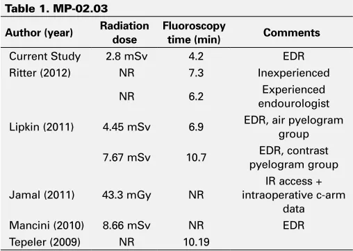
O’Malley, Padraic; Lantz, Andrea G.; Ordon, Michael; Lee, Jason Y. St. Michael’s Hospital, University of Toronto, Toronto, ON, Canada Introduction and Objectives: Percutaneous nephrolithotomy (PCNL) can be associated with large amounts of diagnostic radiation exposure, a topic that has received increasing scrutiny for its potential oncologic risks. Endoscopic-guided PCNL (ePCNL) is a relatively rare approach to PCNL that may have advantages in this regard, compared to standard PCNL tech-niques. This study determines the radiation exposure (fluoroscopy time [FT] and effective dose of radiation [EDR]) related to ePCNL and compares it to the published literature for the standard PCNL technique.
Methods: A retrospective review of all ePCNL procedures performed at our institution from Nov 2011 to Oct 2012 was conducted. All cases were performed by a single surgeon using under-table, C-arm fluoroscopy. Total
Moderated Posters 2: Stones
June 24, 2013, 1600-1800
Table 1. MP-02.03
Author (year) Radiation
dose
Fluoroscopy
time (min) Comments
Current Study 2.8 mSv 4.2 EDR
Ritter (2012) NR 7.3 Inexperienced
NR 6.2 endourologistExperienced
Lipkin (2011) 4.45 mSv 6.9 EDR, air pyelogram group
7.67 mSv 10.7 pyelogram groupEDR, contrast
Jamal (2011) 43.3 mGy NR
IR access + intraoperative c-arm
data Mancini (2010) 8.66 mSv NR EDR Tepeler (2009) NR 10.19
FT and radiation dosage for each case was recorded and the EDR was calculated using standard conversion tables. Pearson correlation was used to assess for covariates significantly correlated with longer FT and higher EDR.
Results: A total of 29 ePCNL cases were reviewed. Mean patient age was 64 ±13 yrs, 45% were male, and mean BMI was 30 ±6.5 kg/m2. Mean
stone size was 3.3 x 2.0 cm with 52% and 11% of stones being partial and complete staghorn calculi, respectively. 49% were upper pole punc-tures, and 61% were supracostal; one patient required 2 tracts. Mean FT was 4.2±3.6 min and mean EDR was 2.8±2.4 mSv. Stone-free rate was 82%; 11% required ancillary procedures. Overall complication rate was 31% with the majority (7/9) being Clavien-Dindo grade 1-2. Longer FT correlated with increased stone size (p=0.002) and the need for ancillary procedures (p=0.005); higher EDR correlated with increased skin-to-stone distance (SSD; p=0.013). Higher BMI did not correlate with FT, EDR or operative time.
Conclusions: The ePCNL technique may be associated with a reduction in radiation dosage when compared to standard PCNL techniques (Table 1), with comparable stone-free and complication rates.
MP-02.04
Complications of Percutaneous Nephrolithotripsy Performed at the University of Alberta
Zemp, Logan; Roy, Joey; Bochinski, Derek; Schuler, Trevor; Wollin, Tim University of Alberta, Edmonton, AB, Canada
Introduction and Objectives: Recently, the Clinical Research Office of the Endourological Society (CROES) established a worldwide percutaneous nephrolithotripsy (PCNL) database reporting clinical outcomes. The clini-cal experience and outcomes for PCNL at the University of Alberta (UofA) have not been previously described. The goal of this study is to ascertain the complications associated with PCNL at our centre and compare these rates to those reported from the CROES database.
Methods: A retrospective cohort study of consecutive PCNL performed by three surgeons at the UofA from February 2011 and February 2012 was performed. The patients’ medical records were reviewed for rel-evant patient, stone and treatment data. Intraoperative and postoperative complications were abstracted and the primary outcome was the 90-day complication rate, graded with the Clavien classification system. Results: A total of 178 patients were treated (102 M/76 F). The average age was 55.1 years. There were 105 right-sided, 67 left-sided, and 6 bilateral cases. Calculi were single, multiple, partial staghorn or complete staghorn in 33.1%, 41.1%, 11.4% and 16.9% respectively. The overall complication rate was 24.7%. Based on the Clavien classification system, these were found to be grade 1 in 21/178 or 11.8%, grade 2 in 12/178 or 6.7%, grade 3 in 9/178 or 5.1%, grade 4 in 1/178 or 0.56%, and grade 5 in 1/178 or 0.56%. Grade 3-4 complications included renal pelvis perforation (5.6%), pleural effusion (5.6%), and pneumothorax (0.6%). The stone free rate was found to be 89.9% and 88.8% of patients were treated with one procedure.
Conclusion: Based on this retrospective cohort study, the complication rate for PCNL at the UofA is comparable to worldwide results. This data will be useful as we endeavor to construct and develop a prospective database to follow these clinical parameters more accurately and improve patient outcomes.
MP-02.05
Are Postoperative Antibiotics Necessary After Uncomplicated Ureteroscopy?
Alsabban, Abdulrahman1; Ross, Michael1; Arsovska, Olga1; Patterson,
Ryan F.1; Eisner, Brian H.2; Kurtz, Michael P.2; Gershman, Boris2; Chew,
Ben H.1; Lange, Dirk1
1University of British Columbia, Vancouver, BC, Canada; 2Department
of Urology, Massachusetts General Hospital, Harvard Medical School, Boston, MA, United States
Introduction and Objectives: The use of perioperative prophylactic anti-biotics is intended to minimize infections resulting from surgical inter-ventions. The AUA Recommendation is for antibiotic prophylaxis for <24
hours. This study sought to evaluate the rate of postoperative urinary infection following ureteroscopy.
Methods: We retrospectively reviewed 153 subjects. Our initial chart review was for 73 subjects. All patients received preoperative and not all patients received postoperative antibiotics. Postoperative urinary tract infection (UTI) was diagnosed only in 2 cases (3.6%). For this initial analysis we excluded patients who had history of recurrent cystitis or pyelonephritis, treatment for positive urine culture within 3 months of planned procedure, and indwelling nephrostomy tube or ureteral stent at the time of procedure. In order to substantiate our findings, we decided to collect more data without exclusions. Additional chart review was performed for 80 subjects who underwent ureteroscopy in Vancouver. Results: 73/80 subjects (91.3%) were confirmed to have received preop-erative antibiotics (Cefazolin n=56), while 7 (8.8%) could not be verified. 41 (51.3%) subjects received postoperative antibiotics while 39 (48.8%) did not. Median age was 52.8+14.5 and mean BMI was 28.6+6.3. Laser lithrotripsy was used in 75 (93.8%), ureteral access sheath in 41 (51.3%), and a basket in 46 (57.5%). Even for the 80 additional subjects without exclusions, only four (5.0%) of the patients developed confirmed post-operative UTIs, two of whom received and the other two did not receive postoperative antibiotics.
Conclusions: Our data suggest that postoperative antibiotics are not nec-essary as the rate of infection after ureteroscopic stone treatment is very low. There was no difference in postoperative infection rates and reducing antibiotic use will limit adverse effects such as drug-induced colitis and antibiotic resistance.
MP-02.06
The Effect of Prone-Flexed Positioning (PFP) on Airway Pressures During Percutaneous Nephrolithotomy (PCNL)
Foell, Kirsten1; Ordon, Michael2; Lantz, Andrea G.2; Pace, Kenneth T.1;
Honey, R. John1
1St. Michael’s Hospital, University of Toronto, Toronto, ON, Canada; 2Division of Thoracic Surgery, Department of Surgery, University of
Toronto, Toronto, ON, Canada
Introduction and Objectives: The prone-flexed position involves 40-60° lumbar flexion and 15° knee flexion, in the prone position. The advantages over the prone position (PP) include increased distance between the tract and the spleen/liver, lowering of the kidneys relative to the ribs, and short-ening of the skin-to-stone distance. However, urologists unfamiliar with the PFP have questioned the effect of abdominal compression on airway pressure and ventilation. We aimed to quantify the effect of positioning on peak airway pressure (PAP), and the relationship between abdominal girth, body mass index (BMI) and PAP.
Methods: Measurements from consecutive patients undergoing PFP PCNL were prospectively recorded from Mar. to Oct. 2012. After intubation and muscle paralysis, the PAP and positive end-expiratory pressure (PEEP) were recorded in the SP (supine position), PP, and PFP. After subtracting PEEP, the PAPs across the 3 positions for each patient were compared with repeated measure ANOVA.
Results: 42 patients (71% male) were included. All PAPs were <35 cmH2O, well below the threshold of 40 cmH2O, which would be an
indication to change position. The mean PAP was higher in the PFP (20.5±0.7cmH2O) than PP (17.2±0.6, p<0.001), which was higher than
the SP (15.4±0.6, p<0.001). The mean BMI was 28.3±5.2kg/m2, and girth
was 101±12cm. Obese (BMI>30, n=15) and non-obese (n=27) patients demonstrated similar mean PAP changes when moving from SP to PFP (p=0.21). On linear regression, increased girth was predictive of elevated PAP in the PFP (p=0.024). No patients required conversion out of the PFP for any reason.
MP-02.07
Best Stent Length Predicted by Simple CT Measurement, Rather than Patient Height
Foell, Kirsten; Lantz, Andrea G.; Ordon, Michael; Lee, Jason Y.; Pace, Kenneth T.; Honey, R. John
St. Michael’s Hospital, University of Toronto, Toronto, ON, Canada Introduction and Objectives: Ureteral stent length is important, as stents that are too long might worsen symptoms, and too short might be at higher risk of migration. We aimed to determine if directly measured ureteral length predicts proper stent positioning, and if patient or radiological parameters correlate with measured ureteral length.
Methods: During stent placement, ureteral length [ureteropelvic junc-tion (UPJ) to ureterovesical juncjunc-tion (UVJ) distance] was measured by endoscopically viewing an open-ended catheter (with its 1-cm markings) emanating from the ureteral orifice. A 22, 24 or 26cm stent was chosen to be at least the measured ureteral length. For ureters >26cm, a 26 cm stent was chosen. Ends of an “ideally positioned” stent were fully curled in the renal pelvis and bladder, without crossing the bladder midline. Rates of ideal stent position were compared between patients with matching stent and ureteral lengths and those with stent lengths differing by ≥1 cm (mismatched). The measured ureteral length was correlated with height, L1-L5 height, and length measured on computed tomography (CT). Results: Of 59 ureters in 57 patients, 39 (66%) were in males, and 25 (42%) were right-sided. Matched stent length was associated with higher rates of ideal stent fit than mismatched (100% vs. 76%, p=0.036). Height was reasonably correlated with L1-L5 height [Spearman correlation coef-ficient (rho)=0.79], though both were poorly correlated with the measured ureteral length (rho=0.18 for height and 0.32 for lumbar height). However, ureteral lengths measured directly and on CT correlated well with each other (rho=0.61, both axial and coronal).
Conclusions: Stents matching directly measured ureteral lengths are asso-ciated with high rates of ideal stent position. CT measurements, rather than height, correlate well with measured length and could be used to choose the appropriate stent length.
MP-02.08 – WITHDRAWN
MP-02.09
Doppler Ultrasound to Predict Stone Fragmentation During Lithotripsy
Whelan, Thomas1; Pugsley, Devin2; McBriarty, Heather1
1Dalhousie University, Saint John, NB, Canada; 2Dalhousie University,
Halifax, NS, Canada
Introduction and Objectives: The ability to assess the degree of stone frag-mentation during shock wave lithotripsy (SWL) is difficult using fluoros-copy or grayscale ultrasound (US). As such, the physician often continues with treatment until the manufacturer’s recommended maximum number of shocks has been delivered. The objective of this study is to determine if the use of Doppler ultrasound (DUS) during lithotripsy can predict stone fragmentation during treatment as the Doppler signal changes.
Methods: Phantom stones were created using a published technique from a commercially available dental plaster. As well, endoscopically removed calculi were used in the study. An in vitro model using ordnance gelatin was used to simulate kidney tissue. Both stone types were embedded in the ordnance gel and treated with the Storz Modulith SLX-F2. The proto-col called for shocks to be delivered at a rate of 120 shocks per minute with an energy level of 8. The DUS signal was recorded at the start and at regular intervals throughout the treatment.
Results: The DUS signal changed during SWL, from thin to thick lines as the stone fragmented. The waveform transition over the treatment appears predictable and reproducible in this fixed model. It is felt to be second-ary to the shock wave hitting multiple fragments of stone, (displayed as a non-uniform thicker pattern), as opposed to a single waveform when the stone is intact.
Conclusions: DUS signal change is shown to be consistent with SWL fragmentation of phantom and human calculi in this in vitro model. We are now studying this to see if the results will be reproducible in vivo.
This may decrease patient treatment times by allowing SWL sessions to be terminated earlier when there is objective evidence of fragmenta-tion. Subsequently, this may provide a safer treatment with fewer shocks, decrease potential complications and lessen our dependence on fluoros-copy and thus limit radiation exposure to patients and providers.
MP-02.10
Physician Related Factors in Extracorporeal Shock Wave Lithotripsy (ESWL) Treatment
Moles, Jon; Watterson, James; Blew, Brian The University of Ottawa, Ottawa, ON, Canada
Introduction and Objectives: ESWL is an effective treatment for uroli-thiasis with both an increased number of shocks and fluoroscopy time correlated with improved stone clearance. Treating urologists often have no prior relationship or follow-up with patients and formal training in ESWL is variable. We reviewed the Ottawa Hospital Lithotripsy logbooks to identify whether a prior physician-patient relationship (self-referral) or endourology fellowship was correlated with treatment parameters known to improve stone clearance. Self-referral was defined as the treating urolo-gist also providing pre and postlithotripsy stone care as opposed to treating another urologist’s patients.
Methods: 1000 consecutive patients referred to the Ottawa Hospital Lithotripsy unit between November 2007 and April 2009 were assessed retrospectively using the lithotripsy treatment logbooks. The number of shocks and fluoroscopy time used as well as treating and referring urolo-gist were identified and a database was created. SAS was used for analysis. Results: Overall 962 patients underwent lithotripsy with the average shock number of 2579 and the average fluoroscopy time of 95s. 251 of 962 (26.1%) were self-referred and 2 of 6 (33.3%) treating urologists had com-pleted endourology fellowships. Overall the self-referred patients received less mean shocks (2470 vs. 2618) but no difference in mean fluoroscopy time (96s vs. 92s) and endourology fellowship trained staff treated with more shocks (2741 vs. 2452). Non-fellowship trained staff treated self-referred patients with less shocks than patients not self-self-referred (2518 vs. 2270) but there was no difference in shocks between those self-referred and not self-referred by the fellowship trained urologists.
Conclusions: At our centre patients referred by other urologists receive a greater number of shocks. This effect was not seen if an endourology fellowship had been completed. Fluoroscopy time was unaffected by fellowship or referral status.
MP-02.11
No Difference in 24-hour Urine Parameters Between Patients with Obstructing and Non-obstructing Urolithiasis Presenting to a Tertiary Referral Centre
Lantz, Andrea G.; Ghiculete, Daniela; Pace, Kenneth T.; Lee, Jason Y.; Honey, R. John
(±5.4). Mean stone size was similar between groups (p=0.898). The rate of hypercalciuric patients was significantly higher in the renal group (17.3% vs. 11.6%, p=0.044) but no other parameters differed between groups. Conclusions: There are few differences in 24-hour urine parameters between obstructive and non-obstructive urolithiasis when location of stone is used as a proxy for obstruction. Only urinary calcium was sta-tistically different between groups but this may be of little clinical sig-nificance. This study may support earlier 24-hour urine evaluation, in order to minimize the “clinic effect”, regardless of urinary obstruction.
MP-02.12
Calcium and Vitamin D Replacement in Vitamin D Inadequate Patients with Previous Urolithiasis
Elkoushy, Mohamed; Lee, Terence; Andonian, Sero McGill University Health Centre, Montreal, QC, Canada
Introduction and Objectives: To assess the impact of maintenance dose vitamin D (VD) and calcium repletion on urinary calcium excretion of vitamin D inadequate (VDI) patients with previous history of urolithiasis. Methods: A retrospective study of VDI patients (VD level < 30 ng/ml) fol-lowed at a tertiary stone clinic from September 2009 to December 2011 was performed. Patients were placed on maintenance dose calcium and VD for abnormal bone mineral density and hyperoxaluria. Metabolic stone work-up including two 24-hour urine collections and serum 25(OH) D, ionized calcium, uric acid and PTH were preformed. Patients with pri-mary hyperparathyroidism or without metabolic work-up were excluded. Results: Fifty-nine patients were included (64.4% males) with a mean age of 53.5 years and 50.8% were recurrent stone-formers. Seven (11.9%) patients were on concomitant thiazide diuretics during calcium and VD repletion. Mean VD and calcium dose supplied were 1150 IU and 895 mg per day. After a mean follow-up period of 8.4 (3-27) months, serum VD significantly increased (22.0 vs. 27.2 ng/ml, p=0.001). However, only 22 (37.3%) patients had normalized VD levels. Mean serum total and ionized calcium levels were comparable pre and post-VD repletion (9.24 vs. 9.32 mg/dL, p=0.43) and (4.64 vs. 4.84 mg/dL, p=0.09), respectively. Despite a significant increase in mean 24-hour urinary calcium excretion after VD repletion (165.6 vs. 217.6 mg/d, p<0.001), it still remained within normal limit. Furthermore, there was no significant difference in 24-hour urinary calcium excretion between those with normal and inadequate VD levels (216 vs. 220 mg/dL, p=0.87). Despite 5 (8.5%) patients developing de novo hypercalciuria, there was no significant difference in the number of patients with hypercalciuria pre- and post-treatment (7 vs. 11, p=0.44). Conclusions: Maintenance doses of calcium and vitamin D do not have significant impact on urinary calcium excretion in VDI patients with his-tory of urolithiasis.
MP-02.13
Prevalence of Urolithiasis and Metabolic Abnormalities of Patients with Surgically Confirmed Primary Hyperparathyroidism
Elkoushy, Mohamed; Yu, Alice; Tabah, Roger; Payne, Richard; Andonian, Sero
McGill University Health Centre, Montreal, QC, Canada
Purpose: To determine the prevalence of urolithiasis in patients with pri-mary hyperparathyroidism (PHPT) and to evaluate the metabolic stone work up before and after parathyroidectomy.
Methods: Institutional Research Ethics approval was obtained. A retro-spective review of proretro-spectively collected data was performed for patients presenting with PHPT to the stone, surgical oncology, and otolaryngology clinics at two tertiary centres from January 2006 to November 2011. Data were collected regarding patient characteristics, the nature of the PHPT (hyperplasia vs. adenoma), presence of urolithiasis together with 24-hour urine collections before and after parathyroidectomy.
Results: Of 333 patients undergoing parathyroidectomy (PTX) during the study period, 255 patients had PHPT (68.2% females) and were included with a mean age of 60.3 (18-91) years. Preoperative renal calcification was detected in 51 (20%) patients; urolithiasis in 48(18.8%) and neph-rocalcinosis in 3 (1.2%) patients. Compared to PHPT patients without
urolithiasis, PHPT patients with urolithiasis were significantly younger (55.9 vs. 61.3 years, p=0.02), less likely to be female (54.2% vs. 71.5%,
p=0.025) and had significantly lower levels of 25-hydroxyl vitamin D (19.7 vs. 23.9 ng/ml, p=0.03). Nine patients (3.5%) had stones post-PTX and were found to have significantly higher post-PTX total serum cal-cium levels when compared to the rest of the cohort. Whereas 62% of stone-formers were hypercalciuric pre-PTX, no patient had hypercalciuria post-PTX (p<0.001). On multivariate regression analysis, male gender (aOR (95%CI): 6.8 (5.3-7.2), p=0.01) and post-PTX total serum calcium levels independently predicted stones post-PTX (aOR (95%CI): 1.48 (1.33--2.12, p=0.02).
Conclusions: A high prevalence of urolithiasis was detected in patients presenting with PHPT. Post-parathyroidectomy stones were associated with male gender and significantly higher serum total calcium levels.
MP-02.14
Retrograde Intrarenal Surgery (RIRS) in the Treatment of Renal Stones, Retrospective Study
Alhasani, Salam
Dubai Health Authority, Dubai Hospital, Dubai, United Arab Emirates Introduction and Objectives: Contemporary treatment of renal calculi includes extracorporeal shock wave lithotripsy, percutaneous nephrostoli-thotomy and retrograde ureteronephroscopy. Success rates for shock wave lithotripsy are reduced for stones greater than 1 cm and/or in patients with anatomical variants. Percutaneous treatment, subjects the patient to increased morbidity. We studied the safety and efficacy of retrograde ureteroscopic treatment of intrarenal calculi.
Materials and Methods: We evaluated 82 stone burdens localized to the lower, middle and upper poles and renal pelvis. Stone burdens were classified as group I: 10 mm or less, group II: 11 to 20mm and group III: greater than 20 mm in largest diameter. Patients with calculi less than 2 cm were treated as outpatients or a one day hospital admission unless concurrent medical conditions required hospitalization. Larger stones and partial staghorn calculi (group III) frequently required 2-stage endoscopic procedures. An acceptable immediate surgical outcome was defined as complete fragmentation reducing the stone burden to dust and 2 mm or smaller fragments.
Results: Endoscopic access and complete stone fragmentation were achieved in 91, 88 and 45% of groups 1, 2 and 3, respectively. After a second treatment the success rate increased to 86% in group 3, with an overall rate of 86%. Of the 19 surgical failures 8 were secondary to inability to access the calyceal stone and 11 were secondary to inability to render the patient stone-free. In 4 of the 19 cases, infundibular stric-tures hindered ureteroscopic access. There were no major complications. Overall stone-free rates with minimum 3-month follow up were 84, 72 and 65% in groups 1, 2 and 3, respectively.
Conclusions: Retrograde ureteronephroscopy is a safe and effective surgi-cal treatment for intrarenal surgi-calculi. RIRS can be a substitute for ESWL or PCNL in selected cases.
MP-02.15
Correlating the Changing Trends in the American Diet to the Increasing Prevalence of Kidney Stones in America
De, Shubha; Liu, Xiaobo; Monga, Manoj
Cleveland Clinic Foundation, Cleveland, OH, United States
Introduction and Objectives: It has been postulated that changing dietary habits may play a role in the prevalence of nephrolithiasis. The United States Department of Agriculture (USDA) annually accounts for all food available for human consumption (adjusting for export, spoilage, waste, etc). Our objective was to evaluate changing trends in the American diet from 1974-2010 and to identify whether caloric intake and the consump-tion of lithogenic foods has been rising.
in stone prevalence. Changes in lithogenic foods were then correlated with increasing stone prevalence. Spearman correlations were performed (p<0.05), using SAS 9.2.
Results: Increased total daily calories (Rho 0.96, CI 0.86-1.00, p<0.001), fat grams (Rho 0.79, CI 0.55-1.00, p<0.001), protein ounces (Rho 0.85, CI 0.67-1.00, p<0.001), and fruit/vegetable (Rho 0.73, CI 0.49-0.97,
p<0.001) correlated strongly with the annual increase in stone prevalence. Specific foods also showed strong correlations with stone prevalence, including dark green vegetables, flour/cereal products, fish/shellfish, corn products (including high fructose corn syrup) and added sugars. Citrus juice was negatively correlated to stone disease (Rho -0.18, CI -0.52-0.17,
p=0.31) though not significant.
Conclusion: Several lithogenic components correlate temporally with the simultaneous increase in stones. Whether these correlations are incidental or causal is difficult to determine. The American diet has clearly changed over the last four decades and further investigation is required to better evaluate how this may impact the national prevalence of nephrolithiasis.
MP-02.16
Comparing the Sensitivity and Specificity of Ultrasound and CT in Kidney Stone Patients by Size, Location, and BMI
De, Shubha; Sarkissian, Carl; Monga, Manoj
Cleveland Clinic Foundation, Cleveland, OH, United States
Introduction and Objectives: Current AUA guidelines recommend follow up with ultrasound imaging (+xray) be performed following endoscopic procedures assessing for silent obstruction and residual fragments. Cited ultrasound sensitivity of 61% and specificity of 97% calculated from an exhaustive literature review.
Our objective was to review patients undergoing ultrasound and CT within 30 days (excluding spontaneously or surgically passed stones), to identify the sensitivity and specificity of stone detection by US at our centre, and the effects of BMI, stone size and position.
Methods: Those diagnosed with stones and having ultrasound followed by CT imaging within 30d were identified. A chart review was performed assessing for stone passage or manipulation between the two studies. We then analyzed the discrepancies between modalities, using the CT as the gold standard reference. Sensitivity and specificity were calculated using SPSS.
Results: Reviewing 198 patients, size positively correlated with sensitivity (>4 mm 75.00%; 0≤2 mm 26.40%; 2≤4 mm 26.10%). Sensitivity was lowest for upper pole (25.71%) compared to mid (53.85%) and lower pole (56.36%) stones. Specificity lowest in stones >4 mm (89.89%) when compared to smaller stones sizes ranging from 0≤2 mm (97.56%) and 2≤4 mm (96.39%). Specificity was greatest for interpolar (95.24%) and lowest for lower pole (91.95%) stones. Increasing BMI lowered sensitivity, while increasing specificity in upper and lower pole stones, and stones >4 mm. Conclusion: Comparing 200 patients’ ultrasound to CT images a specific-ity of 90-96% was found, decreasing with smaller stones and lower pole placement. Sensitivity was lowest for upper pole stones. These values are in keeping with the stated median values from the AUA Guideline, however a heterogeneity is noted with stone and patient characteristics.
MP-02.17
Does Stone Composition Change in Recurrent Stone-formers?
Elkoushy, Mohamed; Lee, Terence; Andonian, Sero McGill University Health Centre, Montreal, QC, Canada
Introduction and Objectives: To assess the prevalence of changes in stone composition of recurrent stone-formers.
Methods: A retrospective review of 303 patients with confirmed stone analysis presenting to a tertiary stone clinic between July 2009 and June 2012. All stones were analyzed at one centralized laboratory using gamma spectroscopy. Stone type was defined as the predominant stone component of at least 60%. Patients were grouped together according to the first predominant stone type on record. In the mixed stone group,
there was no predominant stone composition. Calcium oxalate monohy-drate and dihymonohy-drate were grouped as calcium oxalate (CaOx). Uric acid (UA) stones included uric acid dihydrate and ammonium urate while calcium phosphate, brushite and struvite were grouped as phosphate (Ph) stones. Patient records including operative reports and imaging studies were reviewed. Patients with multiple stone analyses at least 3 months apart were considered recurrent stone-formers and included in the study. Results: The cohort of 303 patients had a mean age of 54.3±30.7 years and 207 (68.3%) were males. The initial stone analysis showed: 145 (47.9%) CaOx, 88 (29%) Ph, 46 (15.2%) UA, 9 (3%) cystine and 15 (5%) mixed. There were 41 (13.5%) recurrent stone-formers with a median time of 404 days. Stone composition changed in 29.3% of recurrent stone-formers. Out of 13 patients with initial CaOx stones, 4 (30.8%) changed to another type (1 mixed, 2 UA, 1 Ph). Out of 11 patients with initial UA stones, 4 (36%) changed to another type (1 mixed and 3 CaOx). Out of 10 patients with initial Ph stones, 1 (10%) changed to CaOx, whereas all 3 patients with mixed stones changed to predominant CaOx, UA and Ph stones (1 patient each). None of the 4 cystinuric patients changed stone composition.
Conclusions: Stone composition may significantly change in subsequent stone analysis in recurrent stone-formers. Therefore, stone analysis is rec-ommended on each stone episode.
MP-02.18
Are There Differences in Metabolic Stone Workup of Patients with Different Stone Compositions?
Elkoushy, Mohamed; Lee, Terence; Andonian, Sero McGill University Health Centre, Montreal, QC, Canada
Introduction and Objectives: To assess differences in metabolic stone workup of patients with different stone compositions.
Methods: A retrospective review of 250 patients presenting between July 2009 and June 2012 to a tertiary stone clinic was performed. All stones were analyzed at one centralized laboratory using gamma spectroscopy. Stone type was defined as the predominant stone component of at least 60%. Patients were grouped together according to the predominant stone type. Calcium oxalate monohydrate and dihydrate were grouped as cal-cium oxalate (CaOx). Uric acid (UA) stones included uric acid dihydrate and ammonium urate while calcium phosphate, brushite and struvite were grouped as Phosphate (Ph) stones. Metabolic stone workup includ-ing two 24-hour urine collections were performed within 6 months of stone analysis.
Results: Of 250 patients, cysteine (n=5) and mixed (n=9) stones were excluded from analysis due to small sample size. Of the remaining 236, the mean age was 55.1± 15.0 years and the mean body mass index was 27.5 ± 5.3. There were significant differences in the distribution of various stone types in men and women. Whereas CaOx stones were the most common stone type in men (57.9%), Ph stones were the most common stone type in women (51.9%) (p<0.001). Men with Ph stones had sig-nificantly higher 24-hour urine calcium and magnesium excretion when compared with men with other stone types. However, serum creatinine was significantly higher in men with UA stones when compared with men with other stone types. Both men and women with Ph stones had significantly higher urine pH when compared with other stone types. All other variables examined were not significantly different among the different stone types.
Conclusions: While CaOx was the most common stone type in men, Ph stones were the most common type in women. The significance of UA stones in men being associated with significantly increased serum creatinine needs to be further investigated in future studies.
MP-02.20
A Global Urolithiasis Management Survey by the Clinical Research Office of the Endourological Society (CROES): Do Urologists Follow the “Golden Rule”?
Roberts, Gregory1; Beiko, Darren1; Razvi, Hassan2; Opondo, Dedan3; de
la Rosette, Jean3
1Queens Urology, Kingston, ON, Canada; 2Division of Urology, University
of Western Ontario, Kingston, ON, Canada; 3Department of Urology, AMC
University Hospital, Amsterdam, Netherlands
Introduction: Urologists aim to practice evidenced-based medicine, yet many clinical decisions are based on a combination of evidence, patient preference and physician experience. We do not know if there is disso-nance between recommendations urologists make to their stone patients and what they would choose if they were the actual patient. The objective of this study was to determine if endourologists follow the Golden Rule - “Do unto others as you would have done unto you”.
Methods: In 2012, CROES sent an email invitation to all members of the international Endourological Society for this 2-part global survey. Members accepting the invitation were sent two identical surveys, six months apart, containing 10 clinical stone scenarios to which there is no clear management consensus. Round 1 asked them to select the proce-dure they would recommend for a patient. Round 2 asked them to select what they would choose if they were the patient. Descriptive statistics were performed on the responses from urologists who participated in both surveys using R-statistical programming software version 2.12.2. Results: 205 members of the Endourological Society accepted the invita-tion to participate in the study. The response rates for the first survey and second survery were 83% (N=172/205) and 81% (N=167), respectively. 157 (76.6%) urologists responded to both surveys. Academic urologists made up 117(74.5%) of respondents, 123(78.3%) completed a fellow-ship in endourology and 27(17.5%) had a past medical history of stones. Consensus was widely lacking between individual experts for each sce-nario, however there was almost absolute congruency (no statistical dif-ferences) with each expert’s recommendation for their patients and their personal choice of treatment modality.
Conclusion: This positive and encouraging global CROES study shows that the Golden Rule is being followed by members of the Endourological Society as they choose to treat others as they would like to be treated.
MP-02.21
A Role of Sonic Hedgehog Signalling in Stent-induced Ureteral Aperistalsis
Janssen, Claudia; Buttyan, Ralph; Fazli, Ladan; Chew, Ben H.; Lange, Dirk Department of Urologic Sciences, University of British Columbia, Vancouver, BC, Canada
Introduction and Objectives: Indwelling ureteral stents disrupt coordi-nated ureteral peristalsis, however the exact molecular mechanisms are not known. Defects in Sonic Hedgehog (Shh) signaling during develop-ment result in hydronephrosis and hydroureter due to a loss of ureteral peristalsis. Furthermore, Shh signaling is also activated during healing and the regeneration of epithelial layers. Indwelling ureteral stents move in response to a patient’s daily activities resulting in urothelial irritation and inflammation. As such we hypothesize that stent-induced disruption of ureteral peristalsis may be the result of alterations in Shh signalling triggered by stent-induced irritation of the developed urothelium. Methods: Upper ureteral segments of unilaterally stented Yorkshire pigs were taken for immunohistochemical analysis for Shh and Gli expres-sion at 2, 4, 7 and 10 weeks post-stent-insertion and compared to the contralateral non-stented sides.
Results: We observed a decrease in Gli1 expression in the ureteral smooth muscle (SM) layers of stented ureters as the degree of inflam-mation increased. Interestingly, this decrease in expression resulted in an increase in Gli1 expression in the SM layers of the contralateral non-stented ureters. Over time, as the degree of inflammation decreased, Gli-1 expression in the SM layers of the stented ureters increased, while that in the contralateral non-stented ureters decreased. Stent removal at week 7 followed by a 3-week recovery period resulted in the recovery of Gli1 expression towards un-stented levels.