e-ISSN: 2278-7461, p-ISSN: 2319-6491
Volume 5, Issue 09 (Oct. 2016) PP:01-10
Optical Thermal Imaging for Breast Cancer Diagnosis
Zubir, N.
1*, Pushpanathan, K.
2*, Mat Noor, N.A.
2*1Department of Biology, Centre for Foundation Studies (CFS), International Islamic University Malaysia
(IIUM), JalanUniversiti, 46350 Petaling Jaya, Selangor
2Department of Biomedical Engineering, Faculty of Engineering, University of Malaya, 50603 Kuala Lumpur,
Malaysia
* These authors contributed equally to this work
Abstract: -Breast cancer has become the most common cancer in women. Fatality could be due to the delay in the intervention and detection of cancer. Mammography, Computated Tomography (CT) scan, Magnetic Resonance Imaging (MRI) and ultrasound are examples of imaging modalities for cancer screening. Nonetheless, considerable demand from the patients for non-invasive, low price, no compression and no ionization have led to the consideration for optical thermal imaging for breast cancer diagnosis. Overdiagnosis and overtreatment using mammography further aided in development of thermal imaging. The fundamental principle of optical thermal imaging relies on physiology such as the distribution of temperature on the skin surface. This article highlights the potential usage of optical thermal imaging, its limitations, advantages and latest concerns pertaining the usage. One of the suggestions is the usage of optical thermal imaging as adjunctive tool to mammography in breast cancer diagnosis. Novel attempts in thermography are also discussed for instance the emerging of rotational thermography replacing the conventional thermography.
Keywords:imaging modalities, thermography, optical thermal imaging, breast cancer, physiology
I.
INTRODUCTION
History
Infrared technology was first used for military purposes during World War 2. Only in the 1950’s after being declassified, infrared imaging technologies were used in the field of medicine [1]. It was found that the temperature of a tumour in the breast was higher compared to normal tissue [2]. It was also found that the temperature of venous blood draining the cancer was higher than blood from artery supplied to the cancer. Only in 1982, Food and Drug Administration certified thermography as a valid diagnosing tool for breast cancer [1].
Working principle of thermal imaging
Temperature distribution on a surface can be determined using thermal imaging. Thermal imaging, also known as thermography, adopts a special thermal camera (infrared camera) to measure the dissipation of thermal radiation [3]. It is non-invasive procedure that involves no radiation which poses no threat to patients. The thermal camera can provide both qualitative and quantitative data. These cameras have a sensitivity to detect temperature difference of 0.025°C. In addition to that, these cameras generate images which displays the distribution of thermal infrared radiation on the surface of objects which can be divided into imaging cameras and measurement cameras where it mostly operates in a passive manner. They observe the scene and detects the thermal radiation emitted by objects. It has to be noted that objects with temperature above absolute zero (-273K) radiate infrared from their surface [1]. According to the Stefan-Boltzmann Law, total radiation emitted by an object is directly proportional to the object’s area, emissivity and the fourth power of its absolute temperature. Humans radiate high level of infrared emission of 0.98 which is close to a perfect black body [4] The wavelengths of the infrared radiation emitted by humans are between 2μm and 20μm [5].Therefore, infrared radiation radiated by human skin can be quantified into accurate value of temperature. Infrared imaging of breast provides vast amount of information on nervous systems, vascular system, and local inflammatory processes.
The growth of cancer cells are exceptionally fast. The cancer cells have higher metabolism rate compared to normal cells [6]. Due to these, the skin temperature of the particular section increases [7]. The issue with thermal imaging is pertaining to temperature difference. If the temperature difference between the objects being analysed and the ambience are small, the contrast in thermal image would be poor [8]. Passive thermography is used for several applications which include the evaluation of tumour development.
static thermal imaging. Dynamic thermal imaging is used when the rate of temperature change cannot be estimated [9].
II
DESIGN AND TECHNIQUE OF OPTICAL THERMAL IMAGING OF BREAST
CANCER DIAGNOSIS
Optical Imaging of Breast cancer diagnosis
A. Handheld Optical device for breast cancer imaging
Diffuse optical imaging is a non-ionizing and non-invasive technique used for breast imaging. It uses near infrared (NIR) light ranging from 650-1000nm. Various research groups have developed hand held optical imaging devices which are portable and convenient for the patient testing. NIR is emitted from a NIR source towards tissue surface. After being scattered by the tissue, the resulting NIR is collected with the help of NIR sensitive detectors. Abnormalities are detected based on the difference in the properties like absorption and scattering between normal and diseased tissue. Contrast can be improved using external fluorescent contrasting agents. Measurement techniques can essentially be divided into three types [10]:
(i) Continuous Wave (CW): The source light intensity is constant with time throughout. However, the light is attenuated as it passes through the tissue due to absorption and scattering phenomena.
(ii) Frequency Domain (FD): This technique uses intensity modulated light which is independent of time. This light is attenuated and becomes phase shifted as it crosses the tissue.
(iii) Time domain (TD): In this technique, time dependent light is used. This light attenuates and broadens as it propagates through the tissue.
The handheld optical imaging device has three major components i.e. the handheld probe, intensified charge couple device (ICCD) and the laser diode source. The handheld probe has a flexible design in order for it to be able to adjust to different curvatures of issue. The handheld probe also has a large imaging area (4cm × 9cm), can simultaneously illuminate six source points and can detect 165 detector points. The probe has three parts and each part is capable of curving to 45˚. This instrumentation is designed to collect CW or FD depending on the requirement. When the equipment is switched to FD mode, signal between 50 and 300 MHz is modulated at the detector end and two frequency synthesizers are attached at the source. CW measurement is used to obtain 2D images in tumour detection. This system allows precise emission and detection of NIR signals at point locations on the tissue surface.
B. Fibre free handheld NIROS
Figure 1. Setup for breast imaging studies consisting of the breast tissue placed in between two transparent plates. [11]
Thermal Imaging of Breast cancer Diagnosis
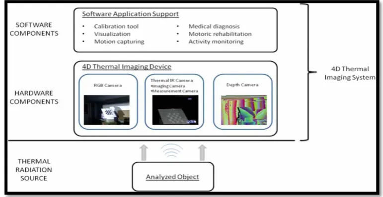
A. Equipment Design for 4D Thermal imaging system
The thermogram consists of two main components namely the hardware which contains four imaging modalities and the software which provides calibration and ensures the system is functional. Imaging system consists of a thermal imaging camera, thermal measurement camera, RGB camera and a depth camera. Depth cameras are devices designed to provide distance measurement in the form of depth maps. The depth camera constitutes the NIR projector and camera paired together. The projector emits light pattern which is visible to the NIR camera. Thermal cameras are essentially of two types namely, imaging cameras and measurement cameras. The measurement cameras are used for measuring temperature and can be calibrated by the manufacturer. On the contrary , imaging cameras produce a colour map of the temperature fields [8].
Figure 2Components of thermogram. [8]
B. Technique of thermal imaging
The imaging system of the thermographic equipment for breast cancer detection has the following components [12]:
a) Camera System: The latest camera system uses focal plane array cameras without cooling and requires less maintenance. Image processing techniques are used and this system provides basic quantitation of the image. Previously, single element detectors which employ an optical mechanical scanning process have been used. Nevertheless, these systems require an inflow of liquid nitrogen for cooling which limits the operation angle of the equipment. Hence, it may induce difficulty in managing the operation.
without the need for external checks. The temperature reference ensures the temperature measurements made by the thermogram are reliable.
c) Mounting imager: It is vital to provide vertical height adjustment of the imager. Previously, photographic tripod stands were used but they caused distortion of the image. Nowadays, studio camera stands are used. They have a stable base with wheels which enables the operator to place it in any reproducible position. The maximum height depends on the position of the patient. Wide angle lenses are mostly used as they reduce the distance between the camera and the patient.
d) Initialization of camera: The camera start up time is short in the modern thermograms. However, in order to obtain quantitative images, it takes 10 minutes to several hours for the camera to produce images in an uncooled system.
e) Image processing: Softwares for thermal imaging that are designed for medical applications are used. Storing images and relevant data is also important for medical thermography.
III
LATEST CONCERNS OF THERMOGRAPHY FOR BREAST CANCER DIAGNOSIS
Thermography is not recognised in Canada [13]. It is suggested that it is used only as a complementary medical device to mammography. FDA has classified thermography as second class medical device [14]. Thermography is unable to localise a tumour or a lesion as the abnormalities found by infrared imaging does not define an area that can be surgically biopsied [15]. In the case of “cold tumour” (false negatives) where metabolic activity is low, interpretation of thermal images was found to be even more difficult [16]. When breast is exposed to cold stress, vascular patterns disappears. The vascular patterns return when stress is removed. These irregularities in vascular patterns causes interpretation of thermography to be rather challenging. This may lead to many false-negatives and false positives.It has to be understood that procedures such as thermography cannot be used to diagnose breast cancer [17]. This is also applied for other techniques such as mammography, ultrasounds and magnetic resonance imaging. These techniques can only provide information of the patients according to the different aspects of the disease. Only biopsy of the breast with histological evaluation would be valid and conclusive diagnostic procedures for breast cancer. Thermography has been widely criticised for its high rate of false negative and false positive. Thermography cannot provide the precise anatomical information of breast or even specify an area which needs to be biopsied [18]. Thermography is a functional test [19]. It needs to be combined with mammography which is an anatomical test [20]. Besides, thermography cannot spot the precise reason responsible for the physiological changes to breast tissue. Hence, thermography can only be used as a risk marker and a complementary technique.
IV
DISCUSSION
The primary advantage of thermography technique lies on its efficiency for women with dense breast. It is suitable for three specific groups which include younger women, women taking hormone therapy and women with fibrocystic changes. About 18% of breast cancer patients are diagnosed in their forties. Younger women who develop breast cancer tend to have faster growth of cancer cells that are more likely to metastasize [21]. Synthetic hormone replacement is found to have an increase in invasive breast cancer [22].Fibrous breast can conceal early cancer especially if no microcalcification is present[23].Approximately 40% of women with fibrocystic disease and abnormal thermogram develop breast cancer within 5 years. On the other hand, women with fibrocystic disease and normal thermogram have less than 3% chance of developing breast cancer. It has to be noted that there are cases of cancer and ductal carcinoma in situ (DCIS) where women’s lives are not threatened. Unfortunately, these cases of cancer and DCIS are also detected through mammography. This eventually leads to overdiagnosis and overtreatment of breast cancer. This causes unnecessary exposure which may lead to the adverse effects which are often linked to cancer treatment [24].Thermal imaging was established as the highest risk marker to detect the presence of breast cancer in the year 1976 at the Third International Symposium on Detection and Prevention of Cancer. It was established that abnormal infrared images has been an indicator for development of breast cancer [25]. In a study carried out by Gautherie (1982) which incorporates 10000 women, it was found that thermography was extremely useful in evaluating risk of cancer [26]. Another study was carried out in the year 1980 which further confirms the importance of thermography in the detection of breast cancer [27]. Approximately 44% of patients with abnormal thermogram were diagnosed with breast cancer after 5 years. It was also found that after being diagnosed in advanced cancer stage using thermogram, there is 24% probability of survival after 3 years [28].
indicating the nature of the tumour compared to the size of the tumour. According to Isard, Sweitzer, & Edelstein (1988), patients with poor thermographic prognostic factors have shorter survival with only 30% surviving at the end of 5 years [31]. The study showed positive correlation against tumour-node-metastasis when compared with the patients’ poor thermographic factor. In a study carried out by Sterns & Zee (1991), out of 214 patients who were diagnosed with breast cancer without distant metastases, 121 of them had thermographic abnormality [32]. They found that patients whose tumours were thermographically abnormal actually had higher proportion of metastatic axillary lymph nodes. But, the overall survival rate and disease-free survival rate are not significantly different for the patients who have no abnormal thermograph..
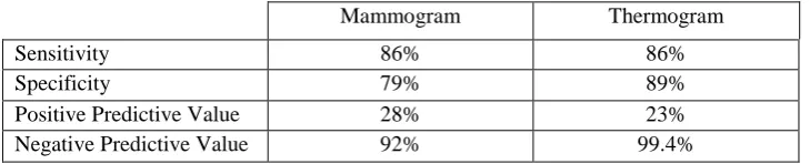
The most commonly used tool to diagnose breast cancer is mammogram. This method is often compared with thermography in terms of sensitivity and also versatility. Mammogram and infrared imaging were compared in a comprehensive manner where a number of aspects were compared and evaluated for examples sensitivity, specificity, positive predictive value, and negative predictive values. The following table (Table 1) shows the comparison between the techniques [33]:
Table 1: Comparison between Mammogram and Thermogram
Mammogram Thermogram
Sensitivity 86% 86%
Specificity 79% 89%
Positive Predictive Value 28% 23%
Negative Predictive Value 92% 99.4%
In a study carried out by Keyserlingk, Ahlgren, Yu, &Belliveau (1998), it was found that the sensitivity for clinical examination, mammography and infrared imaging are 61%, 66% and 83% respectively [34]. However, it was found that when three techniques were combined, the sensitivity increased to a staggering 98%.Thermogram is able to detect the signs of cancer 10 years earlier compared to mammography. This is further established by Kennedy, Lee, &Seely (2009) - 70% of cases, thermography can detect symptoms of breast cancer one year earlier than mammography [35]. Sensitivity of mammography will be reduced when employed for younger people or patients with dense breast tissue [36]. Apart from that, the size of undetectable tumours by mammography is about 1.66 cm while limiting size in thermography is decrease to 1.28 cm [28].
Novel Attempts in Thermography
Rotational thermography is a technique which was designed to overcome the limitations of conventional thermography. In this technique, image of the breast is taken from multiple views to have a proper coverage of the breast. In rotational thermography, specially designed equipment - Mammary Rotational Infrared Thermographic System (MAMRIT) is adopted. The subjects are made to lie down with ventral side down on the MAMRIT with one breast freely suspended into a chamber. A robotic arm fixed with infrared camera integrated inside MAMRIT chamber rotates clockwise around the suspended breast would be able to capture images of the breast from different views. In this technique, texture features which are extracted in the spatial domain will be fed to a support vector machine. Support vector machine (SVM) would classify the breast into normal or malignant. This technique results in classification accuracy of 83.3% [42].
Apart from that, in feature extraction, set of features are defined for efficient analysis and classification of information. Statistical and texture feature are extracted from thermograms in the curvelet domain. Two parameters, number of resolutions and number of angles are used in digital implementation of curvelet transform. Texture of an image is examined by using grey-level co-occurrence matrix (GLCM). GLCM calculates frequency at which pairs of pixels with certain intensities occur in a fixed spatial proximity. Features which are extracted in the curvelet domain from both normal and abnormal images are used to train Support Vector Machine which functions well in pattern recognition. This would be useful for automated classification and the accuracy of 90.91% would be attained [43].
VI
SUGGESTION
Despite being non-invasive, painless, low sensitivity and excessive numbers of false-positive cases which would have resulted in unnecessary invasive assessment of lesion of uncertain malignant potential or highly suspicious of malignancy, thermography as of date is not recommended as primary tool for breast cancer detection. But it was approved by the US FDA as adjunct tool for the diagnosis of breast cancer as mentioned earlier .Challenges faced by previous studies mainly due to limitation on equipment, resolution and sensitivity capabilities. The following suggestions are proposed in order to further improve the technology to make it possible as primary tool to detect malignancy with up to 100% accuracy or as useful adjunct tools to mammogram, with the aim to reduce further invasive procedures.
A. Dynamic Infrared Imaging as an adjunct to Mammogram and compulsory follow up after Biopsy.
Dynamic infrared imaging provides information about active processes and provides information which produce anatomical images that are not associated with active processes [8, 44]. This tool is extremely useful to eliminate the need to perform biopsy on patient that does not have breast cancer. Breast biopsies are traumatic experiences, expose patients to clinical risk, uncomfortable and costly. One of the latest dynamic infrared imaging technologies that has been approved by US FDA is BCS 2100, developed by Computerized Thermal Imaging Incorporated, USA.
The CTI BCS 2100 is a new medical device that has been developed for use as an adjunct to mammography. It is preferably to be followed up rather than undergoing biopsy procedures. This device has the ability to record temperature changes occurring in the breast accurately. It also provides physiological information that allows the characterization of suspicious breast lesions identified from images captured by mammography.
Patients lie on top of a specially designed table in prone position where breast suspends through an opening in the table. The breast is exposed to different temperatures with the aid of climate control system. A series of images are taken using infrared camera which is integrated with the table. The infrared camera captures more than 100 images. Approximately 8,300,000 temperature data points are collected for each breast in 3 minutes. Temperature of a specific point on the breast and also the change in temperature are represented by each pixel of the captured images. A specific algorithm is used for profiling the pixels and the score from the algorithm is ultimately compared against internal threshold. Negative test indicates high probability that the specific area is cancer free. This particular equipment is fast, non-invasive and does not require compression of breast which is painful. There is no ionizing radiation or imaging agents are used in this equipment.
B. Development of Advance and Accurate Image Analysis Algorithm
Figure 3 Infrared Images Processing
Two important steps in this study are the feature extraction and feature analysis. Texture measures smoothness, coarseness and regularity of pixels in an image. These features describe the mutual relationship among intensity values of neighbouring pixels repeated over an area larger than the size of the relationship[45]. There are 2 main classes of texture recognition system which include structural and statistical. These 2 approaches were being used in this study, computed and normalized in order to scale down the values of the computed features before being fed into a classifier. In the classification stage, the normalize features were fed to Support Vector Machine (SVM), a classifier. For classifications, the input data are transformed to high-dimensional feature space with the use of nonlinear kernel functions, so that the transformed data will be more separable compared to original input data. Result of the classifications is shown in Table 2.
This study shows that the accuracy of the diagnostic tool depends on size, quality of training data and the feature chosen as classifier inputs. Discrete Wavelet Transforms (DWT) with biorthogonal, Haar mother wavelets and with neural networks is used to classify the normal and benign thermograms. The images were able to be classified accurately with an efficiency of 86.6% [46]. A new method of thermographic image analysis was proposed using an Independent Component Analysis (ICA) and post-processing of the images resulting from this algorithm [47]. ICA is a subspace projection technique that projects data from a high dimensional space to a lower dimensional space. The proposed method gave a sensitivity of 100% and specificity of 94.7%.
B. Data Acquisition from Larger Pool and Longer Clinical Trial
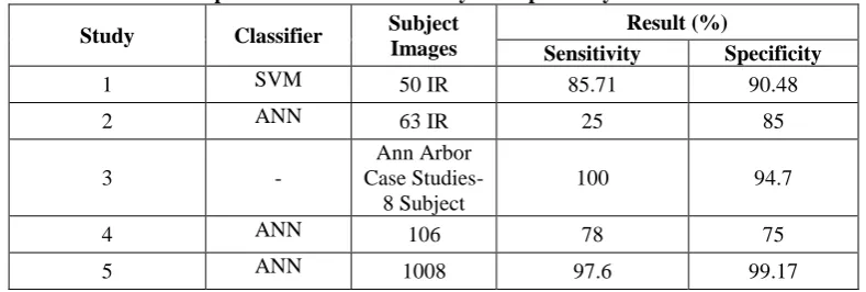
Various studies made on Breast Cancer Detection using Thermography images processing, but most of the studies made with a different data acquisition and some may did not include follow up on the subject’s processed data. Because of these variations of studies and findings, the results are inconclusive to support the use of Thermography as primary tools in breast cancer detection. Table 3 below shows the comparison of data acquisition from various studies.
Table 3: Comparison between Sensitivity and Specificity from Various Studies
Study Classifier Subject
Images
Result (%)
Sensitivity Specificity
1 SVM 50 IR 85.71 90.48
2 ANN 63 IR 25 85
3 -
Ann Arbor Case
Studies-8 Subject
100 94.7
4 ANN 106 78 75
5 ANN 1008 97.6 99.17
Note:
1. “Thermography Based Breast Cancer Detection Using Texture Features and Support Vector Machine”, by Acharya, Ng, Tan, &Sree, 2010, Journal of Medical System.
2. Data obtained from “Digital Infrared thermal Imaging (DITI) of breast lesions: Sensitivity and specificity of detection of primary breast cancers”, by M. Kontos, R.Wilson and I-Fentiman, 2011, Clinical Radiology. 3. “Automated Detection of Breast Cancer in Thermal Infrared Images, Based on Independent Component
4. “The accuracy of digital infrared imaging for breast cancer detection in women undergoing breast biopsy”, by Wishart et al., 2010, European Journal of Surgical Oncology
5. “Evaluation of digital infra–red thermal imaging as an adjunctive screening method for breast carcinoma: A pilot study”, by Rassiwala et al., 2014, International Journal of Surgery
VII CONCLUSION
Thermography does not provide information about morphological structures of breast but it can show functional temperature data and condition of breast tissue’s vessels. Researches support the role of thermography in early diagnosis of abnormal state of breast, which can detect cancer. If abnormal thermograms suggest the abnormal function causing breast cancer, thus it can be provide a chance for early intervention and improve the abnormal function. A combination of clinical examination, mammography, and infrared imaging would provide the highest percentage of survival against breast cancer. Nowadays, none of the current methods can forecast breast cancer for 100%. The only certain diagnostic method is biopsy.
VIII
CONFLICT OF INTERESTS
The authors declare that there is no conflict of interest regarding the publication of this article. None of the authors has a financial relationship with a commercial entity that has an interest in the subject of this paper,
REFERENCE
[1] Bronzino, J. (2006). Medical devices and systems. Boca Raton: CRC/Taylor & Francis.
[2] Lawson, R. (1956). Implications of Surface Temperatures in the Diagnosis of Breast Cancer. Canadian Medical Association Journal, 75(4).
[3] Skala, K., Lipic, T., Sovic, I., &Grubišić, I. (2011). 4D thermal imaging system for medical applications. PeriodicumBiologorum, 113(4).
[4] Hobbie, R. (1997). Intermediate physics for medicine and biology. New York: AIP Press. [5] Bronzino, J. &Diakides, N. (2008). Medical infrared imaging. Boca Raton: CRC Press.
[6] Amoêdo, N., Valencia, J., Rodrigues, M., Galina, A., &Rumjanek, F. (2013). How does the metabolism of tumour cells differ from that of normal cells. Bioscience Reports, 33(6), 865-873. http://dx.doi.org/10.1042/bsr20130066
[7] Xie, W., McCahon, P., Jakobsen, K., & Parish, C. (2003). Evaluation of the ability of digital infrared imaging to detect vascular changes in experimental animal tumours. International Journal of Cancer, 108(5), 790-794. http://dx.doi.org/10.1002/ijc.11618
[8] Skala, K., Lipic, T., Sovic, I., &Grubišić, I. (2011). 4D thermal imaging system for medical applications. PeriodicumBiologorum, 113(4).
[9] Vollmer, M. &Möllmann, K. (2010). Infrared thermal imaging. Weinheim: Wiley-VCH.
[10] Erickson, S., Godavarty, A., Martinez, S., Gonzalez, J., Romero, A., & Roman, M. et al. (2012). Hand-Held Optical Devices for Breast Cancer: Spectroscopy and 3-D Tomographic Imaging. IEEE J. Select. Topics Quantum Electron, 18(4), 1298-1312. http://dx.doi.org/10.1109/jstqe.2011.2170664
[11] Godavarty, A., Rodriguez, S., Jung, Y., & Gonzalez, S. (2015). Optical imaging for breast cancer prescreening. Breast Cancer: Targets and Therapy, 193. http://dx.doi.org/10.2147/bctt.s51702
[12] Ring, E. &Ammer, K. (2015). The technique of infrared imaging in medicine. IOP Publishing.
[13] Health Canada,. (2007). It's Your Health - Mammography. Hc-sc.gc.ca. Retrieved 14 April 2016, from http://www.hc-sc.gc.ca/hl-vs/iyh-vsv/med/mammog-eng.php
[14] Arora, N., Martins, D., Ruggerio, D., Tousimis, E., Swistel, A., Osborne, M., & Simmons, R. (2008). Effectiveness of a non-invasive digital infrared thermal imaging system in the detection of breast cancer. The American Journal of Surgery, 196(4), 523-526. http://dx.doi.org/10.1016/j.amjsurg.2008.06.015 [15] Kennedy, D., Lee, T., &Seely, D. (2009). A Comparative Review of Thermography as a Breast Cancer
Screening Technique. Integrative Cancer Therapies, 8(1), 9-16. http://dx.doi.org/10.1177/1534735408326171
[16] Piana, A. &Sepper, A. (2014). Contemporary Evaluation of Thermal Breast Screening. Pan American Journal of Medical Thermology, 1(2), 93-100. http://dx.doi.org/10.18073/2358-4696/pajmt.v1n2p93-100 [17] FDA,. (2014). Breast Cancer Screening - Thermography is Not an Alternative to Mammography: FDA
Safety Communication. Fda.gov. Retrieved 15 April 2016, from http://www.fda.gov/MedicalDevices/Safety/AlertsandNotices/ucm257259.htm
[19] Fitzgerald, A. &Berentson-Shaw, J. (2012). Thermography as a screening and diagnostic tool: a systematic review. The New Zealand Medical Journal, 125(1351).
[20] Plotnikoff G, Carolyn T. Emerging controversies in breast imaging: is there a place for thermography? Minn Med. Dec 2009;92(12):37-39, 56
[21] Berg, W. (2010). Benefits of Screening Mammography. JAMA, 303(2), 168. http://dx.doi.org/10.1001/jama.2009.1993
[22] Rossouw, J., Anderson, G., & Prentice, R. (2002). Risks and Benefits of EstrogenPlus Progestin in Healthy Postmenopausal Women: Principal Results. Obstetrical & Gynaecological Survey, 57(11), 750-752. http://dx.doi.org/10.1097/00006254-200211000-00019
[23] Gautherie, M. (1983). Thermobiological assessment of benign and malignant breast diseases. American Journal of Obstetrics and Gynaecology, 147(8), 861-869. http://dx.doi.org/10.1016/0002-9378(83)90236-3
[24] National Cancer Institute,. (2014). Mammograms. National Institutes of Health. Retrieved 15 July 2016, from https://www.cancer.gov/types/breast/mammograms-fact-sheet
[25] Amalric, R., Gautherie, M., Hobbins, W., Stark, A., &Thierree, R. (1981). The future of women with isolated abnormal infrared thermogram of the breast. La Nouvelle PresseMédicale, 10(38).
[26] Gautherie, M., Haehnel, P., Walter, J., & Keith, L. (1982). Long-term assessment of breast cancer risk by liquid-crystal thermal imaging. Progress in Clinical And Biological Research, 279-301.
[27] Gautherie, M. &Gros, C. (1980). Breast thermography and cancer risk prediction. Cancer, 45(1), 51-56. http://dx.doi.org/10.1002/1097-0142(19800101)45:1<51::aid-cncr2820450110>3.0.co;2-l
[28] Ohsumi, S., Takashima, S., Aogi, K., &Usuki, H. (2002). Prognostic Value of Thermographically Findings in Patients with Primary Breast Cancer. Breast Cancer Research and Treatment, 74(3), 213-220. http://dx.doi.org/10.1023/a:1016302719017
[29] Gershon-Cohen, J., Hermel, M., & Murdock, M. (1970). Thermography in detection of early breast cancer. Cancer, 26(5), 1153-1156. http://dx.doi.org/10.1002/1097-0142(197011)26:5<1153::aid-cncr2820260527>3.0.co;2-h
[30] Gautherie, M. (1983). Thermobiological assessment of benign and malignant breast diseases. American Journal of Obstetrics and Gynaecology, 147(8), 861-869. http://dx.doi.org/10.1016/0002-9378(83)90236-3
[31] Isard, H., Sweitzer, C., & Edelstein, G. (1988). Breast thermography. A prognostic indicator for breast cancer survival. Cancer, 62(3), 484-488. http://dx.doi.org/10.1002/1097-0142(19880801)62:3<484::aid-cncr2820620307>3.0.co;2-w
[32] Sterns, E. & Zee, B. (1991). Thermography as a predictor of prognosis in cancer of the breast. Cancer, 67(6), 1678-1680. http://dx.doi.org/10.1002/1097-0142(19910315)67:6<1678::aid-cncr2820670633>3.0.co;2-k
[33] Head, J., Lipari, C., & Elliot, R. (1999). Comparison of mammography and breast infrared imaging: sensitivity, specificity, false negatives, false positives, positive predictive value and negative predictive value. In Annual Fall Meeting of the Biomedical Engineering Society. Atlanta: IEEE.
[34] Head, J., Lipari, C., & Elliot, R. (1999). Comparison of mammography and breast infrared imaging: sensitivity, specificity, false negatives, false positives, positive predictive value and negative predictive value. In Annual Fall Meeting of the Biomedical Engineering Society. Atlanta: IEEE.
[35] Kennedy, D., Lee, T., &Seely, D. (2009). A Comparative Review of Thermography as a Breast Cancer Screening Technique. Integrative Cancer Therapies, 8(1), 9-16. http://dx.doi.org/10.1177/1534735408326171
[36] Ring, E. &Ammer, K. (2012). Infrared thermal imaging in medicine. Physiological Measurement, 33(3), R33-R46. http://dx.doi.org/10.1088/0967-3334/33/3/r33
[37] Kapoor, P., Prasad, S., &Patni, S. (2012). Image Segmentation and Asymmetry Analysis of Breast Thermograms for Tumor Detection. International Journal of Computer Applications, 50(9), 40-45. http://dx.doi.org/10.5120/7803-0932
[38] EtehadTavakol, M., Chandran, V., Ng, E., &Kafieh, R. (2013). Breast cancer detection from thermal images using bispectral invariant features. International Journal of Thermal Sciences, 69, 21-36. http://dx.doi.org/10.1016/j.ijthermalsci.2013.03.001
[39] Ahmad, F., Mat Isa, N., Hussain, Z., &Sulaiman, S. (2012). A genetic algorithm-based multi-objective optimization of an artificial neural network classifier for breast cancer diagnosis. Neural Comput&Applic, 23(5), 1427-1435. http://dx.doi.org/10.1007/s00521-012-1092-1
[41] Wan, C., Cao, W., & Cheng, C. (2014). Research of Recognition Method of Discrete Wavelet Feature Extraction and PNN Classification of Rats FT-IR Pancreatic Cancer Data. Journal of Analytical Methods in Chemistry, 2014, 1-6. http://dx.doi.org/10.1155/2014/564801
[42] Francis, S., Sasikala, M., &Saranya, S. (2014). Detection of Breast Abnormality from Thermograms Using Curvelet Transform Based Feature Extraction. J Med Syst, 38(4). http://dx.doi.org/10.1007/s10916-014-0023-3
[43] Francis, S., Sasikala, M., BhavaniBharathi, G., &Jaipurkar, S. (2014). Breast cancer detection in rotational thermography images using texture features. Infrared Physics & Technology, 67, 490-496. http://dx.doi.org/10.1016/j.infrared.2014.08.019
[44] Kontos, M., Wilson, R., &Fentiman, I. (2011). Digital infrared thermal imaging (DITI) of breast lesions: sensitivity and specificity of detection of primary breast cancers. Clinical Radiology, 66(6), 536-539. http://dx.doi.org/10.1016/j.crad.2011.01.009
[45] Acharya, U., Ng, E., Tan, J., &Sree, S. (2010). Thermography Based Breast Cancer Detection Using Texture Features and Support Vector Machine. Journal of Medical Systems, 36(3), 1503-1510. http://dx.doi.org/10.1007/s10916-010-9611-z
[46] Saba, L., Acharya U, R., Guerriero, S., & Suri, J. (2013). Ovarian neoplasm imaging.