291
International Journal of Pharmaceutical Sciences and Drug Research
2017; 9(6): 291-298
Research Article
CODEN (USA): IJPSPP
ISSN: 0975-248X
Development and Validation of RP-HPLC Method for the Simultaneous
Estimation of Ledipasvir and Sofosbuvir in Bulk and Pharmaceutical Dosage
Form
Akshay P. Rote
*, Janardan Alhat, Amol A. Kulkarni
Department of Quality Assurance, Siddhant College of Pharmacy, Sudumbare, Pune-412109, Maharashtra, India
Copyright © 2017 Akshay P. Rote et al. This is an open access article distributed under the terms of the Creative Commons Attribution-NonCommercial-ShareAlike 4.0 International License which allows others to remix, tweak, and build upon the work non-commercially, as long as the author is credited and the new creations are licensed under the identical terms.
ABSTRACT
The day by day new combinations drugs are being introduced in market. Then the multiple therapeutic agents which acts at different sites are used in the management of various diseases and disorders are done. Thus it is necessary to develop methods for analysis with the help of number of analytical techniques which are available for the estimation of the drugs in combination. The analyst were determine the Specific, accurate, simple, selective and stability-indicating RP-HPLC method is developed and validated for simultaneous determination of sofosbuvir and ledipasvir in tablet dosage form. RP-HPLC method was performed on the systronics isocratic HPLC System equipped with SP930 D HPLC pump and dual wavelength UV-VIS detector and C18 column (250
mm × 4.6 mm, 5μm), using the mobile phase (Methanol: Water 83:17 v/v) pH 3.0 with 0.05% acidic acid at a flow rate of 1.0 ml/min, injection volume 20μl and UV detection at 245 nm. This method is validated according to BP, USP and ICH requirements for new methods, which include accuracy, precision, robustness, ruggedness, lod, loq, linearity and range. Linear relationships were obtained in the ranges of 10-50μg/ml and 40-200μg/ml with correlation coefficients of 0.9991 and 0.9994 at Rt value of 7.45 min and 3.50 min for sofosbuvir and ledipasvir respectively. The forced degradation studies as acidity, alkalinity, oxidation and hydrolytic degradation were performed according to ICH guidelines.
Keywords:Sofosbuvir and Ledipasvir, HPLC, Development, Forced degradation, Validation.
DOI: 10.25004/IJPSDR.2017.090602 Int. J. Pharm. Sci. Drug Res. 2017; 9(6): 291-298
*Corresponding author: Mr. Akshay P. Rote
Address: Department of Quality Assurance, Siddhant College of Pharmacy, Sudumbare, Pune-412109, Maharashtra, India Tel.: +91-9271807680
E-mail: [email protected]
Relevant conflicts of interest/financial disclosures: The authors declare that the research was conducted in the absence of any commercial or financial relationships that could be construed as a potential conflict of interest.
Received: 28 September, 2017; Revised: 18 October, 2017; Accepted: 25 October, 2017; Published: 20 November, 2017
INTRODUCTION
Pharmaceutical Analysis plays a vital role in quality assurance and quality control of bulk drugs and their
Int. J. Pharm. Sci. Drug Res. November-December, 2017, Vol 9, Issue 6 (291-298)
amounts of compounds in a sample matter. It is concerned with chemical characterization of matter both quantitative and qualitative. In recent years many analytical techniques have been developed. Analytical method is a particular utilization of a procedure to solve a problem. Analytical instrumentation assumes an imperative part in the production and evaluation of new products and protection of Consumers and the environment. This instrumentation provides the lower detection limits required to assure safe foods, medications, water and air.
Validation of an analytical method is the process by which it is established, by laboratory studies, that the performance characteristics of the method meet the requirements for the intended analytical applications. There are two important reasons for validating assays in the pharmaceutical industry. The first, and by for the most important, is that assay validation is an integral part of the quality control system. The second is that current good manufacturing practice regulation requires assay validation.
Globally, 130-150 millions of people have chronic hepatitis C infection. A significant number of those who are chronically infected will develop liver cirrhosis or liver cancer. Gilead Sciences overcome most common related liver diseases by its Great invention (Harvoni). Harvoni (90 mg ledipasvir/400 mg sofosbuvir) approved by United States FDA. It is indicated for the treatment of chronic HCV genotypes 1, 4, 5, and 6 in adults and also indicated for the treatment of chronic HCV in patients co-infected with HIV. [1-2]
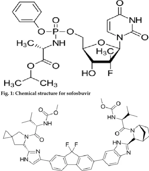
Sofosbuvir is chemically known as (S)-Isopropyl 2-((S)- (((2R,3R,4R,5R)-5-(2,4-dioxo-3,4-dihydropyrimidin- 1(2H)-yl)-4-fluoro-3-hydroxy-4-methyltetrahydrofuran-2-yl)methoxy)-(phenoxy) phosphorylamino) propanoate. It has a molecular formula of C22H29FN3O9P and a molecular weight of 529.45. [2]
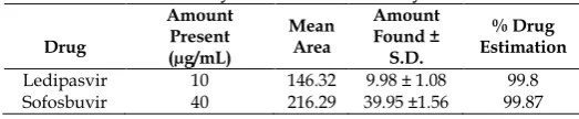
Ledipasvir is chemically known as Methyl [(2S)-1-{(6S)-
6-[5-(9,9-difluoro-7-{2-[(1R,3S,4S)-2-{(2S)-2-[(methoxycarbonyl)amino]-3-methylbutanoyl}
2azabicyclo[2.2.1]hept-3-yl]-1H-benzimidazol-6-yl}-9H- fluoren-2-yl)-1H-imidazol-2-yl]-5-azaspiro[2.4]hept-5-yl}-3-methyl-1-oxobutan-2-yl]carbamate. It has a molecular formula of C49H54F2N8O6 and a molecular
weight of 889.00. [2]
The combination of these two drugs is not official in any pharmacopoeia. Very recently, a limited number of methods have been developed for the individual and simultaneous determination of both drugs. The degradation products of sofosbuvir and ledipasvir under several stress conditions have been determined by HPLC.
MATERIALS AND METHODS Chemicals and Reagents
I. Pure samples: pure samples of sofosbuvir and ledipasvir were obtained as a generous gift samples from the Mylan Pharmaceutical Pvt. Ltd. (India), Mylan
Pharmaceutical Company which is india’s third largest exporter of antiretroviral for HIV/Aids care, hepato care, critical care, onco care and women’s care.
II. Pharmaceutical dosage form: MyHep LVIRtm fixed
dose combination tablets Containing ledipasvir (90mg) and sofosbuvir (400 mg) were manufactured by Mylan Pharmaceutical Pvt. Ltd. (India).
III. Chemicals: all chemicals and reagents of analytical grade and hplc grade were purchased from Merck Chemicals, Mumbai, India.
Fig. 1: Chemical structure for sofosbuvir
Fig. 2: Chemical structure for ledipasvir
Instruments
Chromatographic experimentations were performed using Systronics isocratic HPLC System equipped with SP930 D HPLC pump and dual wavelength UV-VIS detector, Data acquisition and processing was performed using Chemitochrom automation Chromatograph data system software and methods were conducted using an isocratic Reverse phase HPLC techniques. The mobile phase was prepared freshly filtered through 0.45μm membrane filters (Millipore, USA) and sonicated for 30 min, before use in order to degas the mobile phase. A C18 RP-Purosphere column
(5μm, 4.6 mm × 250 mm), Germany was used for analysis. Column was prewashed before the analysis of samples with boiling water and methanol alternately. Selection of Chromatographic Mode
Int. J. Pharm. Sci. Drug Res. November-December, 2017, Vol 9, Issue 6 (291-298)
Selection of Stationary Phase
On the basis of reversed phase HPLC mode and number of carbon present in the molecule (analyte) RP-Purosnosphere C18 (Grace) column (5µm, 4.6 mm × 250
mm), of following configuration was selected for further study. [4]
Selection of Mobile Phase
Ledipasvir and Sofosbuvir are soluble in HPLC grade water and methanol on ultrasonication, insoluble in hexane and are freely soluble in methanol and water, DMSO. Both the drugs on ultrasonication are soluble in methanol, acetonitrile and water mixture. Hence, mixture of methanol: water was used for initial separation. [4]
Selection of Detector and Detection Wavelength UV-Visible detector was selected, as it is reliable and easy to set at the correct wavelength. An overlay spectrum of drugs in methanol water (83:17) was recorded. From the overlay spectrum 245 nm was selected as a wavelength of measurement.
Standard Sample Preparation Ledipasvir and Sofosbuvir [A]
1) An accurately weighed quantity of Ledipasvir 10 mg and 40 mg Sofosbuvir was taken in 25 ml volumetric flask and dissolved in 10 ml Methanol, with the help of ultrasonication for about 10 min. Then the volume was made up to the mark using methanol to get standard stock solution. 1000µgm/ml Ledipasvir and 4000µgm/ml Sofosbuvir--- STOCK.
2) Ledipasvir and Sofosbuvir Standard Solution [A1]: Standard stock Solution [A] 0.1 ml and make vol. With mobile phase 10 ml, = 10µgm/ml Ledipasvir and 40µgm/ml Sofosbuvir
3) Ledipasvir and Sofosbuvir Standard Solution [A2]: Standard stock Solution [A] 0.2 ml and make vol. With mobile phase 10 ml, = 20µgm/ml Ledipasvir and 80µgm/ml Sofosbuvir
4) Ledipasvir and Sofosbuvir Standard Solution [A3]: Standard stock Solution [A] 0.3 ml and make vol. With mobile phase 10 ml, = 30µgm/ml Ledipasvir and 120µgm/ml Sofosbuvir
5) Ledipasvir and Sofosbuvir Standard Solution [A4]: Standard stock Solution [A] 0.4 ml and make vol. With mobile phase 10 ml, = 40µgm/ml Ledipasvir and 160µgm/ml Sofosbuvir
6) Ledipasvir and Sofosbuvir Standard Solution [A5]: Standard stock Solution [A] 0.5 ml and make vol. With mobile phase 10 ml, = 50µgm/ml Ledipasvir and 200µgm/ml Sofosbuvir.
Optimization of HPLC Parameters
Optimizations of HPLC process is to find a set of conditions that adequately separate and enable the quantification of the analytes from the endogenous material with acceptable accuracy, precision, cost, ease and speed. [5]
Optimization of Mobile Phase Strength
Initially methanol and water in different ratios were tried, and then methanol and water at different pH were tried. It was found that in methanol: water 83:17
(v/v), pH 3.0 adjusted using 0.05% acidic acid, this mobile phase at flow rate 1.0 mL/min gave good resolution of peaks with minimum tailing as compared to other mobilephases. [5]
The effect of change in composition on retention time is shown in Table 1.
Optimization of Detection Wavelength
On the basis of overlay spectra different wavelengths were selected. A fixed concentration of analyte mixture was analyzed at selected wavelengths. As per the response of analyte 245 nm wavelength was chosen. At wavelength 245 nm, analyte peak area was greater than that of at other wavelength observed, which is shown in Table 2.
Table 1: Optimization of Mobile Phase Strength S.
No.
Mobile Phase Strength (methanol: water v/v)
tR of Ledipasvir
tR of Sofosbuvir
1 81: 19 7.36 3.35
2 85:15 7.33 3.30
3 83:17 7.45 3.50
4 80:20 7.35 3.43
5 87:23 6.47 4.12
Table 2: Optimization of Detection Wavelength Mobile Phase
Strength (methanol: water v/v)
Wavelength (nm)
Area of Analytes Ledipasvir Sofosbuvir 83:17 244 143.97 212.53 83:17 245 145.17 214.62 83:17 265 146.27 216.41
Table 3: Final Chromatographic Conditions
Chromatographic Mode Chromatographic Condition
Standard Solutions 1000g/mL Ledipasvir and 4000g/mL Sofosbuvir, in mobile phase
HPLC System Younglin ( S.K) Gradient System UV Detector Pump SP930 D HPLC Pump Detector UV 730 D UV-Visible detector Stationary Phase C18 ( Grace) column (250 4.6mm, 5m)
Mobile Phase Methanol :water in ratio of 83:17 v/v pH 3.0 with 0.05% acidic acid Detection Wavelength 245 nm
Flow Rate 1 mL/min
Sample Size 20l
Column Temperature Ambient
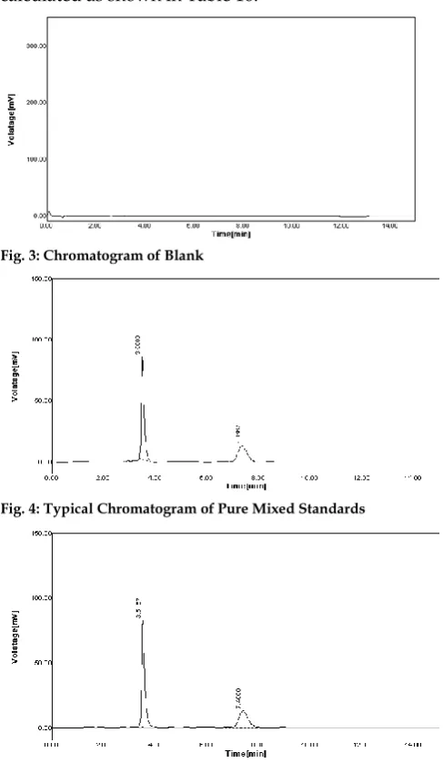
Table 4: Results of Analysis of Mixed Laboratory Standards.
Drug
Amount Present (µg/mL)
Mean Area
Amount Found ± S.D.
% Drug Estimation Ledipasvir 10 146.32 9.98 ± 1.08 99.8 Sofosbuvir 40 216.29 39.95 ±1.56 99.87
Preparation of Mobile Phase
In volumetric flask Methanol: Water (83:17 v/v), pH 3.0, adjusted using 0.05% acidic acid. Then the mobile phase was filtered through 0.45 micron membrane filter paper using suction pump. The content was ultrasonicated for 20 min for degassing.
Preparation of Standard Mixture
Int. J. Pharm. Sci. Drug Res. November-December, 2017, Vol 9, Issue 6 (291-298)
Sofosbuvir (400g/ml). The above solution was filtered through 0.45 Millipore Membrane filter. This solution (1 ml) was diluted to 10 ml using mobile phase to get the Ledipasvir (10g/ml) and Sofosbuvir (40g/ml) solution. The content was ultrasonicated for 20 min. Preparation of Sample Mixture
Twenty tablets were weighed accurately and finely powdered. The tablet powder equivalent to Ledipasvir (1 mg) and Sofosbuvir (4 mg) was weighed accurately. Then it was transferred to a 100 ml volumetric flask containing in methanol: water 83:17 v/v. Then the content was ultrasonicated for 30 min. And volume was made up to the mark using methanol. The above solution was filtered through Whatman filter paper No. 1. This solution was again filtered through 0.45m Millipore Membrane filter. This solution (1ml) was diluted to 10 ml using mobile phase to get Ledipasvir (10g/ml) and Sofosbuvir (40g/ml) solution. The content was ultrasonicated for 20 min.
Procedure [6-7]
Standard (20L) and sample (20L) solutions were injected separately after the equilibrium of stationary phase. The chromatograms were recorded and the response i.e. AUC of major peaks were measured. The content of Ledipasvir and Sofosbuvir were calculated by comparing a sample peak with that of standard. The replicate analysis of Ledipasvir and Sofosbuvir by proposed method showed the content of Ledipasvir and Sofosbuvir as 99.8 and 99.87% respectively (Table 4). The retention time of Ledipasvir and Sofosbuvir were found to be 7.6333 and 3.5167 min respectively (Fig. 1) and the results of the analysis of tablet were given in Table 5. Chromatogram of blank i.e. only mobile phase was also recorded (Fig. 3). The amount of each drug estimated in laboratory mixture was calculated using following formula:
% Label claim = At As X Ds Dt X Ws Wt X A Lc X 100 Where;
At = AUC for sample solution As= AUC for standard solution
Ds = Dilution of standard Dt = Dilution of sample Ws = Weight of standard (mg)
Wt = Weight of sample (mg) Lc = Label claim
A = Average weight
Validation of the Method
The method was validated, in accordance with ICH guidelines (ICH Q2R1), for system suitability, linearity, accuracy, precision, repeatability, ruggedness, robustness, LOD and LOQ.
Linearity
The linearity range of Ledipasvir and Sofosbuvir was evaluated by varying concentrations of standard solutions were injected into HPLC system. The linearity graph was plotted from Fig. 9-10. A calibration curve was constructed for each sample by plotting the peak
area obtained the concentration. The correlation coefficient for the data was calculated as 0.9991 for Ledipasvir and 0.9994 for Sofosbuvir. Linearity results were shown in Table 6-7.
Accuracy
Accuracy of the method was expressed in terms of recovery of added compound at 80%, 100% and 120% level of sample. Mean % recovery and % RSD were calculated and were summarized in Table 8. The Results within 99% to 102% of true concentration of each drug were obtained at each added concentration, indicatingthat the method was accurate.
Precision
Precision studies were carried out using parameters like different days (intraday and inter-day). Results were shown in Table 9. i.e. % RSD is in the range of 0.30-1.78 i.e. less than 2 for different days.
Repeatability
The % RSD of Ledipasvir and Sofosbuvir for Repeatability were calculated as 0.83 and 0.51. Hence low values of % RSD (less than 2) indicate high precision of the method as shown in Table 10.
Robustness
The changes in flow rate, composition of mobile phase and wavelength performed to evaluate the robustness of the method; each parameter selected was varied at three levels. The results indicate that the insignificant differences in peak areas and less variability in retention time and theoretical plates were observed. Results were shown in Table 11-12.
Ruggedness
Ruggedness studies were carried out using different analyst parameter. (Analyst I) and (Analyst II) Results showed that the % RSD is less than 2 for different analyst studies at different days. This study signifies the ruggedness of the method under varying conditions of its performance (Table 13).
Limit of Detection and Limit of Quantitation
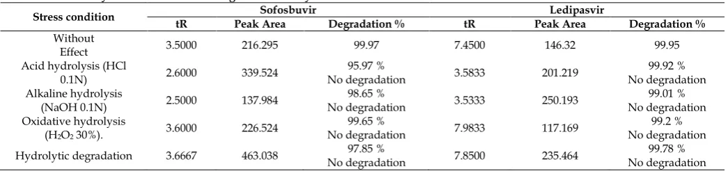
The LOD and LOQ were determined by RP HPLC for Ledipasvir (1.09μg/ml), (3.30μg/ml), and Sofosbuvir (3.36μg/ml), (10.19μg/ml) respectively (Table 14). Forced Degradation Studies [7-9]
The Forced degradation of API was carried out as per ICH guidelines (ICH, Q2B) in acid, base, oxidation, hydrolytic. The results were displayed in Table 16. Acid degradation
Weigh accurately 1 mg of Ledipasvir and 4 mg of Sofosbuvir into 10 ml volumetric flask, dissolve in 10 ml of diluent and 1.0 N aqueous HCL solution and close the volumetric flask by stopper. Keep the solution at room temperature up to 24 hours. Neutralize with 1.0 N aqueous NaOH solution and make up to the mark with the diluent, inject into the chromatographic system and calculate the percent of degradation (Table 16).
Base degradation
Int. J. Pharm. Sci. Drug Res. November-December, 2017, Vol 9, Issue 6 (291-298)
close the volumetric flask by stopper. Keep the solution at room temperature up to 24 hours. Neutralize with 1.0 N aqueous HCl solution and make up to the mark with the diluent, inject into the chromatographic system and calculate the percent of degradation (Table 16).
Peroxide degradation
Weigh accurately 1 mg of Ledipasvir and 4 mg of Sofosbuvir into 10 ml volumetric flask, dissolve in 10 ml of the diluent, add 0.3 ml of 3.0% aqueous H2O2
solution and close the volumetric flask by stopper. Keep the solution at room temperature up to 24 hours. Inject into the chromatographic system and calculate the percent of degradation. No interference was found, although there was some degradation products appeared after H2O2 treatment due to the peak of
hydrogen peroxide. And the degradation percent was calculated as shown in Table 16.
Fig. 3: Chromatogram of Blank
Fig. 4: Typical Chromatogram of Pure Mixed Standards
Fig. 5: Typical Chromatogram of Ledipasvir and Sofosbuvir in Tablet Formulation
Hydrolytic degradation
Weigh accurately 1 mg of Ledipasvir and 4 mg of Sofosbuvir into 10 ml volumetric flask, dissolved in 10 ml of diluent and up to the mark. Add H2O2 and close
the volumetric flask by stopper. Keep the solution at room temperature up to 24 hours. Inject into the chromatographic system and calculate the percent of degradation. No interference was found, although there was some degradation products appeared after H2O2
treatment due to the peak of hydrogen peroxide. And the degradation percent was calculated as shown in Table 16.
Table 5: Analysis of Tablet Formulation (MyHep LVIR - Tablet*- Mylan Pharmaceutical)
S. N o.
Amount Present
(µg/ml) Area
Amount Found (µg/ml)
% Drug Estimation LE
DI SOF
O LED
I
SOF
O LEDI SOFO
LE DI
SOF O 1. 10 40 144.1 215.62 9.97 39.83 99.7 99.57
2. 10 40 144.98 217.758 9.98 39.99 99.8 99.97
3. 10 40 145.2 216.259 9.99 39.95 99.9 99.87
4. 10 40 143.53 216.485 9.93 39.98 99.3 99.95
5. 10 40 145.23 217.354 9.99 39.99 99.9 99.97
MEAN 144.6
08 216.6952 9.972 39.948 SD 0.76 0.86 0.02 0.07
%RSD 0.52 0.40 0.25 0.17
Table 6: Standard Calibration Data of Ledipasvir by RP-HPLC Method
S. No. Concentrations (g/mL) Area Mean (n=3) S.D. %RSD 1 10 146.535 1.93 1.32 2 20 255.030 2.97 1.16 3 30 380.505 3.15 0.83 4 40 483.445 8.59 1.78 5 50 592.555 1.84 0.31
Table 7: Standard Calibration Data of Sofosbuvir by RP-HPLC Method
S. No. Concentrations (g/mL) Area Mean (n=3) S.D. %RSD 1 40 216.689 2.93 1.35 2 80 469.387 4.95 1.05 3 120 736.566 6.58 0.89 4 160 963.558 9.36 0.97 5 200 1210.895 7.83 0.65
RESULTS AND DISCUSSION
Careful evaluation of various parameters influencing analysis is an important aspect for the development of analytical method. The mobile phase was found to be most suitable methanol: water (83:17, v/v) at pH 3.0, adjusted using 0.05 % acidic acid at flow rate 1.0 ml/min give good resolution of peaks with minimum tailing as compared to other mobile phases. The wavelength 245 nm was selected. This system gave good resolution (>1.5 min) and optimum retention time with appropriate tailing factor (<2). The retention time of Ledipasvir and Sofosbuvir was found to be 7.45 and 3.50 min respectively.
Int. J. Pharm. Sci. Drug Res. November-December, 2017, Vol 9, Issue 6 (291-298)
mentioned drugs in their combined dosage formulation without prior separation. [10-15]
Linearity Study
In linearity study combined dosage formulation was found to be linear in range of (10-50g/ml) and (40-200g/ml) with correlation coefficient values 0.9991 and 0.9994 respectively. At selected wavelength 245 nm. [16]
Accuracy (Recovery Studies)
Three replicate injections, each of three different test concentrations in the range of 80%, 100% and 120% of labeled claim of tablet under study yielded the results within 99% to 102% of true concentration of each drug. The results indicated that the method is accurate. [17]
Precision
Precision studies were carried out using parameters like different days and repeatability. Results showed that the % RSD in the range of 0.30-1.78 i.e. less than 2 for different days. [18]
Fig. 6: Calibration Curve for Ledipasvir
Fig. 7: Calibration Curve for Sofosbuvir
Table 8: Standard Addition Techniques for Determination of Ledipasvir and Sofosbuvir
Drug Label Claim (µg/ml) % Added (µg/ml) Amount Total Amount (µg/ml) Amount Recovered (µg/ml) Recovery (%) SD %RSD
LEDI 10
80 8 17.9817.87 7.987.87 99.7498.41 0.88 0.88 18.08 8.08 100.06
100 10 20.00 20.16 10.16 10.0 101.64 100.0 089 0.88 20.20 10.20 100.20
120 12 22.01 21.91 12.01 11.91 101.58 99.26 0.89 0.88 21.11 11.11 99.58
SOFO 40
80 32 71.12 71.99 31.12 30.99 97.25 96.84 0.25 0.25 70.98 30.98 96.81
100 40 79.77 80.16 39.77 40.16 100.40 99.43 0.49 0.49 79.82 39.99 99.97
120 48
87.81 47.81 99.62
0.53 0.53 88.30 48.30 100.63
88.20 48.20 100.40
Table 9: Results of Precision Study
Drug [µg/ml] Conc. Mean Area Amount Found Intra-day S.D. % R.S.D. Mean Area Amount Found Inter-day S.D. % R.S.D.
LEDI 10 30 150.89 377.38 10.30 30.52 1.14 2.68 1.78 0.30 142.80 380.18 30.77 10.02 1.50 2.21 1.55 0.39 50 592.33 49.73 1.97 0.33 589.57 49.47 3.65 0.62
SOFO 120 40 218.34 740.74 123.44 38.85 3.38 3.65 1.67 0.46 223.80 725.68 121.01 40.14 6.28 2.22 0.99 0.87 200 1145.07 194.40 17.28 1.51 1179.89 194.21 12.21 1.04
Repeatability Study of Ledipasvir and Sofosbuvir
Table 10: Results of Repeatability Study of Ledipasvir and Sofosbuvir Drugs (µg/mL) Conc. Area Mean area Amount found SD %RSD
Ledipasvir 20 462.5529 470.1528 465.9851 19.98 3.85 0.83 456.2500
Sofosbuvir 40 256.7151 257.3245 257.7544 39.99 1.31 0.51 259.2235
Robustness
To evaluate the robustness of the method, each parameter selected was varied at three levels. The results indicate that the insignificant differences in peak areas and less variability in retention time and theoretical plates were observed. [18-19]
Ruggedness
Int. J. Pharm. Sci. Drug Res. November-December, 2017, Vol 9, Issue 6 (291-298)
Table 11: Results of Robustness Study of variation (Ledipasvir) Parameters Variation Level Time (min) Retention Area SD %RSD
Flow Rate mL/min
0.9 mL 8.4000 393.35 0.88 0.22 1.0 mL 7.4500 146.32 1.08 0.73 1.1 mL 7.2333 387.11 2.01 0.52
Wavelength 244 nm 245 nm 7.9000 7.4500 146.32 377.4 2.87 1.08 0.76 0.73 246 nm 7.7333 384.01 4.77 1.24 Mobile
Phase
84:16 5.1333 373.0 4.80 1.29 83:17 7.4500 146.32 1.08 0.73 82:18 8.1333 364.76 4.95 1.36
Table 12: Results of Robustness Study of variation (Sofosbuvir) Parameters Variation Level Time (min) Retention Area SD %RSD
Flow Rate mL/min
0.9 mL 3.8000 740.58 2.88 0.38 1.0 mL 3.5000 216.29 1.56 0.72 1.1 mL 4.8887 710.30 5.70 0.80
Wavelength 244 nm 245 nm 3.5833 3.5000 216.29 793.0 4.70 1.56 0.59 0.72 246 nm 3.5333 717.83 9.17 1.28 Mobile
Phase
84:16 3.8167 724.2 6.38 0.88 83:17 3.5000 216.29 1.56 0.72 82:18 5.3333 728.01 10.77 1.48
Table 13: Ruggedness Studies
Drug Claim Label
Amount Found ± S.D. % Label Claim
Analyst I Analyst II Analyst I Analyst II
Ledipasvir 30 29.45 ± 0.66 30.62 ± 0.27 98.16 102.06
Sofosbuvir 120 119.81 ± 0.12 121.73 ± 0.24 99.84 101.44
LOD and LOQ
The LOD and LOQ were determined by RP HPLC for Ledipasvir (1.09μg/ml), (3.30μg/ml), and Sofosbuvir (3.36μg/ml), (10.19μg/ml) respectively. [19]
Table 14: Results of LOD and LOQ by RP-HPLC Method
Sample LOD (µg/mL) LOQ (µg/mL)
Ledipasvir 1.09 3.30
Sofosbuvir 3.36 10.19
System Suitability Parameters
Table 15: System Suitability Test Parameters. System Suitability
Parameters
Proposed Method
Ledipasvir Sofosbuvir
Retention Time (tR) 7.45 3.5 Number of Theoretical Plate 2512.4 3857.2
Tailing Factor 2.0682 1.6667 Resolution Factor (R) 8.4643 0.000
Area [mV*s] 145.1706 214.6290 Area (%) 40.35 59.65
Forced Degradation Studies [18-19]
The forced degradation studies of Sofosbuvir and Ledipasvir tablet formulation was done on stress degradation by hydrolysis under alkaline conditions by using 0.1N NaOH was found to be 99.01% for 24 hours, stress degradation by using 0.1N HCL and product degradation was found to be 99.2% for 24 hours. Oxidative degradation was done by using hydrogen peroxide 99.78% for 24 hours. Hydrolytic degradation was found to be 99.78% for 24 hours. The Sofosbuvir
and Ledipasvir were found to be stable of the condition.
Fig. 8: Chromatogram of Acid degradation
Fig. 9: Chromatogram of Base degradation
Fig. 10: Chromatogram of Peroxide degradation
Fig. 11: Chromatogram of Hydrolytic degradation
ACKNOWLEDGEMENTS
Int. J. Pharm. Sci. Drug Res. November-December, 2017, Vol 9, Issue 6 (291-298)
Table 16: Summary of Results of Forced Degradation Study.
Stress condition tR Peak Area Sofosbuvir Degradation % tR Peak Area Ledipasvir Degradation %
Without
Effect 3.5000 216.295 99.97 7.4500 146.32 99.95 Acid hydrolysis (HCl
0.1N) 2.6000 339.524 No degradation 95.97 % 3.5833 201.219 No degradation 99.92 % Alkaline hydrolysis
(NaOH 0.1N) 2.5000 137.984 No degradation 98.65 % 3.5333 250.193 No degradation 99.01 % Oxidative hydrolysis
(H2O2 30%). 3.6000 226.524
99.65 %
No degradation 7.9833 117.169 No degradation 99.2 % Hydrolytic degradation 3.6667 463.038 No degradation 97.85 % 7.8500 235.464 No degradation 99.78 %
REFERENCES
1. Harvoni (Ledipasvir/Sofosbuvir) Tablets Product Monograph. Gilead Sciences Inc. Foster City, CA 94404 USA, Date of Preparation, 2016.
2. Gilead Files for U.S. Approval of Ledipasvir/Sofosbuvir Fixed-Dose Combination Tablet for Genotype 1 Hepatitis C. Gilead Sciences. 2014.
3. Jeffery GH, Bassett J, Mandham J, Denny RC. Vogel’s Text Book of Quantitative Chemical Analysis. 5th Ed. Longman Scientific and Technical Publication, UK, 1994, pp. 3-4. 4. Watson DG. Pharmaceutical Analysis. 1st ed. Churchill
Livingstone, Edinburgh, 1999, pp. 1-11.
5. Ewing GW. Analytical Instrumentation Handbook. 2nd Ed. Marcel Dekker Inc. New York, 1997, pp. 1-2.
6. Kasture AV, Wadodkar SG, Mahadik KR. A Textbook of Pharmaceutical Analysis. Volume -II. 17th Ed. Nirali Prakashan, Pune, 2007, pp. 148-149.
7. Chatwal GR, Anand SK. Instrumental Methods of Chemical Analysis. 5th Ed. Himalaya Publishing House, New Delhi, 2004, pp. 1.6-1.8.
8. United States Pharmacopeia, Validation of Compedial Methods. Asian ed. Pharmacopeial Convention, Inc., US. Rockville Md, 2009, pp. 734-736.
9. Indian Pharmacopoeia. Govt. of India. Ministry of Health and Family Welfare, Published By the Indian Pharmacopoeia Commission, Ghaziabad, 2007, pp. 247, 747-749.
10. Jeyabaskaran M, Rambabu C, Rajinikanth V. A New RP-HPLC Method Development and Validation of Sofosbuvir in Bulk and Pharmaceutical Dosage Forms. Journal of Pharmacreations. 2014; 1(4):125-133.
11. Vejendla R, Subramanyam CVS, Veerabhadram G. Estimation and Validation of Sofosbuvir in Bulk and Tablet Dosage Form By RP-HPLC. International Journal of Pharmacy. 2016; 6(2): 121-127.
12. Guguloth R, Madhukar, Kanna N, Ravinder A, Sravanthi K. Analytical Method Development and Validation of Sofosbuvir Tablets by RP-HPLC. Journal of Pharma Research. 2016; 5(7): 161-163.
13. Vikas PM, Satyanarayana T, Vinod Kumar D, Mounika E, Sri Latha M, Anusha R, Sathish Y. Development and Validation of New RP-HPLC Method for the Determination of Sofosbuvir in Pure Form. World Journal of Pharmacy and Pharmaceutical Science. 2016; 5(5): 775-781.
14. Madhavi S, Prameela Rani A. Bioanalytical Method Development and Validation for the Determination of Sofosbuvir from Human Plasma. International Journal of Pharmacy and Pharmaceutical Sciences. 2017; 9(3): 35-41. 15. Devilal J, Durgaprasad B, Pal N, Srinivasa Rao A. New
Method Development and Validation for the Determination of Ledipasvir in Bulk Drug Form by Using Reverse Phase HPLC Technique. World Journal of Pharmacy and Pharmaceutical Science. 2016; 5(8): 1312-1321.
16. Rai SY, Prajapati Y, Patni P. Development and Validation of RP-HPLC and UV Spectroscopy Methods for Simultaneous Estimation of Sofosbuvir and Ledipasvir in Their Combined Tablet Dosage Form. Pharma Science Monitor An International Journal of Pharmaceutical Sciences. 2017; 8(2): 369-388.
17. Kothapalli KK, Saisri M, Priyanka M, Subhashini M, Manikanta M. A New Analytical Method Development and Validation for the Simultaneous Estimation of Ledipasvir and Sofosbuvir Using RP-HPLC. Intercontinental Journal of Pharmaceutical Investigations and Research. 2017; 4(1): 142-165.
18. Hassouna MEM, Abdelrahman MM, Mohamed MA. Assay and Dissolution Methods Development and Validation for Simultaneous Determination of Sofosbuvir and Ledipasvir by RP-HPLC Method in Tablet Dosage Forms. J Forensic Sci & Criminal Inves. 2017; 1(3): 555562.
19. Yogendrachari K, Madhu M, Kumar EG, Kumari MV, Naik MC. Analytical Method Development and Validation for Simulta-Neous Determination of Ledipasvir and Sofosbuvir in Tablet Dosage Form By RP-HPLC. Journal of Global Trends in Pharmaceutical Sciences. 2016; 7(3): 3401- 3407. 20. Zaman B, Siddique F, Hassan W. RP-HPLC Method For
Simultaneous Determination of Sofosbuvir and Ledipasvir In Tablet Dosage Form and Its Application To In Vitro Dissolution Studies. Chromatographia. December 2016; 79 (23): 1605–1613.
21. Abdulkareem A, El-Tohamy MF. Validated Capillary Zone Electrophoresis Approach For Simultaneous Separation and Determination of Hepatitis C Sofosbuvir and Ledipasvir In Tablet Dosage Form. World Journal of Pharmaceutical Research. 2017; 6(5):129-147.
22. Rezk MR, Basalious EB, Amin ME. Novel and Sensitive UPLC–MS/MS Method for Quantification of Sofosbuvir in Human Plasma: Application to a Bioequivalence Study. Biomedical Chromatography. 2016; 30(9):1354-1362.
23. Shi X, Zhu D, Lou J, Zhu B, Hu A-R, Gan D. Evaluation of A Rapid Method For The Simultaneous Quantification of Ribavirin, Sofosbuvir and Its Metabolite in Rat Plasma By UPLC–MS/MS. Journal of Chromatography. 2015; 10(02): 353-357.
24. Pan C, Chen Y, Chen W, Zhou G, Jin L, Zheng Y, Lin W, Pan Z. Simultaneous Determination of Ledipasvir, Sofosbuvir and Its Metabolite in Rat Plasma By UPLC–MS/MS and Its Application To A Pharmacokinetic Study. Journal of Chromatography B. 2016; 1008: 255-259.
25. Suryawanshi R, Shinde N, Todkar G. Development and Validation of Simple UV Spectrophotometric Method for the Determination of Ledipasvir in Bulk Form and Stress Degradation Studies. Inventi Rapid: Pharm Analysis & Quality Assurance. 2016; (3): 1-5.
HOW TO CITE THIS ARTICLE: Rote AP, Alhat J, Kulkarni AA. Development and Validation of RP-HPLC