Dissertation zur Erlangung des Doktorgrades der Fakultät für Chemie und Pharmazie der Ludwig-Maximilians-Universität München
Rubisco folding and oligomeric assembly:
Detailed analysis of an assembly intermediate
Amanda Windhof (geb. Starling)
aus
Delaware, Ohio, USA
Acknowledgments
I especially want to thank Prof. Dr. F. Ulrich Hartl and Dr. Manajit Hayer-Hartl for giving me the opportunity to conduct my PhD research in the department. I have learned so much on this journey which would not have been possible without your intellectual support and scientific enthusiasm.
I also want to thank PD Dr. Dr. Winklhofer for being my 2. Gutacherin and of course the other members of my PhD committee: Prof. Dr. Beckmann, Prof. Dr. Vothknecht, Prof. Dr. Gaul, and Prof. Dr. Hopfner.
There are also many people I want to thank for within the department and institute. I am very grateful to Dr. Cuimin Liu for her help and guidance when I first came to this department. She was always open to answer any questions I had. Thank you Dr. Andreas Bracher for your support in many ways, in particular with your crystallographic knowledge and various ideas for developing enhanced methods. I also want to thank Dr. Oliver Müller-Cajar for your never ending passion for science, especially for Rubisco, and for your very insightful discussions.
I am grateful for the lunch conversations with Dr. Yi-chin Tsai and your mass spectrometry expertise. I would also like to thank Thomas Hauser and Mathias Stotz for your valuable discussions and input. Thank you to Evelyn Frey-Royston, Silke Leuze-Bütün, and Andrea Obermayr-Rauter for their administrative support as well as Emmanuel Burghardt, Bernd Grampp, Romy Lange, Nadine Wischnewski, Verena Marcus, Elisabeth Schreil, Albert Ries, Andreas Scaia, and Kevin Gaughan for their invaluable technical support.
Thank you to Dr. A. Young and Prof. Dr. R. Beckmann for your cryo-EM expertise and interpretation. I also want to thank Reinhard Mentele and Elisabeth Weyher-Stingl for technical support as well as the staff of the MPI Animal and Sequencing Facilities. Also, to the rest of the lab, I am very thankful for the help and assistance I have received while being here and all the fun times we have shared.
Contents
1 Summary ______________________________________________________________ 1 2 Introduction ____________________________________________________________ 3 2.1 Protein folding _________________________________________________________ 3
2.2 Molecular chaperones ___________________________________________________ 6 2.2.1 De novo protein folding _______________________________________________ 6 2.2.1.1 Ribosome-associated chaperones ___________________________________ 7 2.2.1.2 Hsp70 system __________________________________________________ 9 2.2.1.3 Chaperonins ___________________________________________________ 11 2.2.1.3.1 E. coli chaperonin system ______________________________________ 11 2.2.1.3.2 Cyanobacterial chaperonin _____________________________________ 14 2.2.1.3.3 Chloroplast chaperonin ________________________________________ 17 2.3 Photosynthesis ________________________________________________________ 20 2.3.1 Light-dependent reactions ____________________________________________ 22 2.3.2 Light-independent reactions __________________________________________ 24 2.3.3 Carbon concentrating mechanisms _____________________________________ 25 2.3.3.1 C4 Plants _____________________________________________________ 26 2.3.3.2 Crassulacean acid metabolism (CAM) photosynthesis __________________ 28 2.3.3.3 Pyrenoid _____________________________________________________ 28 2.3.3.4 Carboxysome __________________________________________________ 29 2.3.4 Ribulose-1,5-bisphosphate carboxylase/oxygenase (Rubisco) ________________ 30 2.3.4.1 Rubisco regulation ______________________________________________ 32 2.3.4.2 Rubisco structure _______________________________________________ 33 2.3.4.3 Rubisco folding and assembly_____________________________________ 36 2.3.4.4 RbcX ________________________________________________________ 38 2.4 Aim of study __________________________________________________________ 42
3 Materials and Methods __________________________________________________ 43 3.1 Materials ____________________________________________________________ 43 3.1.1 Chemicals ________________________________________________________ 43 3.1.2 Reagents and Purification kits _________________________________________ 43 3.1.3 Strains ___________________________________________________________ 43 3.1.4 Plasmids, DNA, and oligonucleotides ___________________________________ 44 3.1.5 Enzymes, proteins, peptides, and antibodies ______________________________ 47 3.1.6 Media ____________________________________________________________ 47 3.1.7 Buffers and Standard Solutions ________________________________________ 48 3.2 Instruments __________________________________________________________ 50
3.3.2.3 TSS transformation _____________________________________________ 53 3.3.3 Plasmid DNA and DNA fragment purification ____________________________ 53 3.3.4 PCR (polymerase chain reaction) ______________________________________ 53 3.3.5 Site-directed mutagenesis ____________________________________________ 54 3.3.6 Restriction digest and DNA ligation ____________________________________ 55 3.3.7 Cloning strategies __________________________________________________ 56 3.4 Protein biochemical and biophysical methods ______________________________ 57 3.4.1 Protein analytical methods ___________________________________________ 57 3.4.1.1 Protein quantification and sequence alignments _______________________ 57 3.4.1.2 SDS-PAGE ___________________________________________________ 57 3.4.1.3 Gradient SDS-PAGE ____________________________________________ 58 3.4.1.4 Native-PAGE _________________________________________________ 59 3.4.1.5 CN Bis-tris-PAGE ______________________________________________ 59 3.4.1.6 Coomassie blue staining of polyacrylamide gels ______________________ 60 3.4.1.7 Western blotting and immunodetection _____________________________ 60 3.4.1.8 Autoradiography _______________________________________________ 61 3.4.1.9 Generation of anti-serum _________________________________________ 61 3.4.1.10 TCA precipitation ______________________________________________ 62 3.4.1.11 SEC-MALS ___________________________________________________ 62 3.4.1.12 Proteolytic digestion and Edman degradation _________________________ 62 3.4.1.13 Mass spectrometry ______________________________________________ 63 3.4.1.14 CD (circular dichroism) spectroscopy _______________________________ 63 3.4.2 Protein crystallization _______________________________________________ 63 3.4.3 Protein expression and purification _____________________________________ 64 3.4.3.1 Syn6301-RbcL8 ________________________________________________ 64
3.4.3.2 Prokaryotic and Eukaryotic RbcL __________________________________ 65 3.4.3.3 Prokaryotic and Eukaryotic RbcS __________________________________ 65 3.4.3.4 Cyanobacterial-RbcX2 ___________________________________________ 66
3.4.3.5 Cyanobacterial-RbcX2(N-6His) ______________________________________ 66
3.4.3.6 At-RbcXI2 ____________________________________________________ 67
3.4.3.7 RbcL8/(RbcX2)8 complex ________________________________________ 67
3.4.3.8 Syn6301-RbcL8/(AnaCA-RbcX(TAG)2(N-6His))8 complexes ______________ 68
3.4.4 Functional analyses _________________________________________________ 69 3.4.4.1 Amber stop codon suppression ____________________________________ 69 3.4.4.2 Site-specific crosslinking ________________________________________ 69 3.4.4.3 Rubisco carboxylation activity assay of E. coli lysate __________________ 69 3.4.4.4 RTS: in vitro translation of Rubisco ________________________________ 70 3.4.4.5 PURE system __________________________________________________ 71 3.4.4.6 CABP synthesis from RuBP ______________________________________ 71 3.4.4.7 Disulfide crosslinking ___________________________________________ 72 3.4.4.8 In vivo expression of cyanobacterial GroEL and GroES in E. coli _________ 72 3.4.4.9 In vivo assembly of RbcL8/(RbcX2)8 complexes in E. coli _______________ 73
3.4.4.10 Analytical gel filtration of RbcL8/(RbcX2)8 complexes _________________ 73
4.2 Role of GroEL/ES and RbcX2 in Rubisco folding and assembly _______________ 77
4.2.1 RbcX2 binding motif ________________________________________________ 79
4.3 Analysis of the RbcL8/(RbcX2)8 interaction ________________________________ 84
4.3.1 Cryo-EM structure of the RbcL8/(RbcX2)8 complex ________________________ 84
4.3.1.1 Examination of the cryo-EM structure ______________________________ 84 4.3.1.2 Crosslinking of RbcL and RbcX ___________________________________ 87 4.3.1.3 Mechanism of RbcX2 during in vitro refolding of RbcL ________________ 90
4.3.1.4 Role of RbcX2 in RbcL8 C-terminal stabilization ______________________ 91
4.3.2 Crystal structure of RbcL8/(RbcX2)8 complex ____________________________ 92
4.3.2.1 RbcL/RbcX interface ____________________________________________ 93 4.3.2.2 Mechanism of RbcX2 function in RbcL dimer assembly ________________ 97
4.3.2.3 The role of RbcS in RbcX2 displacement and Rubisco catalysis _________ 100
4.4 Analysis of eukaryotic Rubisco and refolding components ___________________ 103
5 Discussion ___________________________________________________________ 111 5.1 Role of chaperonins in Rubisco folding ___________________________________ 111
5.2 Highly homologous Rubisco proteins have diverse assembly pathways ________ 113
5.3 Assembly chaperones _________________________________________________ 113 5.3.1 Mechanism of RbcX2-mediated Rubisco assembly _______________________ 115
5.4 Role of RbcL N-terminal region in Rubisco catalysis _______________________ 116
5.5 Role of RbcS in Rubisco catalysis _______________________________________ 117
5.6 Outlook _____________________________________________________________ 117
6 References ___________________________________________________________ 119 7 Appendices __________________________________________________________ 135 7.1 Plasmids constructed in this study _______________________________________ 135
7.2 Primer pairs used for PCR _____________________________________________ 138
7.3 Protein accession numbers _____________________________________________ 140
7.4 Abbreviations ________________________________________________________ 141
7.5 Publications _________________________________________________________ 143
1
Summary
To become biologically active, a protein must fold into a distinct three-dimensional structure. Many non-native proteins require molecular chaperones to support folding and assembly. These molecular chaperones are important for de novo protein folding as well as refolding of denatured proteins under stress conditions. A certain subset of chaperones, the chaperonins, are required for the folding of the enzyme ribulose-1,5-bisphosphate carboxylase/oxygenase (Rubisco); furthermore, correct folding of Rubisco is also aided by the Hsp70 chaperone system.
Rubisco catalyzes the initial step of CO2 assimilation in the Calvin-Benson-Bassham
(CBB) cycle. Unfortunately, this enzyme is extremely inefficient, not only does it exhibit a slow catalytic rate (three CO2 molecules fixed per second per Rubisco) but it also
discriminates poorly between the assimilation of CO2 and O2 to its sugar-phosphate substrate
ribulose-1,5-bisphosphate (RuBP), the latter resulting in loss of photosynthetic efficiency. Due to these inefficiencies, carbon fixation by Rubisco is the rate limiting step of the CBB cycle. Photosynthetic organisms must produce tremendous amounts of Rubisco to alleviate these shortcomings; therefore significant quantities of nitrogen stores are invested in the production of Rubisco making Rubisco the most abundant protein on earth. These drawbacks of Rubisco have important implications in increasing CO2 concentrations and temperatures in
the context of global warming. The ability to engineer a more efficient Rubisco could potentially reduce photosynthetic water usage, increase plant growth yield, and reduce nitrogen usage is plants. However, eukaryotic Rubisco cannot fold and assemble outside of the chloroplast, hindering advancements in creating a more efficient Rubisco.
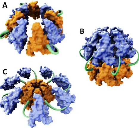
Form I Rubisco, found in higher plants, algae, and cyanobacteria, is a hexadecameric complex consisting of a core of eight ~50 kDa large subunits (RbcL), which is capped by four ~15 kDa small subunits (RbcS) on each end. The discovery of a Rubisco-specific assembly chaperone, RbcX, has lead to a better understanding of the components necessary for the form I Rubisco assembly process. RbcX is a homodimer of ~15 kDa subunits consisting of four α-helices aligned in an anti-parallel fashion along the α4 helix. RbcX2 functions as a stabilizer
of folded RbcL by recognizing a highly conserved C-terminal sequence of RbcL: EIKFEFD, termed the C-terminal recognition motif. As has been demonstrated by studies of cyanobacterial Rubisco, de novo synthesized RbcL is folded by the chaperonins, whereupon RbcX2 stabilizes the folded RbcL monomer upon release from the folding cavity and then
assists in the formation of the RbcL8 core. RbcX2 forms a dynamic complex with RbcL8 and
thereby creating the functional holoenzyme. However, the exact mechanism by which RbcS binding displaces RbcX2 from the RbcL8 core is still unknown. Furthermore, though much
advancement has been made in the understanding of form I Rubisco folding and assembly, an exact and detailed mechanism of form I Rubisco assembly is still lacking. The highly dynamic complex of RbcL/RbcX is critical for the formation of the holoenzyme; however it has hindered attempts to characterize critical regions of RbcL that interact with the peripheral regions of RbcX2. An important observation arose when heterologous RbcL and RbcX2
components interacted; a stable complex could form enabling in depth characterization of the RbcL/RbcX2 interaction.
In the present study, the detailed structural mechanism of RbcX2-mediated cyanobacterial
form I Rubisco assembly is elucidated. To obtain molecular insight into the RbcX2-mediated
assembly process of cyanobacterial form I Rubisco, cryo-EM and crystallographic studies in concert with mutational analysis were employed by taking advantage of the high affinity interaction between RbcL and RbcX2 in the heterologous system (Synechococcus sp.
PCC6301 RbcL and Anabaena sp. CA RbcX2). Structure guided mutational analysis based on
the 3.2 Å crystal structure of the RbcL8/(RbcX2)8 assembly intermediate were utilized to
determine the precise interaction site between the body of RbcL and the peripheral region of RbcX2. From these studies a critical salt bridge could be identified that functions as a
guide-point for correct dimer formation, and it was observed that RbcX2 exclusively mediates
Rubisco dimer assembly. Furthermore, the mechanism of RbcX2 displacement from the
RbcL8 core by RbcS binding was elucidated as well as indications of how RbcS docking on
the RbcL8 core is imperative for full form I Rubisco catalytic function by stabilizing the
enzymatically competent conformation of an N-terminal loop of Rubisco termed the ‘60ies loop’. Finally, initial attempts in in vitro reconstitution of eukaryotic Rubisco are reported along with the characterization of Arabidopsis thaliana RbcX2 binding to the C-terminal
2
Introduction
Proteins are polymers of amino acids linked together by peptide bonds formed between adjacent carboxyl and amino groups. They are necessary for the correct functioning of biological processes in cells from all domains of life. For example these macromolecular molecules are involved in processes such as cellular structure, metabolism, signaling, enzymatic catalysis, immune response, storage, transportation, movement, and regulation.
2.1
Protein folding
In order to become biologically functional, proteins must fold into the proper three-dimensional structure. The physiological function of a protein is dependent on its unique structure as established by the primary, secondary, tertiary, and quaternary structures. The primary structure of a protein is determined by the sequence of amino acids along the polypeptide chain as encoded by the respective gene upon translation. Amino acid composition in the primary structure, not only determines the solubility and ionic properties but also the native structure of the respective protein. The secondary structure arises from formation of α-helical, β-strand/sheets, and loop/turn elements stabilized by hydrogen bonds. The secondary structures combine due to residue interactions, driven in part by the burying of hydrophobic surfaces, to assume the tertiary structure. The tertiary structure is the final overall conformation of a single protein including the formation of α-helical bundles, β-sheets, and/or (α/β) TIM-barrels, which are stabilized by hydrogen bonds, salt bridges, posttranslational modifications, and/or disulfide bond formation. In the tertiary structure, regions that were previously spatially separated in the primary structure come into close contact due to the compaction of the polypeptide chain. The quaternary structure arises from the stable interaction of two or more protein molecules through hydrophobic or polar contacts of side chain residues to form a functional complex. The quaternary structure is stabilized by hydrogen bonds, van-der-Waals contacts, and/or ionic forces.
reactions lead prominently to aggregation products; indicating that the primary sequence alone is not sufficient to drive correct protein folding.
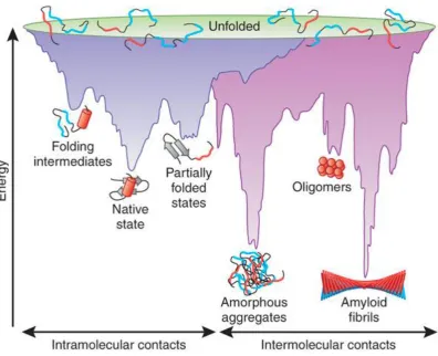
Inherently, the native state of a protein is the most thermodynamically stable, and accordingly protein folding follows the pathway from the high energy extended conformation to the low energy native structure (Frydman 2001). It is well established that protein folding occurs rapidly (<1 s). Therefore demonstrating that protein folding is not a random process, and proteins do not sample all possible conformations before reaching their native structure. The formation of initial structural features leads to the further positioning of successive structural elements; otherwise folding would not proceed in a biologically relevant timeframe (Dobson et al. 1998; Levinthal 1968). As illustrated by the funnel shaped energy landscape representing the free-energy potential during folding of a protein chain (Figure 2.1), there are local energy minima and maxima which must be overcome to obtain the stable, low-energy native state (Hartl and Hayer-Hartl 2009). The ruggedness of the energy landscape indicates that during folding, while the protein may become trapped at local energy minima, proteins adopt conformations/intermediate states with constantly decreasing energy potentials (Jahn and Radford 2005). The final native state is the most stable and energetically efficient structure which can be obtained. Collapse of the elongated non-native protein is initiated by the burial of hydrophobic residues whereupon hydrogen bonds, charge-charge interactions, and van-der-Waals forces contribute to stabilization.
Figure 2.1 Energy landscape of protein folding.
Left side: purple funnel indicates the energy barriers present and intermediates formed during protein folding to the native state. Right side: pink funnel indicates the energy barriers present and intermediates formed when aggregation species are prevalent. Note that overlap occurs between the two surfaces. Intermediates formed during protein folding can lead to the formation of aggregates. Adapted from: (Hartl and Hayer-Hartl 2009)
as Alzheimer’s, Parkinson’s, Huntington’s, and prion disease (Haass and Selkoe 2007). To ensure that aggregation is limited during protein synthesis, folding, and stress conditions, many proteins rely on other macromolecules, chaperones, for assistance to obtain correct native structure.
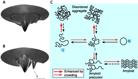
Figure 2.2 Protein aggregation pathways in the cell.
(A) The folding-energy landscape of a small, rapidly folding protein that requires no additional chaperone assistance. (B) The folding-energy landscape of a large protein that requires chaperone assistance to achieve native state due to aggregate formation propensity. A and B adapted from: (Lin and Rye 2006) (C) Red arrows indicate pathway enhancement by cellular crowding. U: newly synthesized unfolded protein chain, I: intermediate, partially folded, N: native protein. Amyloid formation is predicted to be enhanced by molecular crowding, but this has yet to be experimentally proven. Adapted from: (Hartl and Hayer-Hartl 2002)
2.2
Molecular chaperones
The term molecular chaperone was coined in 1978 to describe a particular nuclear protein assisting nucleosome assembly (Laskey et al. 1978). However, the use of this term has broadened to describe a class of macromolecules that supports folding and assembly of non-native proteins without becoming a part of or influencing their final functional conformation (Ellis 1987). Molecular chaperones are important for de novo protein folding, oligomeric assembly, intracellular transport, refolding of stress-denatured proteins, and assist in proteolytic degradation. Here, the involvement of molecular chaperones in protein folding and assembly will be discussed.
2.2.1
De novo
protein folding
As mentioned above, during nascent polypeptide chain synthesis and emergence from the ribosome, extended regions of the chain are exposed to the crowded cellular environment.
A
B
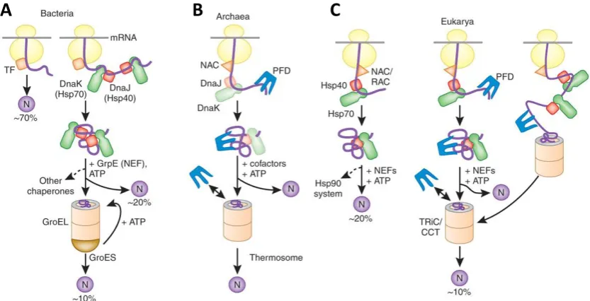
Importantly, many of these exposed regions i.e., hydrophobic side chains, become buried upon formation of the native protein and their transient exposure could lead to unfavorable non-native interactions. To avoid off-pathway, aberrant side reactions during translation, a special class of molecular chaperones specifically interact with nascent chains emerging from the ribosome. This first class of chaperones can be classified as the nascent-chain interacting chaperones, consisting of Ribosome-associated chaperones and the Hsp70 system. Proteins that fail to fold by these specific chaperones are transferred to the downstream chaperones called chaperonins, which provide a sequestered environment for folding. De novo protein folding is ubiquitously present in all three kingdoms of life and is delineated by the initial interaction of specific chaperones with the translating polypeptide chain followed by transfer to downstream chaperones specialized in finalizing the folding process (Figure 2.3) (Hartl and Hayer-Hartl 2009).
Figure 2.3 Model of chaperone-assisted folding of nascent chains in the cytosol.
(A) Bacteria: newly synthesized polypeptide chains interact with Trigger factor (TF) and small proteins, 70% of total, fold to the native state (N) after this interaction. Larger polypeptides, 20% of total, further require the assistance of the ATP-dependent capture and release of the Hsp70 system (DnaK/DnaJ/GrpE). If the native state is still not achieved, 10% of total, these non-native proteins are subsequently transferred to the chaperonin system (GroEL/GroES) for cycles of ATP-dependent encapsulation and release. (B) Archaea: nascent chain-associated complex (NAC), prefoldin (PFD), in some archaeal species DnaK and DnaJ are present, and interact with nascent chains and assist with the transfer to chaperonin, thermosome. (C) Eukarya: NAC most likely interacts with nascent chains but its actual role in folding is not fully understood. 20% of nascent chains interact with ribosome-associated complex (RAC) and the Hsp70 system to obtain their native state, however some are further transferred to Hsp90. 10% of nascent chains are co- or post-translationally transferred to the chaperonin system (TRiC/CCT) from Hsp70 and PFD. Adapted from: (Hartl and Hayer-Hartl 2009)
2.2.1.1
Ribosome-associated chaperones
Ribosome-associated chaperones not only directly interact with the ribosome but also act on nascent chains emerging from the ribosomal exit tunnel. These chaperones are found in all domains of life and include: bacterial Trigger factor, eukaryotic and archaeal nascent chain-associated complex (NAC), and the eukaryotic heat shock protein 70 (Hsp70) system
called the ribosome-associated complex (RAC) (Figure 2.3 B and C) (Hartl and Hayer-Hartl 2009). Bacterial Trigger factor is the best characterized ribosome-associated chaperone; therefore its role in de novo protein folding will be described in further detail.
Figure 2.4 Structure of Trigger factor and Trigger factor interaction with the ribosome.
(A) Ribbon and schematic domain representations of Trigger factor structure left and right panel, respectively (PDB 1W26 (Ferbitz et al. 2004)). Bottom: linear sequence of domain organization. N-terminal tail is red, terminal Arm 1 is green, C-terminal Arm 2 is blue, and Head PPIase domain is yellow. (B) Trigger factor in complex with the ribosomal 50S complex, coloring as in, A. Nascent chain in magenta extending from the exit tunnel is modeled. Location of ribosomal proteins is indicated. A and B Adapted from: (Ferbitz et al. 2004) (C) Model of TF ribosome and substrate binding. 1. Unbound TF is in equilibrium between the monomeric and dimeric states. 2. Conformational change of TF upon ribosomal binding, which may cause activation for substrate interaction. 3. Translating peptide interacts with TF at the ribosomal exit tunnel. 4. After translation polypeptide and TF are released from the ribosome, the polypeptide is either still bound with TF or folded to its native state. Structurally complex proteins are transferred to DnaK for further folding assistance. C Adapted from: (Vabulas et al. 2010)
Trigger factor (TF) is a 48 kDa protein that interacts with the large ribosomal subunit near the exit tunnel in close proximity to ribosomal proteins L23 and L29 with 1:1 stoichiometry (Ferbitz et al. 2004). TF consists of three domains: the N-terminal ribosome-binding domain, the peptidyl-prolyl isomerase (PPIase) domain, and the C-terminal domain. As observed in the crystal structure, the C-terminal domain is located in between the N-terminal and PPIase domains exhibiting two arm-like extensions, Arm 1 and Arm 2 (Figure 2.4 A and B). While all three domains are implicated in binding hydrophobic regions of extended polypeptide chains, the C-terminal arm-like protrusions and the N-terminal domain primarily interact with the extended polypeptide chain (Lakshmipathy et al. 2007). In free solution TF forms a homodimer, however, TF is monomeric when bound to the ribosome (Figure 2.4 C) (Maier et al. 2003). TF interaction with the ribosome is mediated via residues of the N-terminal domain (Lakshmipathy et al. 2007). Ribosomal binding mediates a conformational change in TF, most likely preparing TF for substrate binding (Kaiser et al. 2006). During translation of especially hydrophobic nascent chains, TF binds the ribosome and attaches to the polypeptide followed by subsequent detachment from the ribosome
(Agashe et al. 2004). Substrate binding and release is ATP-independent, additionally binding is mediated by stretches of exposed hydrophobic patches on the extended nascent chain while polypeptide release apparently results from the collapse of the chain and burial of hydrophobic residues (Figure 2.4 C) (Vabulas et al. 2010). Due to TF binding close to the ribosomal exit tunnel, the majority of translating-nascent chains interact with TF and approximately 70% can reach their native state upon release from TF (Figure 2.3 A). However, if the protein does not achieve native state upon dissociation from TF, the non-native proteins are transferred to downstream chaperone systems for further attempts at chaperone-assisted protein folding.
2.2.1.2
Hsp70 system
Hsp70s act downstream of TF and are not only found as constituently expressed forms in the cell but also as highly inducible forms during stress conditions (Hartl 1996). Hsp70 proteins are important for de novo folding of nascent chains and are furthermore involved in protein trafficking and assisting in proteolytic degradation (Bukau and Horwich 1998; Young et al. 2003). The yield of folded protein is enhanced when both TF and the Hsp70 system act on the nascent chain (Agashe et al. 2004). However, Hsp70 and TF are partially functionally redundant. Deletion of TF or Hsp70 alone is not deleterious to cell growth, conversely cells are not viable if both of these proteins are deleted (Teter et al. 1999), thus demonstrating substantial overlap in the subset of substrates which bind to and are assisted in folding by TF and Hsp70. TF and Hsp70 substrate overlap is interesting considering these chaperones display intrinsic mechanistic differences. TF binds and releases substrate independent of nucleotide, whereas Hsp70 substrate interaction is nucleotide regulated. In bacterial systems Hsp70 (DnaK) acts downstream of TF and typically does not bind the ribosome. In the following section, the bacterial Hsp70 system (DnaK/DnaJ/GrpE system) will be discussed in detail as the bacterial system is the best characterized. Homologues of the entire or specific components of the DnaK/DnaJ/GrpE system are found in the cytosol, mitochondria, chloroplast, and endoplasmic reticulum of eukaryotic cells.
and isoleucine residues being preferred (Ruediger et al. 1997). Regions of this character occur approximately every 50-100 amino acids in the majority of protein sequences. In this way, DnaK binds exposed hydrophobic regions preventing non-native contacts, which could occur before the native structure is reached and hydrophobic surfaces are buried. DnaK thereby functions in smoothing the energy landscape to achieve efficient protein folding. DnaK has two conformational forms displaying differential affinities to substrate: the open or closed state (Mayer et al. 2000). In the open conformation ATP is bound and DnaK binds polypeptide substrate with relatively low affinity. The cochaperone Hsp40 (DnaJ), a J-domain protein, attaches to the ATP bound DnaK N-terminus with its N-terminal J-domain. DnaJ functions by increasing the rate of ATP hydrolysis of DnaK, thereby enhancing substrate capture (Szabo et al. 1994). Note that DnaJ can also bind the substrate and transfer it to DnaK (Genevaux et al. 2007). ATP hydrolysis leads to the formation of the ADP-bound closed conformation of DnaK, which binds polypeptide substrate with relatively high affinity (Figure 2.5).
Figure 2.5 Structure of Hsp70 and model of substrate interaction.
Left panel: structure of the DnaK N-terminal ATPase domain with ATP binding site indicated (PDB 1DKG (Harrison et al. 1997)) and the C-terminal peptide binding domain, α-helical lid is indicated in yellow and substrate peptide is indicated in purple and its sequence shown in single-letter code (PDB 1DKZ (Zhu et al. 1996)). Right panel: model of the Hsp70 reaction cycle. 1. Hsp40 delivers substrate to Hsp70 with bound ATP. 2. Hsp40 interaction with Hsp70 accelerates ATP to ADP hydrolysis, Hsp70 in ADP-state results in lid closure and high affinity for substrate. 3. Nucleotide exchange factor (NEF) binds to Hsp70 and catalyzes the release of ADP. 4. ATP binding causes Hsp70 lid to open and low affinity for substrate followed by substrate and NEF release. 5. Released substrate will either fold to native state (N), rebind Hsp70, or be transferred to downstream chaperones. Adapted from: (Hartl and Hayer-Hartl 2009)
repeated with DnaK. Nearly 20% of all E. coli proteins are capable of reaching their native state upon DnaK interaction, however 10% require further folding assistance (Figure 2.3 A) (Teter et al. 1999). Non-native proteins that are unable to reach the native state with the Hsp70 system are transferred to a specialized set of chaperones termed chaperonins, which provide a sequestered environment for folding.
2.2.1.3
Chaperonins
The term chaperonins refers to a class of ubiquitous, essential chaperone complexes. Chaperonins are large, oligomeric proteins (~0.8-1 MDa) consisting of two ring structures stacked back-to-back (Hemmingsen et al. 1988). They exhibit ATPase activity and bind non-native proteins or stress-denatured proteins through hydrophobic interactions. Two families of chaperonins exist in nature as determined by mechanistic and sequence variations: group I and group II (Horwich et al. 2007). Group I chaperonins occur in bacteria (GroEL), chloroplasts (ch-Cpn60), and mitochondria (mt-Hsp60). These chaperonins characteristically contain two seven-membered rings and operate with the lid-like cochaperone Hsp10 (GroES in bacteria, ch-Cpn10 and ch-Cpn20 in chloroplasts, and mt-Hsp10 in mitochondria). In contrast, group II chaperonins occur in the cytosol of archaea (thermosome) and eukaryotes (TRiC/CCT) and are organized as either two eight or nine-membered rings consisting of homooligomeric or heterooligomeric homologous subunits (Frydman 2001). The group II eukaryotic TRiC chaperonin is not stress inducible, in contrast to the group I and thermosome chaperonins (Horwich et al. 2007; Klunker et al. 2003). Furthermore, group II chaperonins do not require a separate cochaperone for their function. Instead, they contain α-helical protrusions that extend from the apical domain of the rings, which behave as a built-in-lid. Both groups of chaperonins exhibit ATP hydrolysis and fold substrate through rounds of encapsulation in an isolated environment followed by release of folded substrate (Gatenby 1992).
2.2.1.3.1
E. coli
chaperonin system
size with complex α/β domain topologies; nevertheless proteins of up to 150 kDa have also been identified as GroEL substrates. Furthermore, proteins that interact with GroEL during folding can be divided into three specific substrate classes (Kerner et al. 2005). Class I substrates maintain the ability to spontaneously fold when diluted from denaturant with low aggregation propensity and exhibit only a slight requirement for GroEL for folding. Class II substrates have higher propensity for aggregation upon dilution from denaturant and chaperones are necessary for folding, but this can be accomplished by either GroEL/ES or the DnaK chaperone system. Class I and class II substrates are generally more abundant proteins with only partial dependence on GroEL for folding. Approximately 85 proteins have been characterized as class III GroEL substrate proteins. Proteins of this class are generally of low abundance in the cytosol and strictly dependent on GroEL/ES for folding. Although these substrates bind DnaK, the DnaK chaperone system is not able to mediate folding. DnaK binds class III substrates and keeps them in a folding competent state (Kerner et al. 2005). The mechanism of GroEL/ES-mediated substrate folding is distinct from the Hsp70 chaperone system in that substrate folding occurs inside an enclosed, globally confined environment.
Figure 2.6 Structure of GroEL/ES and model of GroEL/ES-mediated substrate folding.
(A) Crystal structure of GroEL in complex with GroES and seven ADP bound. Dimensions of the cis and trans-ring and of
the entire complex are indicated. Equatorial, intermediate, and apical domains are labeled for one subunit in the cis-ring as
well as the position of one bound ADP. (PDB 1AON (Xu et al. 1997)). (B) Model of GroEL/ES-mediated substrate folding.
1. Substrate protein is delivered to cis-ring of GroEL by DnaK system. 2. Apical domain movement in an ATP-dependent
manner causes bound substrate stretching. 3. Substrate is encapsulated by GroES in the cavity. 4. Substrate folding in the chaperonin cavity and ATP hydrolysis. 5. Substrate is released into solution either in the native state, or an incompletely folded intermediate state. 6. Incompletely refolded substrate is rebound to GroEL for further cycles of refolding. N: native, I: folding intermediate. Adapted from: (Vabulas et al. 2010)
GroEL is an ~800 kDa tetradecameric complex consisting of ~58 kDa identical subunits. Each subunit has an equatorial domain, an intermediate domain, and an apical domain (Braig et al. 1994). The equatorial domain forms the interface between the two rings and is also the site of nucleotide binding. The intermediate domain is located between the
cis
trans equatorial
intermediate apical
GroES
80 Å
71 Å
184
Å
ADP
equatorial and apical domains and facilitates allosteric communication between these two domains. The flexible apical domain exposes hydrophobic residues and is the binding site for both substrate proteins and GroES. The lid-like cochaperone of GroEL, GroES, is a heptameric ring composed of ~10 kDa identical subunits (Figure 2.6 A) (Xu et al. 1997). The mechanism of GroEL/ES action has been investigated in depth. GroES binding occurs in an ATP-regulated manner and induces conformational shifts in GroEL. Furthermore, binding of GroES to GroEL causes an expansion of the folding cavity and conversion of the cavity from hydrophobic to hydrophilic with an overall net negative charge (Chen et al. 1994). Substrate protein is encapsulated in the GroEL/ES cavity for ~10 s at 25°C, which is the amount of time necessary for ATP hydrolysis in the cis-ring (GroES bound ring). Binding of seven ATPs to the opposite trans-ring causes release of ADP, dissociation of GroES, and release of substrate. Protein that has not reached its final native state can rebind GroEL for further a round of encapsulation and folding (Figure 2.6 B). Intriguingly, GroEL can also support the folding of substrates too large to be enclosed in the cavity by GroES, thereby indicating that large substrates may be folded by the trans-ring of GroEL without encapsulation (Chaudhuri et al. 2001).
The GroEL/ES cavity creates an environment where substrate protein is sequestered from the highly crowded cellular surroundings (Martin 2004). Enclosure of substrate in the cavity reduces the possibility of off-pathway aberrant interactions and causes a smoothing of the energy landscape (Figure 2.2 B) (Brinker et al. 2001). Two divergent theories exist, depicting the mechanism of the GroEL/ES cavity as either passive or active. It has been suggested that the cavity is passive and behaves as an Anfinsen cage signifying that the cavity acts strictly by providing a space for the protein to fold, but folding is determined by the amino acid sequence and intrinsic thermodynamics of the peptide chain (Ellis 1994). In this model the GroEL/ES cavity functions solely by protecting against the crowded cellular environment and thereby impeding aggregation (Apetri and Horwich 2008; Horwich et al. 2009). The passive model assumes that protein folding of the intermediates in the GroEL cage proceeds along a productive folding pathway. However, this model is limited in that it does not address the possibility that during folding, kinetically trapped intermediate states might form that could not further access the native state.
formation of kinetically trapped intermediates through iterative annealing (Lin et al. 2008). It has been demonstrated by a mutant of the model protein maltose-binding protein (MBP) that encapsulation in the GroEL/ES cavity resulted in accelerated refolding rate compared to the spontaneous rate of refolding; furthermore, the folding rate acceleration was not due to prevention of irreversible of aggregates (Chakraborty et al. 2010). The polar nature as well as the size of the folding cavity is extremely important for this process of potentially catalyzing structural annealing as revealed by mutational analyses (Tang et al. 2008). Initial binding and continual rebinding of substrate to GroEL has been shown to induce forced unfolding (iterative annealing) through conformational rearrangement of GroEL upon ATP or GroES binding, thus remodeling kinetically trapped intermediates (Lin et al. 2008; Sharma et al. 2008). It seems likely that the GroEL/ES cage is not only an aggregation prevention cage but may also modulate the rate of folding by providing a sterically confined, polar environment where kinetically trapped intermediates are destabilized.
2.2.1.3.2
Cyanobacterial chaperonin
Detailed studies on the function/mechanism of chaperonin from cyanobacteria have been hindered due to its high degree of instability and tendency to disassociate upon isolation outside of the cell (Kovacs et al. 2001). Interestingly, sequences of complete cyanobacteria genomes have revealed the presence of at least two groEL gene homologues (designated here as Cyan-GroEL1 and Cyan-GroEL2). Cyanobacterial genomes containing two cyan-groEL
homolog genes have only one cyan-groES gene in the genome. However, when three cyan-groEL genes are present there are correspondingly two cyan-groES genes present (Lund 2009). Note that the occurrence of three groEL homologues in the cyanobacterial genome is rare, therefore the former instance will be primarily discussed. The cyan-groEL1 gene is located on a bicistronic operon with cyan-groES (Webb et al. 1990), whereas the operon for
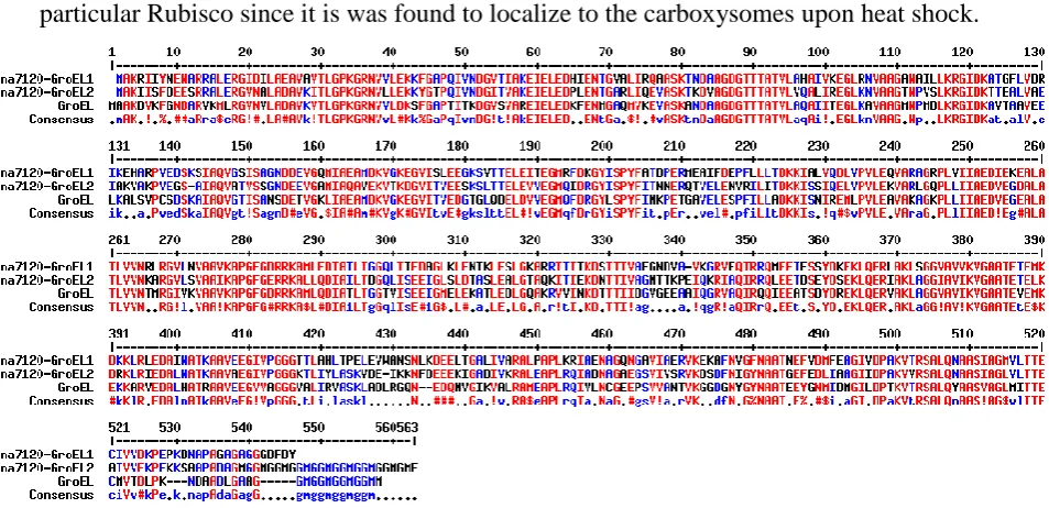
cyan-groEL2 is monocistronic with differential genomic localization (Lehel et al. 1993). Cyan-GroEL1 and Cyan-GroEL2 share up to approximately 65% sequence identity with each other and E. coli GroEL. Examination of the amino acid sequence reveals intriguing differences, especially upon closer inspection of the C-terminal tails (Figure 2.7). E. coli
The cyanobacterial GroEL homologues are both induced upon heat shock; however GroEL1 displays higher fold induction levels than GroEL2. The ability of Cyan-GroEL homologues to functionally replace E. coli GroEL has shown that Cyan-GroEL1 but not Cyan-GroEL2 can overcome the deficiencies in E. coli GroEL defective cells (Furuki et al. 1996). Furthermore, light also plays an important role in the efficiency of heat shock induction; indicating an important role of light for activation of the cyanobacterial heat shock response (Asadulghani et al. 2003). Importantly, Anabaena sp. PCC7120 photosynthetically active cells upon heat shock localized Cyan-GroEL to the thylakoid membrane and carboxysomes (Jager and Bergman 1990; Kovacs et al. 1994). This indicates that GroEL from cyanobacteria is most likely important for the folding of a variety of different proteins and in particular Rubisco since it is was found to localize to the carboxysomes upon heat shock.
Figure 2.7 Sequence alignment of Cyan-GroEL1, Cyan-GroEL2, and E. coli GroEL.
Amino acid alignment of GroEL1 and GroEL2 from Anabaena sp. PCC7120 (Ana7120-GroEL1 and Ana7120-GroEL2) and
GroEL from E. coli (GroEL). Completely conserved residues are shown in red and residues found in two of the sequences are
shown in blue. Black indicates residues which are not conserved. The GGM repeat motif at the ultimate C-terminus is found in GroEL as well as Ana7120-GroEL2, but not in Ana7120-GroEL1. Alignment created using MultAlin.
translated. Accumulation of heat shock proteins enables the refolding of HrcA, which can once again bind to the CIRCE sequence (Liu et al. 2005). Although the CIRCE element is present upstream of many cyan-groEL genes, it may not be the only regulator of the heat shock response or of the transcription of groEL genes in cyanobacteria. Mutational studies recently demonstrated that additional heat-regulatory elements, H-box and K-box, are present in the Anabaena L-31 genome (Rajaram and Apte 2010). The H-box element has been shown to be important for negative heat regulation. The K-box has been identified in groESL
promoter regions in all sequenced Anabaena strains and is involved in positive light-regulation of the heat shock response in these cyanobacterial species. It is very likely that there are other transcriptional regulators of chaperonin expression in cyanobacteria due to the differential induction of the cyan-groEL genes upon heat shock.
Detailed studies of Cyan-GroEL are primarily limited to studies in situ due to the inherent instability of this protein upon purification (Lehel et al. 1992). Cyan-GroEL has a high tendency to dissociate into monomeric or dimeric forms. Recombinant expression of Cyan-GroEL from E. coli has demonstrated the difficulty of purification of cyanobacterial GroEL and caution must be practiced upon interpretation of the results (Huq et al. 2010). Special buffer conditions must be implemented along with high protein concentrations to ensure tetradecameric stability, which can be different for different species. Importantly, once the Cyan-GroEL tetradecamer dissociates, the re-oligomerization of the monomers has been so far not possible (Lehel et al. 1992).
2.2.1.3.3
Chloroplast chaperonin
The occurrence of multiple group I chaperonin genes in an organism is not limited to bacteria, as chloroplast chaperonin possesses two homologous nuclear encoded α and β components (Hemmingsen and Ellis 1986). Moreover, it has been established that the chloroplast chaperonin (ch-Cpn60) is a heterooligomeric complex consisting of seven α and seven β subunits as determined by SDS-PAGE quantification (Hemmingsen et al. 1988; Nishio et al. 1999). This is confounded by the existence of multiple homologues of each α and β subunit as well as several cochaperone homologues within some species e.g., Arabidopsis thaliana. The α and β subunits within a species are homologous but divergent, displaying
approximately 50% sequence identity to each other and E. coli GroEL (Martel et al. 1990). Ch-Cpn60 was first discovered in the early 1980’s during isolation of the enzyme ribulose-1,5-bisphosphate carboxylase/oxygenase (Rubisco) and was termed the Rubisco subunit binding protein (Barraclough and Ellis 1980; Lorimer 2001). However, ch-Cpn60 is not only implicated in Rubisco folding and assembly, but it has also been indicated in association with a variety of proteins in the chloroplast, including imported proteins as well as chloroplast encoded proteins (Lubben et al. 1989). Comparison of the GroEL protein sequence with ch-Cpn60 protein sequence showed that these two proteins were evolutionary homologues with about 46% sequence identity, leading to the assumption that they would also exhibit similar functional behavior in the cell (Hemmingsen et al. 1988).
Chaperonins located in the chloroplast are nuclear encoded and display high sequence homology to Cyan-GroEL2. From phylogenetic studies, it is predicted that during green algae and higher plant evolution, cyanobacteria were taken up by eukaryotic cells through endosymbiosis (Gould et al. 2008). The chloroplast could then be formed through morphogenesis of the cyanobacteria in these early endosymbiotic organisms. Additionally, gene flow from the symbiont to the host resulted in the transfer of the groEL2 and groES
genes to the host nuclear genome correlating with development of protein transfer mechanisms in the host. Gene duplication events of the groEL2 gene most likely produced the homologous α and β subunits now present in photosynthetic eukaryotic organisms (Wastl et al. 1999).
contains two different subunits whose arrangement in the assembled oligomer is still unknown. It has been demonstrated, by recombinant expression, that the ch-Cpn60α subunits are not capable of assembly to the tetradecameric complexes and accordingly maintain a monomeric form (Dickson et al. 2000). Ch-Cpn60β subunits have the propensity to form homo-tetradecameric complexes, however these homooligomers function differently in regard to cochaperone binding compared to ch-Cpn60αβ heterooligomers aiding in the assumption that the heterooligomer is the biologically relevant configuration (Nishio et al. 1999). Additionally, in contrast to GroEL, ch-Cpn60 is less stable and less readily purified especially in the presence of ATP (Hemmingsen and Ellis 1986; Lissin 1995; Musgrove et al. 1987), impeding mechanistic studies.
Figure 2.8 EM structure of ch-Cpn60/ch-Cpn21 complex and ch-Cpn21.
(A) Electron micrograph of pea ch-Cpn60 from chloroplasts incubated with recombinant spinach ch-Cph21 in the presence of ADP. Bullet-shaped (ch-Cpn60 with ch-Cpn21) complexes are indicated with arrows. Scale bar measures 100 nm. (B) Enlargement of bullet-shaped complexes (ch-Cpn60 with ch-Cpn21) top and middle and ch-Cpn60 alone bottom. A and B adapted from: (Viitanen et al. 1995) (C) Electron micrograph of recombinant spinach ch-Cpn21 alone, scale bar is 20 nm. Adapted from: (Baneyx et al. 1995)
There are also important differences in the cochaperones of GroEL and ch-Cpn60; not only is there at least one 10 kDa cochaperone (ch-Cpn10) present in some species, but there are also up to two 20 kDa ch-Cpn60 cochaperones (ch-Cpn21) which have been identified (Bertsch et al. 1992; Koumoto et al. 2001). The ch-Cpn21 cochaperones seemingly evolved from an endoduplication event resulting in the doubling in size from 10 kDa to 20 kDa (Bertsch et al. 1992). Ch-Cpn10, as revealed from in vitro studies, presumably forms a heptamer in solution (Sharkia et al. 2003). The ch-Cpn21 consists of two tandem GroES-like domains, separated by a short linker. Furthermore, the biologically relevant oligomeric configuration of the chloroplast cochaperones is still unknown, but EM pictures of ch-Cpn21 shows a ring-like complex similar to GroES when bound to ch-Cpn60 (Figure 2.8) (Baneyx et al. 1995; Viitanen et al. 1995). In vitro studies have indicated that ch-Cpn21 most likely forms
tetramers (Koumoto et al. 1999), which questions the manner of association of a tetrameric complex to a heptameric ring of ch-Cpn60; various theoretical models of ch-Cpn21 assembly are shown in Figure 2.9 (Weiss et al. 2009). Interestingly, as analyzed by mutational experiments, both domains of the ch-Cpn21 are necessary for its function, however, the N-terminal GroES-like domain appears to have a more prominent role in activity than the C-terminal GroES-like domain (Bertsch and Soll 1995; Bonshtien et al. 2007). Presumably, ch-Cpn21 could have evolved in plants as an adaptation to the various paralogs of chloroplast chaperonin (Boston et al. 1996).
Figure 2.9 Model of co chaperone ch-Cpn21 complex assembly.
Models based on the subunit structure of
Mycobacterium tuberculosis Cpn10 (PDB 1HX5_2). (A) Tetrameric model of ch-Cpn21, N-terminal and C-terminal would bind to the chaperonin. (B) Heptameric model, one domain would bind directly to the chaperonin while the adjacent domain would stack on top of the bound domain. (C) Heptameric model, one domain would bind directly to chaperonin while the adjacent domain extents outwards possibly for interactions with other proteins. Adapted from: (Weiss et al. 2009)
The complexity of the ch-Cpn60 system is best demonstrated in A. thaliana (Hill and Hemmingsen 2001). The A. thaliana genome encodes two ch-Cpn60α and four ch-Cpn60β isotypes. Ch-Cpn60α1 and ch-Cpn60α2 share approximately 60% sequence identity, while the three of the four Cpn60β subunits share up to 90% sequence identity. The fourth ch-Cpn60β retains 60% sequence identity to the other three. In addition, there are also several homologues of cochaperone: one 21 kDa Cpn21) cochaperone and two 10 kDa (ch-Cpn10) cochaperones. These cochaperones have also been identified in pea (Schlicher and Soll 1996) and spinach (Bertsch and Soll 1995). Additionally, two homologues of ch-Cpn21 have been identified in Chlamydomonas reinhardtii, ch-Cpn21 and ch-Cpn23 (Weiss et al. 2009). The cochaperone homologues exhibit 50% sequence identity to each other; furthermore ch-Cpn21 displays 50% identity within its own GroES-like domains. The identification of several genes for ch-Cpn60 and cochaperone raises many questions that remain to be elucidated. For instance, it is unknown whether there are functional/mechanistic differences between these chaperonin complexes, or if the homologues are advantageous for substrate specificity or differential substrate affinity. Recent studies focused on substrate
A
B
specificity of the ch-Cpn60α and β paralogs in A. thaliana have demonstrated that the low abundant ch-Cpn60β4 is crucially necessary for the folding of the NdhH protein, which is a protein associated with the NADH dehydrogenase-like (NDH) complex in chloroplasts (Peng et al. 2011). This result suggests that the different ch-Cpn60 subunits display different specificities for certain substrates, which could explain the occurrence of more than one homolog of chaperonin subunits in the chloroplast.
A proteomics study analyzing A. thaliana stroma was recently conducted, which included Native-PAGE electrophoresis coupled with mass spectrometry (Peltier et al. 2006). In this analysis the α and β paralogs ch-Cpn60α2, ch-Cpn60β1, ch-Cpn60β2, and ch-Cpn60β3 were identified in the isolated tetradecameric complex; the presence of Cpn60α1 and ch-Cpn60β4 in the complex could not be detected. Interestingly, cochaperones were also not detected in the complex; however ch-Cpn21 and ch-Cpn10 were instead isolated alone in complexes of 150-170 kDa. The cochaperone ch-Cpn21 was found to be the most highly expressed chaperonin component in the chloroplast (Peltier et al. 2006). The dissociation of cochaperone from ch-Cpn60 could possibly be explained by the absence of additional nucleotide during the isolation process. This does not, however, account for the exceedingly high protein levels of cochaperone in the chloroplast in relation to chaperonin levels, which could signify possible additional, divergent roles of this protein (Weiss et al. 2009). Indeed, ch-Cpn21 has been indicated in seed development for Vitis vinifera, Nicotiana benthamiana, and Lycopersicon esculentum (Hanania et al. 2007). It is highly probable that the chloroplast chaperonin system mechanistically functions similar to GroEL/ES, and is necessary for the proper folding of a variety of chloroplast proteins, most importantly Rubisco.
2.3
Photosynthesis
Life on earth is dependent on photosynthesis. Photosynthesis is the transfer of solar energy into physiologically usable chemical energy converting an electron donor, usually water, and atmospheric CO2 into carbohydrates (Horton et al. 2002). Organisms capable of
photosynthesis, phototrophs, include prokaryotes: e.g., cyanobacteria and purple bacteria as well as eukaryotes: diatoms, algae, and plants. During oxygenic photosynthesis, which is found in eukaryotes and some prokaryotes, water is used as the electron donor resulting in the release of O2.
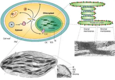
cyanobacteria into eukaryotic cells (Xiong et al. 2009). The thylakoid membrane, located on the inside of chloroplasts, is highly folded and usually arranged as flattened-stacked vesicles called grana. The machinery necessary for the capture of solar energy and conversion to chemical energy (light-dependent reactions) is embedded in the thylakoid membrane, including: photosystems I and II, electron carrier chains, chloroplast ATP-synthetase, light harvesting complexes, as well as other complexes. The aqueous phase inside the thylakoid membrane is the lumen, while the aqueous phase outside the thylakoid membrane is the stroma. The stroma contains the soluble proteins and molecules necessary for the reduction of atmospheric carbon into organic carbon (light-independent reactions).
Figure 2.10 Structure of the chloroplast.
Schematic diagram of a plant cell with zoom-in of an electron micrograph of a chloroplast from Pisum sativum. The
chloroplast contains three membrane systems: the outer envelope (OE), the inner envelope (IE), and the thylakoid membrane. The chloroplast also contains three aqueous phases: the thylakoid lumen, the stroma, and the inter envelope space (IES). Some nuclear encoded proteins must be imported to their correct cellular compartment, which is accomplished by signal transit peptides shown in yellow. In the case of nuclear encoded chloroplast proteins this occurs through two translocons: TOC and TIC, located in the OE and IE, respectively. The right panel illustrates the thylakoid organization. Granal membranes (segments of the thylakoid membrane that do not contact the stroma) are enriched with photosystem II (blue) and
the cytochrome b6f complex (purple) while stromal membranes (segments of the thylakoid membrane that do contact the
stroma) are enriched with photosystem I (red) and ATP synthetase (green). N: nucleus, M: mitochondria, P: peroxisomes, PM: plasma membrane. Adapted from: (Soll and Schleiff 2004)
Prokaryotic phototrophs, for instance cyanobacteria, also carry out oxygenic photosynthesis, thus releasing O2 as the byproduct. Cyanobacteria, the predecessors of
where pigment molecules such as phycocyanin are attached. Conversely, anoxygenic photosynthetic species e.g., purple sulfur bacteria, usually only contain one photosystem and use hydrogen sulfide and not water as an electron donor resulting in the release of sulfur from the organism. Photosystems occurring alone do not have the oxidative strength to remove electrons from water resulting in the necessity of these organisms to use alternative electron donors for photosynthesis (Leslie 2009; Tavano and Donohue 2006). Furthermore, anoxygenic photosynthetic species contain different pigment molecules which are termed bacteriochlorophyll.
Figure 2.11 Cyanobacteria cell structure.
Immunofluorescence microscopy of
Synechococcus sp. PCC7942 cells. Thylakoid membrane is shown in red and location of Rubisco in the carboxysomes is shown in green.
Adapted from: (Savage et al. 2010)
2.3.1
Light-dependent reactions
As mentioned above, the complexes involved in the light-dependent reactions are located in the thylakoid membrane. During the light-dependent reactions solar energy is converted to ATP and NADPH, which are necessary as energy sources and reductive equivalents during the light-independent reactions. Photons of light are captured by chlorophyll or other accessory pigment molecules. Chlorophyll absorbs light through a network of conjugated double bonds; upon light absorption a ground-state electron is energized to a higher molecular state. Some pigments are located within two specific complexes in conjunction with other proteins and molecules, which are collectively called photosystems.
(Umena et al. 2011)). The Z-scheme schematically illustrates the movement of electrons and reduction potentials of the electron carrier components, which create the high energy molecules ATP and NADPH (Figure 2.12 A). Light energy, which is channeled from antennae pigments and light harvesting complexes and further passed to the reaction center, is used for the electron flow from the photosystems through protein transfer chains. In this process NADP is reduced to NADPH. The transfer of electrons is not only necessary for NADP reduction, but also results in the translocation of protons through the thylakoid membrane into the lumen (Figure 2.12 B). The proton gradient created between the lumen and stroma is then utilized by the chloroplast ATP-synthetase for the photophosphorylation of ADP to ATP (Graciet et al. 2004).
Figure 2.12 Light-dependent reactions in photosynthesis.
(A) Z-scheme of electron transport
from H2O to NADP in
photosynthesis. Antenna complexes (colored spheres) absorb photons of light and the absorbed energy is transferred to PSII (P680) and PSI (P700). The manganese center of
P680 removes electrons from H2O
which are transferred to the P680 reaction center. The excited P680 transfers the electrons to pheophytin
(Pheo) then plastoquinone (QA and
QB), which are then passed over a
chain of intermediate electron
carriers (cytochrome b6f complex
(Cyt) and plastocyanin (PC)), and finally to the reaction center of P700. The electron flow from PSII to PSI
results in H+ being transferred to the
lumen resulting in a proton-motive force which is used by ATP-synthetase for ATP formation. P700 is excited by light and electrons are transferred to ferredoxin (FD) where NADP is reduced to NADPH. Adapted from: (Orr and Govindjee 2010) (B) Diagram of electron and
H+ transfer in the chloroplast.
Plastoquinones (PQ), blue arrows: electron transfer that results in reduction, orange arrows: electron transfer that results in oxidation.
Electron transfer from H2O to PSII
and PSI results in the movement of H+ into the lumen as well as the reduction of NADP resulting in the formation of ATP and NADPH, respectively. ATP and NADPH are utilized by the light-independent
reaction of photosynthesis for
carbohydrate synthesis. Adapted
from: (Peltier et al. 2010)
2.3.2
Light-independent reactions
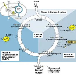
The NADPH and ATP derived from the light-dependent reactions in the thylakoid membrane are necessary for carbon assimilation in the light-independent reactions through the Calvin-Benson-Bassham (CBB) cycle (Horton et al. 2002). The CBB cycle occurs in the stroma of the chloroplast of eukaryotes or cytosol of bacteria and is divided into three phases (Figure 2.13): carbon fixation, reduction (carbohydrate synthesis), and regeneration of the CO2 acceptor (ribulose-1,5-bisphosphate (RuBP)). The carbon fixation reaction is catalyzed
by the enzyme ribulose-1,5-bisphosphate carboxylase/oxygenase (Rubisco). The five carbon sugar substrate of Rubisco, RuBP, is carboxylated, and cleavage of which results in the formation of two molecules of 3-phosphoglycerate (3-PGA). Subsequently, in the reduction phase, 3-PGA is ATP-dependently phosphorylated to 1,3-bisphosphoglycerate followed by NADPH-dependent reduction to form glyceraldehyde-3-phosphate (G3P). G3P is either used for the synthesis of carbohydrates or is kept in the CBB cycle and used for the regeneration of the carbon acceptor, RuBP. The regeneration phase also relies on ATP as an energy source and involves the formation of three- to seven-carbon sugars through transaldolase and transketolase reactions. Overall during the carboxylation of six five carbon sugar substrates, the CBB cycle consumes 18 ATP molecules and oxidizes 12 NADPH molecules for the formation of two G3P molecules and regeneration of 6 molecules of RuBP.
Figure 2.13 Calvin-Benson-Bassham cycle.
Phase 1 carbon fixation: CO2 is fixed by the
action of Rubisco into the five carbon substrate molecule RuBP and the six carbon intermediate is rapidly cleaved into two molecules of 3-phosphoglycerate (3-PGA). Phase 2 reduction: 3-PGA is phosphorylated, mediated by ATP hydrolysis, and further reduced by oxidation of NADPH resulting in
the production of
2.3.3
Carbon concentrating mechanisms
During the initial phase of the light-independent reactions, Rubisco not only carboxylates RuBP, but Rubisco also oxygenates RuBP when O2 is in the active site initiating
a side reaction to photosynthesis called photorespiration (Bauwe 2011; Foyer et al. 2009; Somerville and Ogren 1982). The affinity of Rubisco for CO2 (Km: 9 µM) is greater than for
O2 (Km: 350 µM), however the atmospheric concentration of O2 is 500 times higher than CO2
resulting in a significant amount of O2 also diffusing to the Rubisco active site leading to
oxygenation of RuBP with approximately every fourth Rubisco catalyzed reaction cycle (Peterhansel and Maurino 2011). Oxygenation of RuBP results in the formation of a five carbon intermediate, which is rapidly cleaved to one molecule of 3-PGA (three carbon molecule) and one molecule of 2-phosphoglycolate (PPG) (two carbon molecule). 3-PGA is utilized by the CBB cycle to regenerate the five carbon Rubisco substrate RuBP, whereas PPG must undergo modifications in the glycolate pathway involving reactions through the peroxisome and mitochondria before returning to the CBB cycle in a usable form i.e., 3-PGA (Figure 2.14). The recycling of PPG to 3PGA during photorespiration through two other cellular compartments leads to a net loss of a carbon atom and consumption of ATP.
Figure 2.14 Photorespiration cycle.
Photorespiration is initiated by the oxygenation of RuBP by Rubisco resulting in a five carbon intermediate that is cleaved to the three carbon 3-phosphoglycerate (3-PGA) and the two carbon 2-phosphoglycolate (PPG). 3-PGA is utilized in the CBB cycle to regenerate substrate RuBP while PPG transfers to the peroxisome where it is further processed and transferred to the mitochondria before the atoms are in a usable form and return to the CBB cycle. Phosphoglycolate phosphatase (PGP), glycolate oxidase (GOX), serine-glyoxylate aminotransferase (SGT), glutamate-glyoxylate aminotransferase (GGT), glycine decarboxylase (GDC), serine hydroxymethyltransferase (SHMT), peroxisomal hydroxypyruvate reductase (HPR1), glycerate kinase (GLYK), catalase (CAT), hydroxypyruvate reductase (HPR2), peroxisomal malate dehydrogenase (pMDH), glutamine synthetase (GS2), ferredoxin-dependent glutamate synthase (GOGAT). Adapted from: (Bauwe 2011)
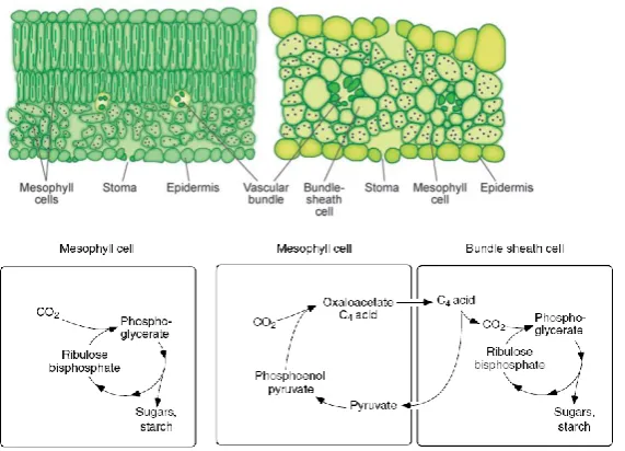
Terrestrial plants possess three photosynthetic pathways: the C3, the C4, and the Crassulacean acid metabolism (CAM) pathway. The C3 pathway (e.g., in rice, wheat, and barely) is the most common, and as described above atmospheric CO2 is fixed in the CBB
cycle by Rubisco, which is localized in the chloroplast of mesophyll cells (West-Eberhard et al. 2011). In contrast, the C4 pathway (e.g., maize, sugarcane, and sorghum) and the CAM pathway (e.g., pineapple, agaves, and orchids) display mechanisms of carbon concentrating within the plant either by physical or temporal separation, respectively. Carbon concentrating mechanisms are not only a feature of some plants, they are also observed in other species such as algae (pyrenoid) and cyanobacteria (carboxysomes).
2.3.3.1
C4 Plants
C4 photosynthesis arose from environmental conditions facilitating increased tendencies for Rubisco to behave as an oxygenase i.e., higher temperatures and reduced CO2