Original Research Article.
Comparison of Hemodynamic Stability of 2% Lignocaine and
0.2% Ropivacaine in Chest Tube Removal in
Post Cardiac Surgery Patients
Nancy Chaudhary
1*, Gaurav Goyal
2, Dharam Das Jethava
3, Durga Jethava
31*PG Resident, 2Associate Professor, 3Professor,
Department of Anaesthesia, Mahatma Gandhi Medical College, Jaipur, Rajasthan, India.
ABSTRACT
Background: Recovery from cardiac surgery depends on the
preoperative clinical state of patient, type of surgery, and postoperative analgesia. A chest tube insertion is done to preserve hemodynamic stability and cardiopulmonary function after cardiac surgery by ensuring fluid and air drainage from the pleural, pericardial or mediastinal cavities. The aim of the present study is to compare hemodynamic stability of 2% lignocaine and 0.2% ropivacaine in Chest Tube Removal in Post Cardiac Surgery Patients.
Materials and Methods: This prospective randomized double
blinded comparative study was conducted involving 60 patients (30 in each group) posted for coronary artery bypass grafting at Mahatma Gandhi Hospital, Jaipur. The cardiac monitor was attached with proper recording of hemodynamic parameters such as invasive blood pressure, ECG, arterial oxygen saturation. All the data was arranged in a tabulated form and analysed using SPSS software. Chi square test and Student’’s
t test were used for the analysis of data. P value of less than 0.05 was considered significant.
Results: There were 14 patients in group C and 15 patients in group S that had ASA grade 2. There were 16 patients in group
C and 15 patients in group S had ASA grade 3. The mean heart rate value at 0 minutes in Group C and Group S was 98.87 and 90.20 respectively. There was a significant difference between the two as the p value was less than 0.05.
Conclusion: Both drugs were found to be safe to use as we
noted no adverse reactions in our patients. The dose of fentanyl used in our study did not cause respiratory depression and sedation.
Keywords: Chest Tube, Lignocaine, Mediatinal, Pain.
*Correspondence to:
Dr. Nancy Chaudhary,
PG Resident, Department of Anaesthesia,
Mahatma Gandhi Medical College, Jaipur, Rajasthan, India.
Article History:
Received: 18-11-2017, Revised: 02-12-2017, Accepted: 27-12-2017
Access this article online
Website:
www.ijmrp.com
Quick Response code
DOI:
10.21276/ijmrp.2018.4.1.018
INTRODUCTION
Postoperative pain in cardiac surgery patients is a multidimensional phenomenon. Skin incision, tissue retraction and surgical dissection are source of nociceptive stimuli common to all surgical procedures.1 However, the presence of additional chest tubes, inserted to cause lung expansion and drain surgical fluids, add to post-operative discomfort in patients undergoing cardiac surgery. Since the insertion of chest drain is associated with increased discomfort and pain for the patient due to mechanical irritation of the heart and pericardium2, early extraction of chest tubes after coronary artery bypass graft surgery in case of no significant drainage, is recommended as an effective measure to progress towards healing.3 Multimodal analgesia has been shown to be more effective than any single method of pain reduction.3 Regional anaesthesia in combination with systemic analgesics
has been shown to have favourable patient outcomes and decreased duration of hospital stay. The effectiveness of combined use of infiltration of lignocaine 2 % along with
intravenous fentanyl has been found to be safe leading to profound reduction in pain.4 Ropivacaine, which is a long acting amide local anaesthetic agent5, has reduced potential for central nervous system and cardiovascular toxicity. It causes very less myocardial suppression6, which is beneficial for use in post cardiac surgery patients, who are more prone to develop arrhythmias. Therefore, local infiltration with ropivacaine during chest tube removal, with prior opioid administration appears to be an effective analgesic technique. The aim of the present study is to compare hemodynamic stability of 2% lignocaine and 0.2% ropivacaine in Chest Tube Removal in Post Cardiac Surgery Patients.
MATERIALS AND METHODS
50.5 49
0 10 20 30 40 50 60
Group C Group S
M
ea
n
board and all the subjects were informed about the study and a written consent was obtained from all. Patients included in the study were those who underwent left internal mammary artery (LIMA) and two vein off pump cardiac bypass surgery through median sternotomy technique and had two mediastinal chest tube drains inserted intraoperatively by cardiac surgeons. Patients with combined surgery or Non standard surgery having other than midline sternotomy were excluded from the study. Any patient who had received analgesic drugs within 4 hours of study which may confound the result analysis were excluded from the study. Each
patient’s nurse was asked to prepare the patient for chest tube
removal post operatively after a period of 24 hours. No analgesic was administered before duration of four hours of performing the procedure. All patients were kept in post cardiac surgery recovery unit. The cardiac monitor was attached with proper recording of hemodynamic parameters such as invasive blood pressure, ECG,
arterial oxygen saturation. In addition to intravenous fentanyl, patients received either lignocaine 2% infiltration (group C) or ropivacaine 0.2% (group S), 6 ml around each of the chest drains inserted. (1 pleural and 2 mediastinal) A total of 18 ml infiltration was done with a 22G needle and a 20ml syringe surrounding all the three chest tubes. Severity of pain was recorded via visual analogue scale obtained from patient. These recordings were made at various intervals: t(-5m): during administration of fentanyl and infiltration with local anaesthetic, t(0m) at 0minutes – during chest tube removal, followed by recordings at 2 minutes t(2m), 5 minutes t(5m), 10 minutes t(10m), 20 minutes t(20m), 30 minutes t(30m), 1hour t(1h), 4 hours t(4h), 24 hours t(24h) after chest tube removal. All the data was arranged in a tabulated form and analysed using SPSS software. Chi square test and Student t test were used for the analysis of data. P value of less than 0.05 was considered significant.
Table 1: Distribution of subjects according to ASA grade.
Group C Group S P value
Grade 2 14 15 0.8
Grade 3 16 15
Total 30 30
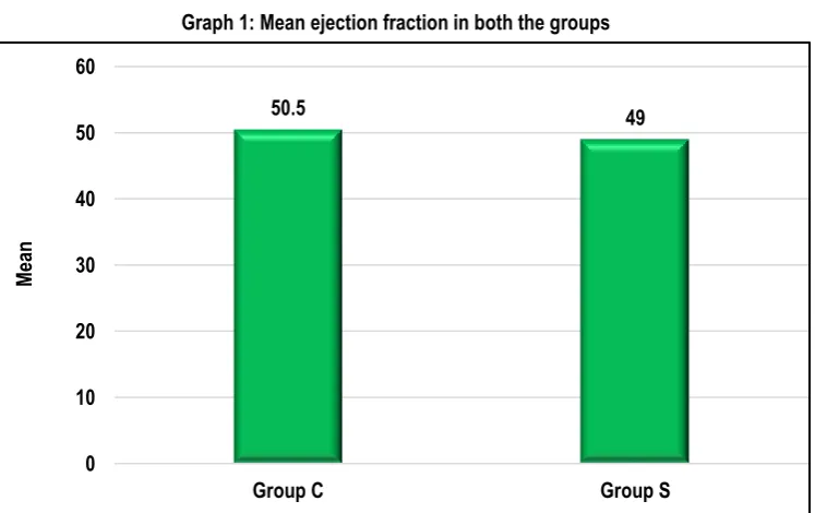
Graph 1: Mean ejection fraction in both the groups
RESULTS
Table 1 shows the distribution of subjects according to ASA grade. There were 14 patients in group C and 15 patients in group S that had ASA grade 2. There were 16 patients in group C and 15 patients in group S had ASA grade 3. There was no significant difference between the two groups as the p value was more than 0.05.
Graph 1 shows the mean ejection fraction in both the groups. Mean Ejection Fraction % in group C was 50.50±6.34 and 49±8.14 in group S. On applying chi square test the p value was more than 0.05 and hence no significant difference between the two groups.
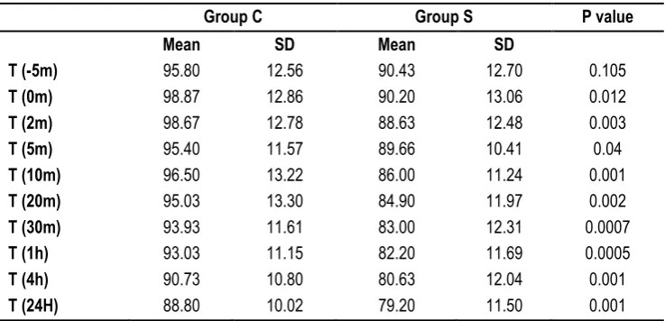
Table 2 shows the Heart rate at different time intervals. The mean value at 0 minutes in Group C and Group S was 98.87 and 90.20
respectively. There was a significant difference between the two as the p value was less than 0.05. The mean value at 2 minutes in Group C and Group S was 98.67 and 88.63 respectively. There was a significant difference between the two as the p value was less than 0.05.
0 20 40 60 80 100 120 140
T (-5m) T (0m) T (2m) T (5m) T (10m) T (20m) T (30m) T (1h) T (4h) T (24H)
M
ea
n
Group C Group S
0 10 20 30 40 50 60 70 80
T (-5m) T (0m) T (2m) T (5m) T (10m)T (20m)T (30m) T (1h) T (4h) T (24H)
M
ea
n
Group C Group S
Table 2: Heart rate at different time intervals
Group C Group S P value
Mean SD Mean SD
T (-5m) 95.80 12.56 90.43 12.70 0.105
T (0m) 98.87 12.86 90.20 13.06 0.012
T (2m) 98.67 12.78 88.63 12.48 0.003
T (5m) 95.40 11.57 89.66 10.41 0.04
T (10m) 96.50 13.22 86.00 11.24 0.001
T (20m) 95.03 13.30 84.90 11.97 0.002
T (30m) 93.93 11.61 83.00 12.31 0.0007
T (1h) 93.03 11.15 82.20 11.69 0.0005
T (4h) 90.73 10.80 80.63 12.04 0.001
T (24H) 88.80 10.02 79.20 11.50 0.001
Graph 2: Systolic blood pressure at different time intervals
0 10 20 30 40 50 60 70 80 90 100
T (-5m) T (0m) T (2m) T (5m) T (10m) T (20m) T (30m) T (1h) T (4h) T (24H)
M
e
an
Group C Group S
Graph 4: Mean arterial pressure at different time intervals
Graph 2 shows the systolic blood pressure at different time intervals. The mean value at 0 minutes in Group C and Group S was 129.37 and 123.80 respectively. There was no significant difference between the two as the p value was more than 0.05. The mean value at 2 minutes in Group C and Group S was 128.47 and 122.23 respectively. There was no significant difference between the two as the p value was more than 0.05. The mean value at 10 minutes and 20 minutes in Group C was 123.87 and 122.53 respectively. The mean value at 10 minutes and 20 minutes in Group S was 119.30 and 117.60 respectively. There was no significant difference between the two. The mean value at 1 hour in Group C and Group S was 119.67 and 115.63 respectively. At 24 hours the heart was 114.07 in Group C and 111.90 in Group S. There was no significant difference between the two groups.
Graph 3 shows the diastolic blood pressure at different time intervals. The mean value at 0 minutes in Group C and Group S was 74.27 and 70.73 respectively. There was no significant difference between the two as the p value was more than 0.05. The mean value at 2 minutes in Group C and Group S was 72.23 and 70.40 respectively. There was no significant difference between the two as the p value was more than 0.05. The mean value at 10 minutes and 20 minutes in Group C was 70.70 and 70.17 respectively. The mean value at 10 minutes and 20 minutes in Group S was 68.77 and 68.63 respectively. There was no significant difference between the two. The mean value at 1 hour in Group C and Group S was 69.57 and 66.93 respectively. At 24 hours the heart was 67.93 in Group C and 65.80 in Group S. There was no significant difference between the two groups. Graph 4 shows the mean arterial pressure at different time intervals. The mean value at 0 minutes in Group C and Group S was 92.63 and 88.42 respectively. There was no significant difference between the two as the p value was more than 0.05. The mean value at 2 minutes in Group C and Group S was 90.98 and 87.68 respectively. There was no significant difference between the two as the p value was more than 0.05. The mean value at 10 minutes and 20 minutes in Group C was 88.42 and
87.62 respectively. The mean value at 10 minutes and 20 minutes in Group S was 85.61 and 84.96 respectively. There was no significant difference between the two. The mean value at 1 hour in Group C and Group S was 86.27 and 83.17 respectively. At 24 hours the heart was 83.31 in Group C and 81.17 in Group S. There was no significant difference between the two groups.
DISCUSSION
Puntillo in 2004, observed in his study that pleural chest tube removal is more painful than mediastinal chest tube.7 Nonsteroidal anti-inflammatory drug such as ketorolac is not indicated post cardiac surgery patients, who are on anti-platelet agents because of risk of development of coagulopathy, gastric ulcers and nephrotoxicity. Carson in 1994, compared four analgesic means used in patients for chest tube removal in a prospective, randomized, controlled multiple-group comparison, in 80 adult patients who underwent heart surgery and who had two mediastinal chest tubes.7 Before chest drain removal, subjects were medicated with either of the four groups: intravenous morphine or intravenous morphine and subfascial lidocaine hydrochloride, or intravenous morphine and subfascial normal saline or subfascial lidocaine. There was no significant difference between scores (p = 0.8948) on analysis of variance. The percentage of comments rated as ‘not bad’ for groups 1, 2, 3, 4
depression of cardiac contractility. It is used for local anaesthesia including infiltration, nerve block, epidural and intrathecal anaesthesia. In a double-blind placebo-controlled study conducted by Moffitt in 2001 on 18 healthy volunteers, ropivacaine was used for local infiltration in skin surgery.9 Four concentrations of ropivacaine (1, 2, 5, and 7.5 mg/ml) were injected intradermally. Pinprick sensation was used to assess the onset and duration of anaesthesia. Pain during infiltration of ropivacaine was compared with lidocaine 2% + epinephrine 1:80,000. It concluded that ropivacaine has a rapid onset and long duration of action. Mean time elapsed to regain full sensation was recorded as 773 minutes for 7.5 mg/ml and 692 minutes for 5 mg/ml. Ropivacaine was less painful to inject, even at its maximum strength, than lidocaine with epinephrine.10 Lesser variation in heart rate was seen in infiltration with ropivacaine than lignocaine, during chest tube removal and the intervals following it. But both the groups had similar fluctuations in systolic, diastolic and mean blood pressure. Our study was conducted on patients who underwent coronary artery bypass grafting in our institute, which limited our sample size to a smaller number. We also had elaborate exclusion criteria, which was important in order to avoid complications and to be able to assess the sedation scores and analgesia appropriately. The limitations of our study were testing similar local infiltrations in multiple settings like general wards and intensive care units could help us in confirming the effectiveness of both these drugs in combination with intravenous fentanyl during chest tube removal. We also did not compare the use of non-pharmacological interventions for pain relief during chest tube removal, which could have proven to be cost effective.
CONCLUSION
Both drugs were found to be safe to use as we noted no adverse reactions in our patients. The dose of fentanyl used in our study did not cause respiratory depression and sedation. Thus, clinicians may adopt this multi modal analgesia strategy as one of the pharmacological interventions for effective analgesia during chest tube removal in post cardiac surgery patients.
REFERENCES
1. Mueller XM, Tinguely F, Tevaearai HT, Revelly JP, Chioléro R, von Segesser LK. Pain location, distribution, and intensity after cardiac surgery. 2000;118(2):391-6.
2. Sessler CN, Varney K.Patient-focused sedation and analgesia in the ICU. 2008 Feb;133(2):552-65.
3. Kelet H, Dahl JB. The value of “multimodal” or “balanced analgesia” in postoperative pain treatment. Anesth Analg.
1993;77(5):1048-56. .
4. Gaurav Goyal, Varun Chhabra, Premraj. Comparison of two analgesia techniques for pain management during chest tube removal after cardiac surgery. Journal of Evolution of Medical and Dental Sciences. 2015; Vol. 4, Issue 82, October 12; Page: 14306-14312.
5. Owen MD, Dean LS. Ropivacaine. Expert Opin Pharmacother 2000; 1(2): 325-36.
6. Reutsch YA, Boni T, Borgeat A. From cocaine to ropivacaine: the history of local anaesthetic drugs. Current Topics in Medicinal Chemistry 2001; 1: 175-182.
7. Puntillo KA, Ley SJ. Appropriately timed analgesics control pain due to chest tube removal. Am J Crit Care. 2004;13(4):292-301. 8. Carson MM, Barton DM, Morrison CC, Tribble CG. Managing pain during mediastinal chest tube removal.1994 Nov-Dec;23(6):5005.
9. Mohammed Abd Al Jawad, Ihab Ali, Hoda Shokri, Mohammed S. Shorbagy Systemic versus local analgesia for chest drain removal in post cardiac surgery patients: The taming of a beast, the Egyptian Society of Cardio-Thoracic Surgery, 2017;ISSN 1110-578X,
10. Moffitt DL, De Berker DA, Kennedy CT, Shutt LE Assessment of ropivacaine as a local anesthetic for skin infiltration in skin surgery. Dermatol Surg. 2001; May;27(5):437-40.
[
Source of Support: Nil.
Conflict of Interest: None Declared.
Copyright: © the author(s) and publisher. IJMRP is an official publication of Ibn Sina Academy of Medieval Medicine & Sciences, registered in 2001 under Indian Trusts Act, 1882. This is an open access article distributed under the terms of the Creative Commons Attribution Non-commercial License, which permits unrestricted non-commercial use, distribution, and reproduction in any medium, provided the original work is properly cited.